您的位置: 首页 > 2023年5月 第38卷 第5期 > 文字全文
2023年7月 第38卷 第7期11
目录

新发缺血性脑卒中患者的视网膜黄斑区OCT特征

OCT characteristics of retinal macular region in patients with new-onset ischemic stroke

来源期刊: 眼科学报 | 2023年5月 第38卷 第5期 414-421 发布时间:2023-05-24 收稿时间:2023/5/24 16:20:34 阅读量:11327
作者:
关键词:
神经节细胞层缺血性脑卒中光学相干断层扫描
ganglion cell layer ischemic stroke optical coherence tomography
DOI:
10.12419/j.issn.1000-4432.2023.05.08
收稿时间:
 
修订日期:
 
接收日期:
 
目的:测量黄斑区视网膜神经节细胞层和内界膜-视网膜色素上皮层厚度,分析其与新发缺血性脑卒中的相关性。方法:采用前瞻性研究,选择2021年12月至2022年3月在潍坊医学院附属医院体检的健康者30例(60眼)为对照组(A组);选择同期收治且年龄匹配的新发缺血性脑卒中患者30例(60眼)为卒中组(B组)。比较两组黄斑区神经节细胞层(ganglion cell layer,GCL)和内界膜-视网膜色素上皮层(inner limiting membrane-Retinal pigment epithelium,ILM-RPE)厚度及平均GCL厚度/ILM-RPE平均容积厚度(GCL/ILM-RPE厚度比)。结果:共纳入120只眼,A、B组分别60只眼。A组的平均GCL为(83.5±4.7)μm,B组的平均GCL厚度为(78.7±10.5)μm,组间比较差异有统计学意义(P<0.001)。A组的GCL最小厚度为(80.4±4.4)μm,B组的GCL最小厚度为(70.7±16.3)μm,组间比较差异有统计学意义(P<0.001)。A组的ILM-RPE中心子区厚度为(247.5±19.8)μm,B组的ILM-RPE中心子区厚度为(241.6±36.3)μm,组间比较差异无统计学意义(P=0.393)。A组的ILM-RPE平均容积厚度为278.4±9.8μm,B组的ILM-RPE平均容积厚度为(278.5±15.3)μm,组间比较差异无统计学意义(P=0.448)。A组的GCL/ILM-RPE厚度比为0.300 3±0.012 2,B组的GCL/ILM-RPE厚度比为0.286 2±0.028 6,组间比较差异有统计学意义(P<0.001)。结论:缺血性脑卒中影响GCL厚度和GCL/ILM-RPE厚度比, GCL厚度和GCL/ILM-RPE厚度比与新发缺血性脑卒中可能有关。
Objective: To measure the thickness of retinal ganglion cell layer and inner limiting membrane-retinal pigment epithelium layer in the macular region, and analyze its correlation with new-onset ischemic stroke. Methods: By using a prospective study, 30 healthy subjects (60 eyes) who underwent physical examination in Affiliated Hospital of Weifang Medical University from December 2021 to March 2022 were selected as the control group (Group A). Thirty age-matched patients with new-onset ischemic stroke admitted at the same time (60 eyes) were selected as the stroke group (Group B). The ganglioncelllayer (GCL) and innerlimitingmembrane-Retinalpigmentepithelium (ILM-RPE) were compared between the two groups. It included the thickness and mean GCL thickness/Mean Volume thickness of ILM-RPE (GCL/ILM-RPE thickness ratio). Results: A total of 120 eyes were included, with 60 eyes in group A and B. The average GCL thickness in group A was (83.5±4.7) μm, and the average GCL thickness in group B was (78.7±10.5) μm,and the difference between groups was statistically significant (P<0.001). The minimum thickness of GCL in group A was (80.4±4.4) μm, and the minimum thickness of GCL in group B was (70.7±16.3) μm, showing statistical significance(P<0.001). The thickness of the central subregion of ILM-RPE in group A was (247.5±19.8) μm, and the thickness of the central subregion of ILM-RPE in group B was (241.6±36.3) μm, and the difference was not statistically significant between two groups (P=0.393). The mean volumetric thickness of ILM-RPE in group A was (278.4±9.8) μm, and the mean volumetric thickness of ILM-RPE in group B was (278.5±15.3) μm, and the difference was not statistically significant (P=0.448). The GCL/ILM-RPE thickness ratio of group A was (0.300 3±0.012 2), and that of group B was (0.286 2±0.028 6), showing a statistical difference between groups (P<0.001). Conclusion: Ischemic stroke affects the GCL thickness and the thickness ratio of GCL/ILM-RPE, which may be related to new-onset ischemic stroke.
脑卒中是常见脑血管疾病,目前为世界范围内主要致死性疾病之一,特点为高发病率、高死亡率和高致残率[1]。缺血性脑卒中(ischemic stroke)的发病率较高,占脑卒中总数的60%~70%,高危因素包括年龄、高血压、糖尿病、血脂异常、吸烟和体重指数等[2]。缺血性脑卒中是神经急症,易复发且致残率高,早期诊断和干预对改善患者预后具有重要价值,许多研究发现眼缺血事件对缺血性脑卒中有一定的预测价值[3-4]。作为颈内动脉的终末性分支,解剖学将眼底动脉划分到小动脉范畴,可反映全身小血管的病变情况,与高血压、冠心病、糖尿病、脑卒中的关系已得到证实[5]。朱玉等[6]发现眼底动脉直径变化又与脑卒中风险存在较高相关性。Steele等[7]的研究发现对大鼠进行大脑中动脉栓塞后,短期内同侧眼动脉血流量降低近90%,同侧视网膜神经节细胞(retinal ganglion cells,RGCs)密度也明显减少。低灌注所导致的缺血、缺氧会损伤RGCs[8]。因此我们假设RGCs更易受到缺血性脑卒中的影响,且神经节细胞层(ganglion cell layer,GCL)与视网膜全层,即内界膜(inner limiting membrane)至视网膜色素上皮层(retinal pigment epithelium)的厚度比更能体现出神经节细胞层的厚度变化。本研究使用光学相干断层扫描(optical coherence tomography,OCT)检测新发缺血性脑卒中患者和正常对照组之间的GCL厚度、ILM-RPE厚度和平均GCL厚度/ILM-RPE平均容积厚度(GCL/ILM-RPE厚度比),并进行比较,现报告如下。

1 对象与方法

1.1 研究对象

本研究为前瞻性研究,根据《赫尔辛基宣言》进行。获得潍坊医学院附属医院医学研究伦理委员会批准(批准号:WYFY2021KY131)。该研究已在中国临床试验注册中心注册[ChiCTR2100054128]。获得所有参与者的知情和同意。选择2021年12月至2022年3月潍坊医学院附属医院体检的健康者30例(60眼)为对照组(A组);同期收治的且年龄匹配的新发缺血性脑卒中患者30例(60眼)为卒中组(B组)。

1.2 纳入标准和排除标准

1.2.1 纳入标准
B组:①确诊为新发的缺血性脑卒中;②颅脑MRI示有新鲜脑梗死灶;③年龄45~75岁;④患者有正常的言语交流能力,能正确清晰地表达自己的观点和想法;⑤有高血压者用药后,收缩压<140 mmHg(1 mmHg=0.133 kPa),舒张压<90 mmHg;⑥受试者自愿加入本研究,签署知情同意书,依从性好。
A组:①年龄45~75岁;②无全身疾病的健康人;③受试者自愿加入本研究,签署知情同意书,依从性好,配合随访。
1.2.2 排除标准
B组:①最佳矫正视力<0.5;②眼压>21 mmHg;③有影响眼底的眼科疾病,如青光眼、高度近视、糖尿病性眼底病变等;④有眼部手术、外伤史;⑤患有危及生命的严重系统性疾病;⑥凝血功能异常;⑦患有糖尿病;⑧收缩压≥140 mmHg,舒张压≥90 mmHg;⑨患者无正常的言语交流能力。
A组:①最佳矫正视力<0.5;②眼压>21 mmHg;③有脑血管疾病;④患有眼科疾病;⑤有眼部手术、外伤史;⑥患有全身疾病;⑦凝血功能异常;⑧年龄不符。

1.3 研究方法

收集受试者临床资料,包括一般情况如性别、年龄、心率、血压、最佳矫正视力、等效球镜度数、眼压;血清低密度脂蛋白(low density lipoprotein, LDL);颈部彩色多普勒超声(彩超)结果。
所有受试者的检查均在上午进行,采用Cirrus HD-OCT 500(Software version 8.0)进行扫描,无需扩瞳,嘱受试者下颌置于颌托,固视光标,检查按标准方法进行。所有受试者均使用Macular Cube模式,信号强度≥7的扫描纳入分析。黄斑模块包含了512个水平B扫描,每个B型超声扫描由128个A型超声扫描构成,测量范围为6 mm×6 mm。GCL厚度测量指标包括平均值、最小值、上方区域、鼻上区域、鼻下区域、下方区域、颞下区域和颞上区域(图1)。视网膜厚度(实际为ILM-RPE厚度值),测量指标包括双眼黄斑区视网膜平均厚度、 黄斑中心凹视网膜厚度及黄斑部9个分区的视网膜厚度(图2)。以上均由同一经验丰富的检查人员进行3次检查,不同区域厚度取三次结果的平均值。收集卒中组颈部10血管(双侧颈总、颈内外、椎、锁骨下动脉)彩超检查结果,以颈部10血管彩超的检查结果显示纤维帽连续为板块稳定组,纤维帽不连续为板块不稳定组。
图1 一例缺血性脑卒中患者黄斑区神经节细胞分析的OCT图像
Figure1 OCT image of ganglion cell analysis in macular area of a patient with ischemic stroke
图2 一例健康人黄斑区视网膜的OCT图像
Figure2 OCT image of the macular retina in a healthy human

1.4统计学方法

    采用SPSS 9.4统计学软件进行数据分析,计量资料以 (x±s )表示。采用独立样本t检验、χ2检验比较A组与B组间的性别、年龄、心率、血压、最佳矫正视力、等效球镜度数、眼压、LDL、颈动脉狭窄等基线资料。采用独立样本t检验分析了颈部彩超所示动脉斑块的板块稳定性与GCL和ILM-RPE 厚度的统计学差异。采用独立样本t检验比较A组与B组间GCL、ILM-RPE厚度及GCL/ILM-RPE厚度比。所有分析均以P< 0.05为差异有统计学意义。

2 结果

2.1 基线资料

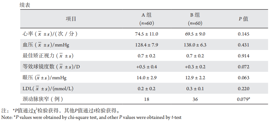
本研究共包括120眼。A组和B组各有60眼。两组之间在性别、年龄、心率、血压、最佳矫正视力、等效球镜度数、眼压、LDL等基线资料方面比较差异均无统计学意义(P均>0.05),见表1,另外,排除了颈动脉斑块的稳定性对实验结果的影响(P>0.05),见表2。

表 1 正常对照组和缺血性脑卒中组的基本资料

Table1 Basic data of normal control group and ischemic stroke group

20230629160101_9942.png

表 2 颈部彩超所示动脉斑块的板块稳定性与 GCL 和 ILM-RPE 厚度的比较

Table2 Comparison of plate stability of carotid plaque with GCL and ILM-RPE thickness as shown by cervical color ultrasound

2.2 GCL厚度

A组的平均GCL厚度为(83.5±4.7)μm,B组的平均GCL厚度为(78.7±10.5)μm,比较差异有统计学意义(P<0.001),见表3。A组的GCL最小厚度为(80.4±4.4)μm,B组的GCL最小厚度为(70.7±16.3)μm,组间比较差异有统计学意义(P<0.001),见表3。

表 3 正常对照组和缺血性脑卒中组神经节细胞层和 ILM-RPE 的比较

Table3 Comparison of ganglion cell layer and ILM-RPE between normal control group and ischemic stroke group

2.3 ILM-RPE厚度

A组的ILM-RPE中心子区厚度为(247.5±19.8)μm,B组的ILM-RPE中心子区厚度为(241.6±36.3)μm,组间比较差异无统计学意义(P=0.393),见表3。A组的ILM-RPE平均容积厚度为(278.4±9.8)μm,B组的ILM-RPE平均容积厚度为(278.5±15.3)μm,组间比较差异无统计学意义(P=0.448),见表3。

2.4 GCL/ILM-RPE厚度比

A组的GCL/ILM-RPE厚度比为0.3003±0.0122,B组的GCL/ILM-RPE厚度比为0.2862±0.0286,组间比较差异有统计学意义(P<0.001),见图3。
图3 每组神经节细胞层(GCL)内界膜-视网膜色素上皮层(ILM-RPE)厚度比(箱式图)
Figure3 Ganglion cell layer (GCL)
从下到上的数值分别代表最小值、25%分位数、中位数、75%分位数和最大值。红点代表每组的平均值。A组和B组的GCL/ILM-RPE厚度比比较差异有统计学意义(P< 0.001)。
inner boundary membrane-retinal pigment upper cortex (ILM-RPE) thickness ratio of each group (box diagram).

3 讨论

近年来中国脑卒中的发生率总体呈上升趋势,而死亡率逐渐下降,残障和失能等后遗症成为更突出的问题,为个人和社会带来了沉重的医疗负担[9]。故早期预防缺血性脑卒中的发生对患者预后具有重要的临床意义。研究发现[10-11],脑卒中患者同侧颈内动脉出现变窄情况后,眼底血管也随之出现异常改变。这些变化可以通过多种方法观察到,包括眼底照相、OCT和荧光血管造影。特别是OCT的技术发展允许通过高分辨率图像对视网膜结构进行更详细的分析。
目前的研究证实,眼底动脉硬化分级可以判断患者颅内血管病变程度,对早期发现脑血管疾病患者有重要的临床价值[6,12],这些研究采用了免散瞳眼底照相机行双眼视网膜血管照相检查。然而,本研究选用了OCT测量新发缺血性脑卒中患者的视网膜细微结构并进行分析。首先,本研究测量了ILM-RPE的厚度,以确定新发缺血性脑卒中患者眼底血管的改变是否对视网膜厚度产生影响。眼动脉是颈内动脉和颈外动脉之间的交通支,脑部供血不足时部分颈外动脉的血液通过眼动脉流入颈内动脉,在眼动脉处表现为与正常血流方向相反的逆向血流[13]。眼动脉逆流能够改善脑组织灌注,但对于眼部而言是一种“窃血”现象,可能会导致眼部血流减少和紊乱[14]。本研究结果表明,与健康对照组相比,卒中组ILM-RPE中心子区厚度无明显变化;ILM-RPE平均容积厚度无明显变化。说明新发缺血性脑卒中患者眼底血供的改变,不足以影响视网膜的厚度。Anjos[15]等发现缺血性脑卒中患者的RNFL变薄,Park[16]等的研究结果表明,缺血性脑卒中组双眼RNFL变薄.另有大鼠的大脑中动脉阻塞诱导视网膜缺血的试验显示,其组织病理学改变为视网膜神经节细胞和视神经损伤[17]。而RNFL主要由视网膜神经节细胞的轴突在视神经结合之前组成[18]。本研究结果表明,与正常对照组相比,卒中组的平均GCL厚度、GCL最小厚度降低。此外,本研究还对GCL/ILM-RPE厚度比进行了分析,因为这更能代表GCL相对于视网膜全层的厚度变化。与正常对照组相比,卒中组GCL/ILM-RPE厚度比较小。这与之前的研究结果有相通之处,由此推测新发脑卒中引起的眼底供血改变,影响了对缺血缺氧更为敏感的GCL。现已有研究证实,在脑卒中发生之前,已有颈动脉、脑动脉血流动力学改变[19-20]。多种视网膜血管性疾病在眼底血管发生异常改变之前,视网膜神经成分已有损伤,特别是RNFL[21]。视网膜神经成分对缺血缺氧的感知更灵敏。因此,GCL厚度及GCL/ILM-RPE厚度比与新发缺血性脑卒中有相关性。通过OCT寻找缺血性脑卒中患者的眼底特征的方式有希望为脑血管疾病研究提供新的“窗口”。
这项研究有几个局限性,第一,样本量较小,且本研究纳入的受试者年龄为45~75岁的中老年人,年龄跨度较大,可能存在选择偏倚。第二,本研究卒中组仅考虑纳入新发缺血性脑卒中,没有对卒中区域范围和评级和用药情况进行分析,可能影响结果。
尽管有上述局限性,本研究也有优点。首次对脑卒中患者的GCL/ILM-RPE厚度比进行分析,结果显示与正常对照组相比,卒中组的GCL/ILM-RPE厚度比存在差异,但仍需进一步大样本量的前瞻性研究来证明这一点。
综上所述,卒中患者的ILM-RPE层厚度与正常人相近,而卒中组的GCL厚度和GCL/ILM-RPE厚度比均降低,因此对于有可干预性危险因素的人群,包括高血压、糖尿病、血脂异常、心脏病、吸烟、酒精摄入、饮食、超重或肥胖、体力活动不足、心理因素等[22],可进行早期的干预和治疗,降低缺血性脑卒中发病的概率。

利益冲突

所有作者均声明不存在利益冲突。

开放获取声明

本文适用于知识共享许可协议(Creative Commons),允许第三方用户按照署名(BY)-非商业性使用(NC)-禁止演绎(ND)(CC BY-NC-ND)的方式共享,即允许第三方对本刊发表的文章进行复制、发行、展览、表演、放映、广播或通过信息网络向公众传播,但在这些过程中必须保留作者署名、仅限于非商业性目的、不得进行演绎创作。详情请访问:https://creativecommons.org/licenses/by-nc-nd/4.0/
1、Johnston SC, Mendis S, Mathers CD. Global variation in stroke burden and mortality: estimates from monitoring, surveillance, and modelling[ J]. Lancet Neurol, 2009, 8(4): 345-354.Johnston SC, Mendis S, Mathers CD. Global variation in stroke burden and mortality: estimates from monitoring, surveillance, and modelling[ J]. Lancet Neurol, 2009, 8(4): 345-354.
2、Lackland DT, Roccella EJ, Deutsch AF, et al. Factors influencing the decline in stroke mortality: a statement from the American Heart Association/American Stroke Association[ J]. Stroke, 2014, 45(1): 315-353.Lackland DT, Roccella EJ, Deutsch AF, et al. Factors influencing the decline in stroke mortality: a statement from the American Heart Association/American Stroke Association[ J]. Stroke, 2014, 45(1): 315-353.
3、Helenius J, Arsava EM, Goldstein JN, et al. Concurrent acute brain infarcts in patients with monocular visual loss[ J]. Ann Neurol, 2012, 72(2): 286-293.Helenius J, Arsava EM, Goldstein JN, et al. Concurrent acute brain infarcts in patients with monocular visual loss[ J]. Ann Neurol, 2012, 72(2): 286-293.
4、Benavente O, Eliasziw M, Streifler JY, et al. Prognosis after transient monocular blindness associated with carotid-artery stenosis[ J]. N Engl J Med, 2001, 345(15): 1084-1090.Benavente O, Eliasziw M, Streifler JY, et al. Prognosis after transient monocular blindness associated with carotid-artery stenosis[ J]. N Engl J Med, 2001, 345(15): 1084-1090.
5、Ong YT, Wong TY, Klein R, et al. Hypertensive retinopathy and risk of stroke[ J]. Hypertension, 2013, 62(4):706-711.Ong YT, Wong TY, Klein R, et al. Hypertensive retinopathy and risk of stroke[ J]. Hypertension, 2013, 62(4):706-711.
6、朱玉, 李海燕, 王瑜. 分析眼底动脉硬化分级和腔隙性脑梗死患 者脑血管改变相关因素[ J]. 中华养生保健, 2020, 38(9): 48-49.
朱玉, 李海燕, 王瑜. 分析眼底动脉硬化分级和腔隙性脑梗死患 者脑血管改变相关因素[ J]. 中华养生保健, 2020, 38(9): 48-49.
7、Steele EC Jr, Guo Q, Namura S. Filamentous middle cerebral artery occlusion causes ischemic damage to the retina in mice[ J]. Stroke, 2008, 39(7): 2099-2104.Steele EC Jr, Guo Q, Namura S. Filamentous middle cerebral artery occlusion causes ischemic damage to the retina in mice[ J]. Stroke, 2008, 39(7): 2099-2104.
8、林莉, 王玮. 视网膜神经节细胞损伤机制及神经保护药物研究 进展[ J]. 医学综述, 2009, 15(24): 3745-3748.
Lin L, Wang W. Retinal ganglion cells injury mechanisms and neuroprotective drug research[ J]. Med Recapitul, 2009, 15(24): 3745- 3748.
林莉, 王玮. 视网膜神经节细胞损伤机制及神经保护药物研究 进展[ J]. 医学综述, 2009, 15(24): 3745-3748.
Lin L, Wang W. Retinal ganglion cells injury mechanisms and neuroprotective drug research[ J]. Med Recapitul, 2009, 15(24): 3745- 3748.
9、Wang W, Jiang B, Sun H, et al. Prevalence, incidence, and mortality of stroke in China: results from a nationwide population-based survey of 480 687 adults[ J]. Circulation, 2017, 135(8): 759-771.Wang W, Jiang B, Sun H, et al. Prevalence, incidence, and mortality of stroke in China: results from a nationwide population-based survey of 480 687 adults[ J]. Circulation, 2017, 135(8): 759-771.
10、陈旭, 鲁晓华, 刘兰英, 等. 腔隙性脑梗死与非腔隙性脑梗死的危 险因素异同性分析[ J]. 西部医学, 2012, 24(2): 319-320.
Chen X, Lu XH, Liu LY, et al. Diering risk factor proles lacunar and nonlacunar ischemic strokes[ J]. Med J West China, 2012, 24(2): 319- 320.
陈旭, 鲁晓华, 刘兰英, 等. 腔隙性脑梗死与非腔隙性脑梗死的危 险因素异同性分析[ J]. 西部医学, 2012, 24(2): 319-320.
Chen X, Lu XH, Liu LY, et al. Diering risk factor proles lacunar and nonlacunar ischemic strokes[ J]. Med J West China, 2012, 24(2): 319- 320.
11、纪文洋, 蔡美卿. 颈动脉斑块检测与高血压、腔隙性脑梗死的 相关性[ J]. 中国老年学杂志, 2014, 34(13): 3754-3755.
Ji WY, Cai MQ Correlation between carotid plaque detection and hypertension and lacunar cerebral infarction[ J]. Chin J Gerontol, 2014, 34(13): 3754-3755.
纪文洋, 蔡美卿. 颈动脉斑块检测与高血压、腔隙性脑梗死的 相关性[ J]. 中国老年学杂志, 2014, 34(13): 3754-3755.
Ji WY, Cai MQ Correlation between carotid plaque detection and hypertension and lacunar cerebral infarction[ J]. Chin J Gerontol, 2014, 34(13): 3754-3755.
12、宋伟琼, 周小平, 邝国平, 等. 脑梗死患者颈动脉病变与眼底动脉 硬化的相关性研究[ J]. 国际眼科杂志, 2017, 17(11): 2151-2153.
Song WQ, Zhou XP, Kuang GP, et al. Study on the correlation between carotid artery lesion and fundus arteriosclerosis in patients with cerebral infarction[ J]. Int Eye Sci, 2017, 17(11): 2151-2153.
宋伟琼, 周小平, 邝国平, 等. 脑梗死患者颈动脉病变与眼底动脉 硬化的相关性研究[ J]. 国际眼科杂志, 2017, 17(11): 2151-2153.
Song WQ, Zhou XP, Kuang GP, et al. Study on the correlation between carotid artery lesion and fundus arteriosclerosis in patients with cerebral infarction[ J]. Int Eye Sci, 2017, 17(11): 2151-2153.
13、Hofmeijer J, Klijn CJ, Kappelle LJ, et al. Collateral circulation via the ophthalmic artery or leptomeningeal vessels is associated with impaired cerebral vasoreactivity in patients with symptomatic carotid artery occlusion[ J]. Cerebrovasc Dis, 2002, 14(1): 22-26.Hofmeijer J, Klijn CJ, Kappelle LJ, et al. Collateral circulation via the ophthalmic artery or leptomeningeal vessels is associated with impaired cerebral vasoreactivity in patients with symptomatic carotid artery occlusion[ J]. Cerebrovasc Dis, 2002, 14(1): 22-26.
14、Klijn CJ, Kappelle LJ, van Schooneveld MJ, et al. Venous stasis retinopathy in symptomatic carotid artery occlusion: prevalence, cause, and outcome[ J]. Stroke, 2002, 33(3): 695-701.Klijn CJ, Kappelle LJ, van Schooneveld MJ, et al. Venous stasis retinopathy in symptomatic carotid artery occlusion: prevalence, cause, and outcome[ J]. Stroke, 2002, 33(3): 695-701.
15、Anjos R, Vieira L, Costa L, et al. Macular ganglion cell layer and peripapillary retinal nerve fibre layer thickness in patients with unilateral posterior cerebral artery ischaemic lesion: an optical coherence tomography study[ J]. Neuroophthalmology, 2016, 40(1): 8-15.Anjos R, Vieira L, Costa L, et al. Macular ganglion cell layer and peripapillary retinal nerve fibre layer thickness in patients with unilateral posterior cerebral artery ischaemic lesion: an optical coherence tomography study[ J]. Neuroophthalmology, 2016, 40(1): 8-15.
16、Park HY, Park YG, Cho AH, et al. Transneuronal retrograde degeneration of the retinal ganglion cells in patients with cerebral infarction[ J]. Ophthalmology, 2013, 120(6): 1292-1299.Park HY, Park YG, Cho AH, et al. Transneuronal retrograde degeneration of the retinal ganglion cells in patients with cerebral infarction[ J]. Ophthalmology, 2013, 120(6): 1292-1299.
17、Lee JY, Castelli V,Bonsack B, et al. Eyeballing stroke: Blood flow alterations in the eye and visual impairments following transient middle cerebral arter y occlusion in adult rats. Cell Transpla nt.2020;29:963689720905805.Lee JY, Castelli V,Bonsack B, et al. Eyeballing stroke: Blood flow alterations in the eye and visual impairments following transient middle cerebral arter y occlusion in adult rats. Cell Transpla nt.2020;29:963689720905805.
18、Chronopoulos A, Schutz JS. Central retinal artery occlusion—a new, provisional treatment approach[ J]. Surv Ophthalmol, 2019, 64(4): 443-451.Chronopoulos A, Schutz JS. Central retinal artery occlusion—a new, provisional treatment approach[ J]. Surv Ophthalmol, 2019, 64(4): 443-451.
19、张硕, 马燕, 冯娟. 超微血流成像对进展性缺血性卒中的判断价 值[ J]. 中国脑血管病杂志, 2016, 13(8)393-397.
Zhang S, Ma Y, Feng J. Judgemental value of superb microvascular imaging for progressive ischemic stroke[ J]. Chin J Cerebrovasc Dis,2016, 13(8)393-397.
张硕, 马燕, 冯娟. 超微血流成像对进展性缺血性卒中的判断价 值[ J]. 中国脑血管病杂志, 2016, 13(8)393-397.
Zhang S, Ma Y, Feng J. Judgemental value of superb microvascular imaging for progressive ischemic stroke[ J]. Chin J Cerebrovasc Dis,2016, 13(8)393-397.
20、史小华. 颈部血管超声对脑卒中功能早期改变的评估价值[ J]. 神经损伤与功能重建, 2015, 10(4): 375-376.
Shi XH. Evaluation value of cervical vascular ultrasound in early changes of stroke function[ J]. Neural Inj Funct Reconstr, 2015, 10(4): 375-376.
史小华. 颈部血管超声对脑卒中功能早期改变的评估价值[ J]. 神经损伤与功能重建, 2015, 10(4): 375-376.
Shi XH. Evaluation value of cervical vascular ultrasound in early changes of stroke function[ J]. Neural Inj Funct Reconstr, 2015, 10(4): 375-376.
21、袁晓萌,张稚平,石艳梅. 视网膜神经纤维层的定量评估在视网 膜疾病中的应用[ J]. 眼科学报,2023,38(3):253-259.
Yuan XM, Zhang ZP, Shi YM, et al. Application of quantitative assessment of retinal nerve fiber layer in retinal diseases[ J]. Eye Science, 2023, 38(3): 253-259.
袁晓萌,张稚平,石艳梅. 视网膜神经纤维层的定量评估在视网 膜疾病中的应用[ J]. 眼科学报,2023,38(3):253-259.
Yuan XM, Zhang ZP, Shi YM, et al. Application of quantitative assessment of retinal nerve fiber layer in retinal diseases[ J]. Eye Science, 2023, 38(3): 253-259.
22、《中国脑卒中防治报告》编写组. 《中国脑卒中防治报告 2019》概要[ J]. 中国脑血管病杂志, 2020, 17(5): 136-144.
Compilation team of the “China Stroke Prevention and Treatment Report”. Brief report on stroke prevention and treatment in China, 2019[ J]. Chin J Cerebrovasc Dis, 2020, 17(5): 136-144.
《中国脑卒中防治报告》编写组. 《中国脑卒中防治报告 2019》概要[ J]. 中国脑血管病杂志, 2020, 17(5): 136-144.
Compilation team of the “China Stroke Prevention and Treatment Report”. Brief report on stroke prevention and treatment in China, 2019[ J]. Chin J Cerebrovasc Dis, 2020, 17(5): 136-144.
1、山东省自然科学基金(ZR2020MH173)。
This work was supported by Natural Science Foundation of Shandong Province (ZR2020MH173).()
上一篇
下一篇
其他期刊
  • 眼科学报

    主管:中华人民共和国教育部
    主办:中山大学
    承办:中山大学中山眼科中心
    主编:林浩添
    主管:中华人民共和国教育部
    主办:中山大学
    浏览
  • Eye Science

    主管:中华人民共和国教育部
    主办:中山大学
    承办:中山大学中山眼科中心
    主编:林浩添
    主管:中华人民共和国教育部
    主办:中山大学
    浏览
推荐阅读
出版者信息
目录