您的位置: 首页 > 2023年8月 第38卷 第8期 > 文字全文
2023年7月 第38卷 第7期11
目录

超脉冲C02激光治疗眼睑肿物的疗效与安全性评估

Efficacy and safety assessment of ultrapulse carbon dioxide laser in treating eyelid tumors

来源期刊: 眼科学报 | 2023年8月 第38卷 第8期 541-550 发布时间:2023-09-28 收稿时间:2023/9/28 16:36:57 阅读量:5756
作者:
关键词:
超脉冲CO2激光眼睑肿物眼睑缘治疗效果并发症
ultrapulse CO2 laser eyelid lesions eyelid tumors treatment efficacy complications
DOI:
10.12419/2309150002
收稿时间:
 
修订日期:
 
接收日期:
 
目的:评估超脉冲二氧化碳(CO2)激光治疗不同类型眼睑肿物的疗效和安全性。方法:纳入50例眼睑肿物患者,其中男12例、女38例。患者年龄4~84岁。肿物类型包括眼睑色素痣、睑黄瘤、分裂痣、眼睑疣等,其中25例累及眼睑灰线,10例肿物直径>10 mm。所有患者接受超脉冲CO2激光治疗,并进行术后随访。治疗效果通过术后数码照片评估,同时记录术后1个月并发症发生情况。结果:50例眼睑肿物总体治愈率为92%,有效率达到100%。4例眼睑色素痣在治疗后1个月内复发。术后并发症主要包括轻微倒睫(5例)、睫毛稀疏部分缺失(4例)和瘢痕增生及色素沉着(4例),未出现其他严重并发症。结论:对于眼睑肿物,特别是睑缘肿物及大肿物,超脉冲CO2激光是一种更为精确、微创、安全有效的治疗方法,可作为眼睑肿物治疗的优选方案。
Objective: To evaluate the efficacy and safety of ultrapulse carbon dioxide (CO2) laser in the treatment for various types of eyelid tumors. Methods: A total of 50 patients, including 12 males and 38 females,with eyelid tumors were included in the study The age range is  from 4 to 84 years, with an average age of 37.9±20.0 years. The tumors found in our study include eyelid pigmented nevus, xanthelasma, divided nevus, and molluscum. Among them, 25 cases involved the gray line of the eyelid,and 10 cases had a tumor diameter greater than 10 mm. All patients underwent ultrapulse CO2 laser treatment and postoperative follow-up. The treatment outcomes were assessed through digital photos, and complications were recorded one month after surgery. Results: The total cure rate of the 50 cases of eyelid tumors in our study was 92%, with the effective rate reaching 100%. 4 cases of eyelid pigmented nevi recurred within one month after treatment, while all other patients were cured. Postoperative complications mainly included minor trichiasis (5 cases), partial sparse to absent eyelashes (4 cases), and hypertrophic scar with hyperpigmentation (4 cases). No other serious complications were reported in our study. Conclusions: For eyelid tumors, especially eyelid margin and larger tumors, the ultrapulse CO2 laser is a more precise, minimally invasive, safe and effective treatment method. It can be used as a preferred treatment option for eyelid tumors, and should be promoted widely in clinical practice.

眼睑作为人体颜面部的重要结构,不仅在维持眼表生理和保护眼球方面起到关键作用,还在颜面部的视觉美观中占据核心地位[1]。眼睑肿物是眼睑常见疾病之一,尤其是睑缘肿物,其不仅影响美观,还会阻挡视线、摩擦角膜等,影响患者的生活质量,严重者存在癌变可能,若不及时治疗甚至可危及患者生命[2-4]。近年来,眼睑肿物的发生率呈现上升趋势,其治疗受到临床的高度关注。因此,及时准确地诊断、合理有效地治疗眼睑肿物对改善患者预后至关重要。
手术切除为眼睑肿物的传统治疗方法,然而对于累及睑缘的肿物,手术可能会导致睑缘成角畸形、切迹、眼睑内翻、眼睑外翻、瘢痕、组织缺损等不良后果[5];对于病灶直径超过10 mm的肿物,存在传统手术切除后须行皮瓣移植术而带来的一系列并发症[6]。随着医学技术的进步,非手术治疗尤其是超脉冲二氧化碳(CO2)激光,逐渐应用在眼睑肿物的治疗中[7-8]
目前已有许多研究探讨超脉冲CO2激光治疗眼睑肿物的疗效[8-11],但局限于眼睑皮肤面及睑缘小肿物的研究。对于累及灰线的睑缘肿物以及直径超过10 mm以上的大眼睑肿物,目前笔者尚未见文献报道。评估超脉冲CO2激光在治疗睑缘肿物以及大肿物的效果及并发症具有重要的意义[12]。因此,本研究深入探讨超脉冲CO2激光治疗不同类型眼睑肿物的效果,尤其是睑缘肿物及大肿物,以提供更全面的临床指导和决策支持。
为系统评估超脉冲CO2激光治疗眼睑肿物的疗效与安全性,本文回顾性分析2021年6月至2023年6月收治的50例患者。所有病例均接受超脉冲CO2激光治疗,并进行了随访疗效评估及并发症记录,现报道如下。

1 对象与方法

1.1 研究对象

本研究回顾性选择2021年6月至2023年6月在中山眼科中心眼整形与泪道科就诊并被确诊为眼睑肿物的患者作为研究对象。多因影响视野、局部不适或美容需求前来治疗。每例患者在治疗前均签署超脉冲CO2激光治疗知情同意书、照片使用协议。本研究方案遵循《赫尔辛基宣言》,经中山大学中山眼科中心伦理委员会批准(批件号:2023KYPJ286),研究对象知情同意并签署知情同意书。
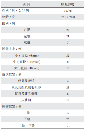
共纳入50例患者,其中男12例、女38例,年龄范围4~84岁,平均年龄(37.9±20.0)岁。肿物类型:眼睑色素痣25例,睑黄瘤6例,分裂痣3例,黄色肉芽肿2例,眼睑疣3例,血管瘤2例,眼睑脂溢性角化病2例,汗管瘤2例,眼睑恶性黑色素瘤1例,眼睑包涵囊肿3例,眼睑纤维瘤样组织增生1例。
病灶仅累及灰线(未累及睫毛根部)2例,同时累及灰线及睫毛根部23例,仅累及睫毛根部(未累及灰线)6例,皮肤面肿物19例。肿物大小直径>10 mm 10例,6~10 mm 8例,直径<6 mm 32例。病灶位置仅累及上眼睑17例,仅累及下眼睑26例,同时累及上下眼睑7例。见表1。

表 1 50 例眼睑肿物患者人口学特征与临床信息
Table 1 Demographic characteristics and clinical data of 50 patients with eyelid lesions

20230928165301_5409.png

1.2 病例纳入与排除标准

纳入标准:1)临床确诊为眼睑肿物;2)无光过敏、出血或凝血障碍病史;3)能够按时复诊和接受随访。
排除标准:1)目标区域存在感染者;2)对利多卡因过敏者;3)瘢痕体质或皮肤修复能力较差者;4)妊娠或处于哺乳期者;5)近期有严重传染病、皮肤病活动期或有心脑血管、肝、肾、内分泌和造血系统等严重疾病者;6)有恶性肿瘤或免疫疾病史者。

1.3 设备

超脉冲CO2点阵激光仪(Lumenis Ultrapulse Encore Laser)由美国科医人公司生产,激光波长:10 600 μm。

1.4 治疗方法

治疗前先对病变部位照相,然后根据病变的具体情况,设置激光参数:脉冲能量200 mJ,连续1个脉冲,功率2.0 W,持续时间10 ms,光斑直径0.2~1.0 mm可调。治疗开始时先行2.5%利多卡因乳膏表面麻醉或2%利多卡因注射液局麻浸润麻醉,对累及睑缘肿物用利多卡因滴眼液行眼表表面麻醉。予聚维酮碘溶液消毒。累及睑缘及睑结膜的病灶在治疗开始前放置涂有妥布霉素眼膏的角膜保护板,将病变组织轻轻捏起,按皮肤面-睑缘-睑结膜的顺序,每次治疗范围不超过6 mm×6 mm,激光沿着病灶根部烧灼,在烧灼过程中,使用消毒棉签蘸取生理盐水擦去汽化不尽的碳化组织,直至肉眼可见的病变组织完全清除。如创面有渗血,用棉签按压5 min。对累及睑结膜肿物,治疗结束佩戴角膜接触镜。尽可能将切除的组织进行组织病理学检查。治疗结束后治疗部位照相,并立即涂抹妥布霉素凝胶。向患者交代注意事项及用药方法(保持创面干燥卫生,涂抹妥布霉素凝胶及小牛血清去蛋白提取物眼用凝胶每天2次),预约复诊时间或下次治疗时间,观察15 min后患者离开。

1.5 观察指标

包括超脉冲CO2激光治疗眼睑肿物疗效评估和术后并发症情况评估。

1.6 疗效评估标准

疗效的评估由2名未参与本研究的整形科医师完成,主要依据患者术前和术后的数码照片来判断眼睑肿物的治疗效果。评估标准如下:
治愈指病灶消失,术后1个月无复发;有效指病灶消失,术后1个月有复发;无效指病灶无改变。有效率=(治愈例数+有效例数)/总例数×100%;治愈率=治愈例数/总例数×100%。

1.7 术后并发症评估

常见的并发症有:瘢痕增生牵拉,睑缘成角畸形或切迹,眼睑外翻,眼睑内翻,眼睑退缩,色素脱失,色素沉着,睫毛脱失或乱睫,泪小点损伤,溢泪,肿物残留复发等。肿物切除部位出现以上情况定义为该病例出现并发症。术后1个月复查时,由2名未参与本研究的整形科医师依据患者的数码照片,判断是否出现并发症。二者不一致时由更高年资医生判定。

1.8 统计学分析

采用SPSS 23.0软件进行统计分析。计数资料用频数和率表示,组间比较用X2检验。P<0.05为差异有统计学意义。

2 结果

2.1 疗效分析

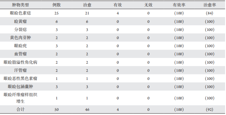
50例患者中,治愈46例、有效4例、无效0例,总有效率100%,总治愈率90%,见表2。4例眼睑色素痣患者在治疗1个月内复发,其余类型肿物未见复发。

表 2 50 例眼睑肿物超脉冲 CO2 激光治疗效果 [n(%)]
Table 2 Treatment effcacy of ultrapulse CO2 laser of 50 eyelid lesions[n(%)]

20230928165343_2446.png

2.2 术后并发症与不良反应

50例患者术后通常会有一过性烧灼面充血、肿胀和轻度灼痛,均能耐受。术后治疗区域出现轻微倒睫5例,睫毛稀疏部分缺失4例,少许瘢痕增生及色素沉着4例,肿物原位复发4例。无感染、泪小点损伤等严重并发症发生。
根据肿物大小以及肿物是否累及睫毛根部进行分组:倒睫及睫毛部分缺失仅发生于累及睫毛根部的肿物(P=0.005);而累及睫毛根部的肿物发生瘢痕增生的比例(0%)低于未累及睫毛根部的肿物(19%)(P=0.014)。大肿物瘢痕增生比例(30%)高于中小肿物(3%)(P=0.004);在肿物均累及睫毛根部的情况下,大肿物倒睫及睫毛部分缺失的比例(67%)高于中小肿物(22%)(P=0.034)。见表3。

表 3 不同亚组眼睑肿物激光治疗并发症比较 [n(%)]
Table 3 Probability of treatment complications among subgroups of eyelid lesions[n(%)]

20230928162723_6401.png

2.3 术后病理

术后送病理检查共32例,结果显示眼睑色素痣25例、眼睑疣3例、眼睑恶性黑色素瘤1例、眼睑纤维瘤样组织增生1例、黄色肉芽肿1例、眼睑脂溢性角化病1例。其余18例未取材原因包括睑黄瘤诊断明确(6例)、血管瘤激光汽化后无法取材(2例)、病灶较小汽化后无足够剩余标本等(10例)。

2.4 典型病例

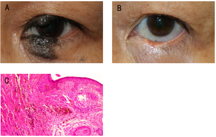
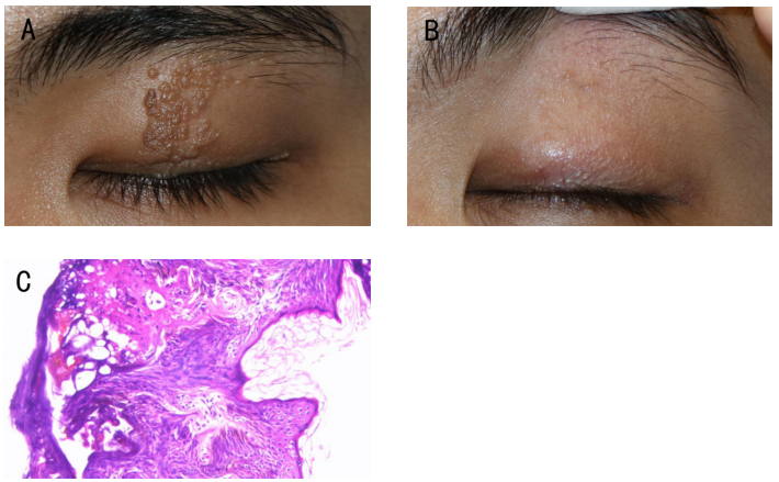
见图1
20230928162910_5513.png
图1 患者女,52岁,左眼眼睑分裂痣激光治疗前后眼外观对比图及激光切除肿物病理检查图
Figure 1 Divided nevus (OS) before and after laser treatment and pathological examination in a 52-year-old female
注:(A)激光治疗前,左眼上下睑黑色肿物,邻近泪小点,累及睑结膜,边界清楚;(B)第7次激光治疗后1个月可见睫毛部分缺失,未见肿物复发,未见明显瘢痕,未见内外翻,睑缘平滑;(C)激光切除物病理诊断:左眼脸色素痣。病理切片:复层鳞状上皮下见痣细胞巢及散在痣细胞,细胞极性存在,细胞无明显异型。(HE染色,×200)
Notes: (A) Before the laser treatment: pigmented nevus located at the upper and lower eyelid margins of the left eye, adjacent to the lacrimal punctum and extending to the eyelid conjunctiva. (B) One month after the seventh laser treatment: evidence of partial sparse eyelashes, absence of lesion recurrence, no scar hyperplasia, and a smooth eyelid margin without signs of entropion or ectropion. (C) Pathological diagnosis: pigmented nevus OS. Pathological findings: scattered nevus cells beneath the stratified squamous epithelium, with preserved cellular polarity and no discernible cellular atypia. (HE staining, ×200)
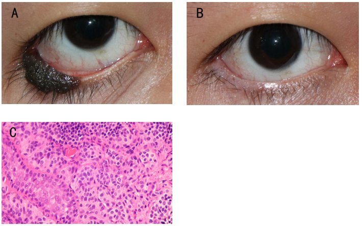
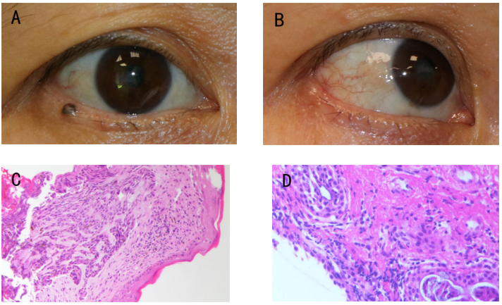
见图2
20230928163226_3551.png
图2 患者男,25岁,左眼下睑色素痣激光治疗前后眼外观对比图以及激光切除肿物病理检查图
Figure 2 Pigmented nevus (OS) before and after laser treatment and pathological examination in a 25-year-old male
注:(A)激光治疗前,下睑外侧沿着睑缘生长黑色肿物,大小约12 mm×5 mm,边界清楚,累及睫毛根部及睑结膜;(B)第四次激光治疗后1月,未见肿物复发,未见瘢痕增生,未见内外翻,治疗区域睫毛部分缺失,睑缘平滑;(C)激光切除物病理诊断:右眼眼睑色素痣。病理切片:激光切除物病理镜下所见激光切除物病理镜下所见复层鳞状上皮下见痣细胞巢及散在痣细胞,细胞极性存在,细胞无明显异型。(HE染色,×200)
Notes: (A) Before the laser treatment: a black lesion grew along the eyelid margin on the lateral side of the lower eyelid, about 12 mm×5 mm in size, with a clear boundary, involving the root of eyelashes and conjunctiva. (B) One month after the fourth laser treatment: absence of lesion recurrence, no scar hyperplasia, a smooth eyelid margin without signs of entropion or ectropion, and partial sparse eyelashes in the treatment area. (C) Pathological diagnosis: pigmented nevus OS. Pathological findings: scattered nevus cells beneath the stratified squamous epithelium, with preserved cellular polarity and no discernible cellular atypia. (HE staining, ×200)
见图3
20230928163415_7627.png
图3 患者女,48岁,双眼上睑黄色瘤激光治疗前后眼外观对比图
Figue 3 Xanthelasma (OU) before and after laser treatment in a 48-year-old female
注:(A1&A2)激光治疗前,双眼上睑内侧扁平隆起的黄色病灶,边缘清晰,右眼病灶大小约10 mm×6 mm,左眼病灶大小约15 mm×6 mm;(B1&B2)第5次激光治疗后1个月,未见肿物复发,少许瘢痕增生及色素沉着。
Notes: (A1 & A2) Before the laser treatment: there were flat yellow lesions on the inner side of the upper eyelid of both eyes with clear edges. The size of the lesions in the right eye was about 10 mm× 6 mm, and that in the left eye was about 15 mm× 6mm. (B1 & B2) One month after the fifth laser treatment: there was no lesion recurrence, minimal scar hyperplasia and pigmentation.
见图4
20230928163513_4355.png
图4 患者男,18岁,左眼上睑疣激光治疗前后眼外观对比图以及激光切除肿物病理检查图
Figure 4 Eyelid molluscum (OS) before and after laser treatment and pathological examination in an 18-year-old male
(A)激光治疗前,上睑自睑缘到眉毛根部,垂直病灶,密集颗粒样,范围约8 mm×20 mm;(B)第3次激光治疗后1年,未见肿物复发,未见瘢痕增生,未见乱睫;(C)激光切除物病理诊断:左眼眼睑疣。病理切片:激光切除物病理镜下所见见增生、变性的复层鳞状上皮,上皮角化不全、角化过度,少数细胞胞浆可见空泡,核稍增大,组织挤压变性严重,细胞形态欠清。(HE染色,×200)
(A) Before the laser treatment: a dense granular lesion extending vertically from the eyelid margin to the eyebrow root, ranging about 8 mm×20 mm. (B) One month after the third laser treatment: no evidence of lesion recurrence, scar hyperplasia, or trichiasis. (C) Pathological diagnosis: eyelid molluscum OS. Pathological findings: proliferative and degenerative changes in the stratified squamous epithelium. The epithelium showed incomplete keratinization alongside hyperkeratosis. (HE staining, ×200)
见图5
20230928163552_4046.png
图5 患者女,48岁,右眼下睑肿物激光治疗前后眼外观对比图以及激光切除肿物病理检查图
Figure 5 Malignant melanoma (OD) of the eyelid before and after laser treatment and pathological examination in a 48-year-old female
(A)激光治疗前,下睑睫毛根部黑色肿物,边界清楚,大小约2 mm×2 mm,未累及灰线;(B)激光治疗后1个月,未见肿物复发,未见瘢痕增生,未见内外翻,治疗区域睫毛部分缺失,睑缘平滑;(C)激光切除病理诊断(HE染色,×200):右眼睑恶性黑色素瘤;(D)激光治疗后1个月行病灶区域手术控制性切除,病理镜下可见均为纤维组织增生、变性,少量淋巴细胞浸润,未见明显肿瘤组织。(HE染色,×200)
(A) Before the laser treatment: black lesion located at the root of the eyelashes of the lower eyelid, about 2 mm×2 mm in size, and not involved gray line. (B) One-month post-laser treatment: no recurrence of the lesion, no evidence of scar hyperplasia, no signs of entropion or ectropion, and partial sparse eyelashes in the treated area. (C) Pathological diagnosis (HE staining, ×200): malignant melanoma OD. (D) One-month after laser treatment: a controlled surgical excision of the lesion area was performed. Histological examination revealed fibrous tissue proliferation and degeneration, with minimal lymphocytic infiltration and no evident tumor tissue. (HE staining, ×200)

3 讨论

眼睑肿物的治疗原则包括完整去除病变并保护眼睑功能[13]。睑缘肿物的手术切除创面较大、眼睑全层切除后容易产生如睑缘缺损、睑缘错位等并发症[14-16],影响眼部外观及其部分功能。相比之下,超脉冲CO2激光具有直接汽化靶组织,而不产生明显碳化的特点,治疗前无需体检,治疗时出血少,组织消融精确,原位愈合,操作时长短,治疗后直接开放用药,无需包眼,更容易被患者接受[17-18]。特别是对于与泪小点相邻的病变,激光治疗能够避免损伤泪小点,确保治疗的安全性[12]
对于面积较大的眼睑肿物,超脉冲CO2激光治疗技术与传统外科手术相比,也具有独特的优势与效果。特别是当肿块直径超过10 mm时,单纯的手术切除可能导致较大范围的组织缺损,往往需要进一步的皮瓣植入或眼睑重建等手术进行修复。这不仅增加了手术的难度及皮瓣与周围组织存在差异等潜在风险,还可能导致术后恢复期延长及治疗费用增加。超脉冲CO2激光治疗能通过分次治疗,精准地消除肿块,减少瘢痕的形成,具有较好的美容效果。
本研究显示,超脉冲CO2激光治疗眼睑肿物取得了良好的治疗效果,但该术式仍然存在一定的并发症风险[19]。这些并发症包括乱睫倒睫、瘢痕形成牵拉、色素沉着、肿物复发等。本研究中,肿物的位置、大小及深度是影响术后并发症发生率的重要因素。乱睫倒睫及睫毛部分缺失仅发生于累及睫毛根部的肿物,原因为当肿物累及睫毛根部,治疗可能损伤毛囊,导致乱睫倒睫或部分缺失;而对比未累及睫毛根部的肿物,累及睫毛根部肿物出现瘢痕比例较低,原因可能为睑缘结构特殊,是皮肤与黏膜交界,相对皮肤发生瘢痕风险较小。此外,大肿物发生瘢痕增生的概率高于中小肿物,而且,当肿物累及睫毛根部的情况下,肿物越大发生乱睫倒睫的比例越大,主要原因可能为:肿物病变范围较广或较深时,激光烧灼范围大,治疗时间长,对组织的损伤大,术后瘢痕形成或毛囊损失的风险较高。这提示临床医生在治疗这类肿物时应格外注意,制定合理的治疗方案,并与患者沟通。本研究中出现一例肿物激光治疗后病理检查为恶性黑色素瘤,这提示所有治疗需尽可能取病变组织做病理学检查,当发现有恶性病变时,需再行控制性切除,保证患者病情得到及时诊疗。
激光治疗眼睑肿物时的注意事项包括:如肿物存在恶性疑虑,应优先考虑手术治疗。其次,治疗过程中激光的能量不宜过高。对于较大范围的病变治疗中不应追求一次去除,多次治疗能够减少组织损伤并降低并发症的发生风险,达到更满意的治疗效果。完成治疗后,为避免感染,伤口需要及时消毒并保持干燥,避免强行去除结痂以防止伤口撕裂。为避免治疗后色素沉着,治疗后应避免强光照射并涂抹防晒产品。在激光治疗眼睑肿物时,避免损伤周围组织及角膜至关重要。为了避免对周围组织造成热损伤,手术前应在肿物病灶基底部位注射利多卡因或普鲁卡因。除了麻醉作用外,注射会使眼睑水肿,利用液体的吸热和散热效果为周围组织提供保护,并为术后组织的修复创造有利条件。在操作过程中,放置保护角膜的角板,确保在治疗过程中不会发生角膜的意外损伤[20-21]。累及睑结膜的肿物,治疗结束后建议佩戴角膜接触镜保护角膜。
本研究为单中心回顾性研究,样本量较小,随访时间短,未进行随机对照研究,结果尚需进一步扩大样本多中心验证。未来研究将采用前瞻性对照设计,扩大样本量并延长随访时间,对不同类型及不同部位眼睑肿物的疗效与安全性进行系统评估,为CO2激光的临床应用提供更可靠的证据支持。综上所述,对于眼睑肿物,特别是睑缘肿物及大肿物,超脉冲CO2激光提供了一种更为精确、微创、安全、有效的治疗方法,可作为眼睑肿物治疗的优选方案,值得临床推广。

利益冲突

所有作者均声明不存在利益冲突

开放获取声明

本文适用于知识共享许可协议(Creative Commons),允许第三方用户按照署名(BY)-非商业性使用(NC)-禁止演绎(ND)(CC BY-NC-ND)的方式共享,即允许第三方对本刊发表的文章进行复制、发行、展览、表演、放映、广播或通过信息网络向公众传播,但在这些过程中必须保留作者署名、仅限于非商业性目的、不得进行演绎创作。详情请访问:https://creativecommons.org/licenses/by-nc-nd/4.0/。
1、Downie LE, Bandlitz S, Bergmanson JPG, et al. CLEAR - Anatomy and physiology of the anterior eye[J]. Cont Lens Anterior Eye, 2021, 44(2): 132-156.Downie LE, Bandlitz S, Bergmanson JPG, et al. CLEAR - Anatomy and physiology of the anterior eye[J]. Cont Lens Anterior Eye, 2021, 44(2): 132-156.
2、Herwig-Carl%20MC%2C%20L%C3%B6ffler%20KU.%20Eyelid%20tumors%3A%20clinical%20aspects%20of%20ophthalmic%20pathology%5BJ%5D.%20Klin%20Monbl%20Augenheilkd%2C%202018%2C%20235(7)%3A%20776-781.%0AHerwig-Carl%20MC%2C%20L%C3%B6ffler%20KU.%20Eyelid%20tumors%3A%20clinical%20aspects%20of%20ophthalmic%20pathology%5BJ%5D.%20Klin%20Monbl%20Augenheilkd%2C%202018%2C%20235(7)%3A%20776-781.%0A
3、Goto H, Yamakawa N, Komatsu H, et al. Epidemiological characteristics of malignant eyelid tumors at a referral hospital in Japan[J]. Jpn J Ophthalmol, 2022, 66(4): 343-349.Goto H, Yamakawa N, Komatsu H, et al. Epidemiological characteristics of malignant eyelid tumors at a referral hospital in Japan[J]. Jpn J Ophthalmol, 2022, 66(4): 343-349.
4、Gniesmer S, Sonntag SR, Schiemenz C, et al. Diagnosis and treatment of malignant eyelid tumors[J]. Ophthalmologie, 2023, 120(3): 262-270.Gniesmer S, Sonntag SR, Schiemenz C, et al. Diagnosis and treatment of malignant eyelid tumors[J]. Ophthalmologie, 2023, 120(3): 262-270.
5、Liu J, Sun J, Wang Z, et al. Treatment of divided eyelid nevus with orbicularis oculi myocutaneous flap: report of 17 cases[J]. Ann Plast Surg, 2020, 85(6): 626-630.Liu J, Sun J, Wang Z, et al. Treatment of divided eyelid nevus with orbicularis oculi myocutaneous flap: report of 17 cases[J]. Ann Plast Surg, 2020, 85(6): 626-630.
6、Torto FL, Losco L, Bernardini N, et al. Surgical treatment with locoregional flaps for the eyelid: a review[J]. Biomed Res Int, 2017, 2017: 6742537.Torto FL, Losco L, Bernardini N, et al. Surgical treatment with locoregional flaps for the eyelid: a review[J]. Biomed Res Int, 2017, 2017: 6742537.
7、Zhao S, Duan J, Zhang J, et al. Evaluation of meibomian gland function after therapy of eyelid tumors at palpebral margin with super pulse CO2 laser[J]. Dis Markers, 2022, 2022: 8705436.Zhao S, Duan J, Zhang J, et al. Evaluation of meibomian gland function after therapy of eyelid tumors at palpebral margin with super pulse CO2 laser[J]. Dis Markers, 2022, 2022: 8705436.
8、Zhang J, Duan J, Gong L. Super pulse CO2 laser therapy for benign eyelid tumors[J]. J Cosmet Dermatol, 2018, 17(2): 171-175.Zhang J, Duan J, Gong L. Super pulse CO2 laser therapy for benign eyelid tumors[J]. J Cosmet Dermatol, 2018, 17(2): 171-175.
9、Rentka A, Grygar J, Nemes Z, et al. Evaluation of carbon dioxide laser therapy for benign tumors of the eyelid margin[J]. Lasers Med Sci, 2017, 32(8): 1901-1907.Rentka A, Grygar J, Nemes Z, et al. Evaluation of carbon dioxide laser therapy for benign tumors of the eyelid margin[J]. Lasers Med Sci, 2017, 32(8): 1901-1907.
10、富秋涛, 魏宁, 孟辉, 等. 超脉冲CO2激光治疗睑缘分裂痣疗效观察[J]. 激光生物学报, 2019, 28(1): 80-83.
Fu QT, Wei N, Meng H, et al. Efficacy of ultrapulse CO2 laser treatment divided nevus of the eyelid[J]. Acta Laser Biol Sin, 2019, 28(1): 80-83.
富秋涛, 魏宁, 孟辉, 等. 超脉冲CO2激光治疗睑缘分裂痣疗效观察[J]. 激光生物学报, 2019, 28(1): 80-83.
Fu QT, Wei N, Meng H, et al. Efficacy of ultrapulse CO2 laser treatment divided nevus of the eyelid[J]. Acta Laser Biol Sin, 2019, 28(1): 80-83.
11、曾颖, 罗益金, 占魁. CO2激光治疗睑缘色素痣的临床观察[J]. 应用激光, 2020, 40(4): 768-771.
Zeng Y, Luo YJ, Zhan K. Clinical observation on palpebral margin melanocytic naevus with super pulsed CO2 laser treatment[J]. Appl Laser, 2020, 40(4): 768-771.
曾颖, 罗益金, 占魁. CO2激光治疗睑缘色素痣的临床观察[J]. 应用激光, 2020, 40(4): 768-771.
Zeng Y, Luo YJ, Zhan K. Clinical observation on palpebral margin melanocytic naevus with super pulsed CO2 laser treatment[J]. Appl Laser, 2020, 40(4): 768-771.
12、Mao Z, Lin BY, Huang YD, et al. Microscopic treatment of benign eyelid margin lesions with ultrapulse carbon dioxide (CO2) laser[J]. J Cosmet Laser Ther, 2021, 23(7-8): 184-187. Mao Z, Lin BY, Huang YD, et al. Microscopic treatment of benign eyelid margin lesions with ultrapulse carbon dioxide (CO2) laser[J]. J Cosmet Laser Ther, 2021, 23(7-8): 184-187.
13、Varde MA, Murali KV, Wiechens B. Surgical treatment of eyelid tumors[J]. HNO, 2018, 66(10): 743-750.Varde MA, Murali KV, Wiechens B. Surgical treatment of eyelid tumors[J]. HNO, 2018, 66(10): 743-750.
14、Then SY, Malhotra R. Superiorly hinged blepharoplasty flap for reconstruction of medial upper eyelid defects following excision of xanthelasma palpebrum[J]. Clin Exp Ophthalmol, 2008, 36(5): 410-414.Then SY, Malhotra R. Superiorly hinged blepharoplasty flap for reconstruction of medial upper eyelid defects following excision of xanthelasma palpebrum[J]. Clin Exp Ophthalmol, 2008, 36(5): 410-414.
15、王越, 李洋, 侯志嘉, 等. 眼睑分裂痣的新分类法及整复手术效果 [J] . 中华眼科杂志, 2022, 58(9) : 676-681.
Wang Y, Li Y, Hou ZJ, et al. A new classification method of eyelid divided nevi and the effect of plastic surgical treatment[J]. Chin J Ophthalmol, 2022, 58(9): 676-681.
王越, 李洋, 侯志嘉, 等. 眼睑分裂痣的新分类法及整复手术效果 [J] . 中华眼科杂志, 2022, 58(9) : 676-681.
Wang Y, Li Y, Hou ZJ, et al. A new classification method of eyelid divided nevi and the effect of plastic surgical treatment[J]. Chin J Ophthalmol, 2022, 58(9): 676-681.
16、Chin JKY, Yip W, Young A, et al. A six-year review of the latest oculoplastic surgical development[J]. Asia Pac J Ophthalmol, 2020, 9(5): 461-469. Chin JKY, Yip W, Young A, et al. A six-year review of the latest oculoplastic surgical development[J]. Asia Pac J Ophthalmol, 2020, 9(5): 461-469.
17、Cho HJ, Lee W, Jeon MK, et al. Staged mosaic punching excision of a kissing nevus on the eyelid[J]. Aesthetic Plast Surg, 2019, 43(3): 652-657. Cho HJ, Lee W, Jeon MK, et al. Staged mosaic punching excision of a kissing nevus on the eyelid[J]. Aesthetic Plast Surg, 2019, 43(3): 652-657.
18、Al-Niaimi F. Ultrapulsed CO2 ablation in the treatment of xanthelasma palpebrarum: high satisfaction treatment with low recurrence[J]. J Dermatolog Treat, 2022, 33(2): 1116-1118. Al-Niaimi F. Ultrapulsed CO2 ablation in the treatment of xanthelasma palpebrarum: high satisfaction treatment with low recurrence[J]. J Dermatolog Treat, 2022, 33(2): 1116-1118.
19、Li D, Lin SB, Cheng B. CO2 laser treatment of xanthelasma palpebrarum in skin types III-IV: efficacy and complications after 9-month follow-up[J]. Photobiomodul Photomed Laser Surg, 2019, 37(4): 244-247.Li D, Lin SB, Cheng B. CO2 laser treatment of xanthelasma palpebrarum in skin types III-IV: efficacy and complications after 9-month follow-up[J]. Photobiomodul Photomed Laser Surg, 2019, 37(4): 244-247.
20、Krupa Shankar D, Chakravarthi M, Shilpakar R. Carbon dioxide laser guidelines[J]. J Cutan Aesthet Surg, 2009, 2(2): 72-80. Krupa Shankar D, Chakravarthi M, Shilpakar R. Carbon dioxide laser guidelines[J]. J Cutan Aesthet Surg, 2009, 2(2): 72-80.
21、van Gemert MJC, Bloemen PR, Wang WY, et al. Periocular CO2 laser resurfacing: severe ocular complications from multiple unintentional laser impacts on the protective metal eye shields[J]. Lasers Surg Med, 2018, 50(10): 980-986.van Gemert MJC, Bloemen PR, Wang WY, et al. Periocular CO2 laser resurfacing: severe ocular complications from multiple unintentional laser impacts on the protective metal eye shields[J]. Lasers Surg Med, 2018, 50(10): 980-986.
上一篇
下一篇
其他期刊
  • 眼科学报

    主管:中华人民共和国教育部
    主办:中山大学
    承办:中山大学中山眼科中心
    主编:林浩添
    主管:中华人民共和国教育部
    主办:中山大学
    浏览
  • Eye Science

    主管:中华人民共和国教育部
    主办:中山大学
    承办:中山大学中山眼科中心
    主编:林浩添
    主管:中华人民共和国教育部
    主办:中山大学
    浏览
推荐阅读
出版者信息
目录