您的位置: 首页 > 2024年3月 第39卷 第3期 > 文字全文
2023年7月 第38卷 第7期11
目录

微创玻璃体切割术后急性眼内炎的临床分析

Clinical analysis of endophthalmitis after minimally invasive vitrectomy

来源期刊: 眼科学报 | 2024年3月 第39卷 第3期 113-119 发布时间: 收稿时间:2024/7/2 11:38:43 阅读量:956
作者:
关键词:
微创玻璃体切割术眼内炎危险因素临床分析
minimally invasive vitrectomy endophthalmitis risk factors clinical analysis
DOI:
10.12419/24031902
收稿时间:
2024-02-23 
修订日期:
2024-02-29 
接收日期:
2024-03-15 
目的:分析23G/25G微创玻璃体切割术后发生眼内炎的危险因素。方法:回顾性分析2014年6月—2023年5月于中国人民解放军中部战区总医院行微创玻璃体切割术后,临床诊断为眼内炎患者(开放性眼外伤患者除外)的临床特征。结果:纳入8 955例行微创玻璃体切割术患者,其中11例微创玻璃体切割术后患眼发生眼内炎,发生率为0.12%。平均年龄(60.8±7.6)岁。11例其中,5例(45.4%)患者合并糖尿病;原发眼疾构成情况:黄斑疾病7例(63.6%)、增殖期糖尿病性视网膜病变继发玻璃体积血2例(18.2%),视网膜裂孔继发玻璃体积血1例(9.1%)、视网膜脱离1例(9.1%);术中联合行白内障手术3例(27.3 %);术毕8例(72.7%)患眼玻璃体腔填充无菌空气,3例(27.3%)填充平衡盐溶液,术毕所有患者均未缝合巩膜穿刺切口;术后低眼压3例(27.3 %)。术后发生眼内炎的时间为(2.8±1.1)d。11例患者经过局部和全身抗感染治疗后炎症控制不佳,均再次行玻璃体切割术联合术中配置万古霉素液灌注,其中9例术毕玻璃体腔填充硅油,术后所有眼内炎得到控制,10例(91.0%)患者最终矫正视力有所提高。结论:微创玻璃体切割术后,免缝合的巩膜切口可能是病原微生物侵入眼内导致眼内炎的潜在途径。尤其要重视黄斑手术中玻璃体不全切除引起巩膜切口处发生玻璃体束综合征可能是术后发生眼内炎的危险因素之一。


Objective: To analyze risk factors for endophthalmitis occurred after 23G/25G minimally invasive vitrectomy. Methods: Retrospective analysis of the clinical characteristics of patients with endophthalmitis (except patients with open eye trauma) after minimally invasive vitrectomy in General Hospital of Central Theater Command(Wuhan,430064) from June 2014 to May 2023. Results: This study included 8,955 patients, of which 11 cases occurred endophthalmitis after minimally invasive vitrectomy, with an incidence rate of 0.12%. The average age was (60.8±7.6) years, and 5 patients (45.4%) were complicated with diabetes; The composition of primary eye diseases: 7 cases (63.6%) of macular disease, 2 cases (18.2%) of vitreous hemorrhage secondary to proliferative diabetic retinopathy and 1 case (9.1%) vitreous hemorrhage secondary to retinal fissure, 1 case (9.1%) of retinal detachment; During the operation, 3 cases (27.3%) underwent combined cataract surgery; After the operation, 8 cases (72.7%) were filled with sterile air in the vitreous cavity of affected eye, the other 3 cases (27.3%) were filled with equilibrium liquid,and sclera puncture incision was not sutured in all patients; 3 cases (27.3%) had low intraocular pressure after operation. The time for postoperative endophthalmitis to occur after operation was 2.8±1.1day. 11 patients had poor inflammation control after local and systemic anti-inflammatory treatments, and all underwent vitrectomy combined with intraoperative injection of vancomycin solution. Among them, 9 patients were filled with silicone oil in the vitreous cavity after the surgery. After the operation, all the endophthalmitis were controlled and final corrected visual acuity of 10 patients improved. Conclusions: Minimally invasive vitrectomy and suture-free scleral incision may be a potential way for pathogenic microorganisms to invade the eye and cause endophthalmitis. Particular attention should be paid to the ‘Vitreous Wick Syndrome’ at the scleral incision caused by incomplete vitrectomy in macular surgery, which may be one of the risk factors for postoperative endophthalmitis.

文章亮点

1. 关键发现

    通过分析 8 955 例 23G/25G 微创玻璃体切割术后发生眼内炎的病例,发现免缝合的巩膜切口及玻璃体不全切除可能是术后发生眼内炎的危险因素之一。

2. 已知与发现

   免缝合的巩膜切口是微创玻璃体切割术的患者发生眼内炎的危险因素。
   黄斑手术中玻璃体不全切除致巩膜切口处发生玻璃体束综合征可能是发生眼内炎的危险因素。
   玻璃体切割术联合硅油填充是治疗眼内炎的有效手段。

3. 意义与改变

   为眼内炎的预防与治疗提供了临床方法。

       眼内炎是细菌或真菌对玻璃体和(或)房水的感染,是最具破坏性的眼部感染,可能在症状出现后数小时或数天内导致受感染眼发生不可逆转的视力损[1-2]。玻璃体切割术自20世纪70年代初引入临床以来,已成为应用广泛的玻璃体视网膜外科手术方式,是治疗视网膜脱离、玻璃体积血、黄斑裂孔等多种玻璃体视网膜疾病的重要方式[3]。免缝合23G/25G微创玻璃体切割术(minimally invasive vitrectomy,MIVS)由于其炎症反应轻、恢复时间快等优势,在临床中广泛应[4]。然而,由于经结膜入路和手术的无缝线性质,微创玻璃体切割术后眼内炎发生率升高已引起关注[5]。因此,本文对2014年6月—2023年5月在我院行MIVS术后的8 955例患者进行回顾性分析,其中11例(11眼)发生眼内炎,文章总结其临床特征,以期为临床工作提供借鉴。

1 对象和方法

1.1 对象

       收集2014年6月—2023年5月就诊,并在我院行MIVS术后发生急性眼内炎患者(11例11眼)的病历资料。纳入标准:MIVS术后符合眼内炎的临床诊断标准:1)不明原因患眼畏光、疼痛、流泪;2)视物模糊,视力迅速下降至手动、光感;3)眼压正常或降低;4)角膜后弹力层皱襞,角膜后壁沉淀物 (keratic precipitats,KP) 明显;5)前房浮游细胞增多,房水闪光明显。前房纤维素性渗出,前房积脓;6)眼底红光反射消失,双目间接检眼镜下不能辨认视盘;7)超声影像学检查:可见均匀一致点状致密玻璃体混浊物;8)房水或玻璃体的微生物学检查检出病原菌。排除标准:1)开放性眼外伤;2)内源性眼内炎、葡萄膜炎或既往有眼部感染疾病史。

1.2 方法

      收集患者病历资料,回顾性分析相关临床资料,包括性别、年龄、原发疾病、糖尿病史、手术方法、填充物,缝合情况、术后眼压情况、晶体状态、发生眼内炎时间、培养结果、治疗方法、治疗过程中视力变化等情况。本研究已通过中国人民解放军中部战区总医院伦理委员会审查([2024]075-01),在研究实施前已取得患者知情同意。

1.3 统计学方法

       采用正态性检验计量资料是否符合正态分布,符合正态分布采用(x±s)表示,非正态分布采用(P 25, P 75)表示,计数资料采用(%)表示。

2 结果

2.1 眼内炎的发生情况

     2014年6月—2023年5月共有8 955例患者于我院行MIVS,术后发生眼内炎11例,发病率为0.12%。

2.2 发生眼内炎病例的原发疾病

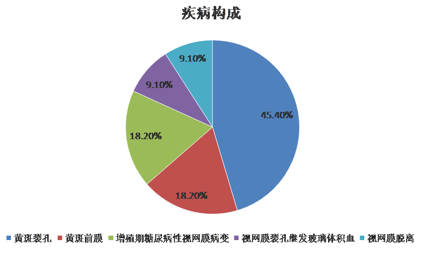
    在MIVS术后发生眼内炎的11例患者中,男4例、女7例;左眼7例,右眼4例;年龄为49~71岁,平均年龄为(60.8±7.6)岁;全身情况:5例患者(45.5%)患有糖尿病;原发眼疾构成情况:黄斑手术7例(63.6%),其中黄斑裂孔5例(45.4%)、黄斑前膜2例(18.2%)、玻璃体积血3例(27.3 %),2例为增殖期糖尿病性视网膜病变,当中视网膜裂孔继发玻璃体积血1例、视网膜脱离1例(9.1%),见图1。

图1 11例眼内炎原发疾病构成图 Figure 1 Composition of primary diseases of 11 patients with endophthalmitis

图1 11例眼内炎原发疾病构成图 Figure 1 Composition of primary diseases of 11 patients with endophthalmitis

2.3 眼内炎发生前的基本情况

     因病情需要,3眼(27.3 %)联合行白内障手术;8眼(72.7%)于玻璃体腔填充无菌空气,而另3眼为平衡盐溶液填充;手术均顺利,术毕观察巩膜切口密闭性良好,均未行缝合;术后3例(27.3 %)患者出现低眼压[眼压≤10 mmHg(1 mmHg=0.133 kPa)]。见表1。

2.4 眼内炎发生的情况及处理

       术后发生眼内炎时间为3(1, 5)d。11例患者中,4例以眼痛为主要症状,4例为眼痛伴明显视力下降,3例患者局部症状不明显。眼内炎发生后视力均下降为手动至指数,主要表现为角膜水肿、房闪、角膜后沉着物、前房积脓伴玻璃体腔混浊,所有患者眼部B超均提示玻璃体腔弥漫性点状混浊。 11例患者均接受经验性全身(头孢他啶、左氧氟沙星注射液)及局部(结膜下注射地塞米松、玻璃体腔注射万古霉素)抗感染治疗后,病情控制欠佳,最终行玻璃体切割术(灌注液中配置质量浓度为0.01 g/L的万古霉素),其中9例填充硅油,1例填充无菌空气,另1例填充灌注液。2例患者因先行玻璃体腔注射万古霉素,未取得玻璃体腔液,余9例患者均抽取玻璃体腔液进行培养,其中2例检出表皮葡萄球菌、1例检出屎肠球菌、6例检出阴性。最终11例患者炎症得以控制,其中10例患者视力较治疗前提高,1例视力改善不明显。见表2。

表1 11例微创玻璃体切割术后发生眼内炎患者的基本情况

Table 1. Basic condition of 11 patients with endophthalmitis after minimally invasive vitrectomy


序号

性别

年龄/岁

眼别

初步

糖尿病

手术过程

填充物

缝合

低眼压

人工晶体

术前矫正视力

诊断

1

53

VH

PPV+Phaco+IOL

灌注液

指数

2

55

MH

PPV+ILM翻转覆盖

空气

0.04

3

49

VH

PPV

空气

指数

4

60

MH

PPV+ILM翻转覆盖

空气

指数

5

65

MH

PPV+ILM翻转覆盖+Phaco+IOL

空气

0.05

6

60

ERM

PPV+ MMP

灌注液

0.1

7

65

VH

PPV

空气

指数

8

68

ERM

PPV+ MMP

灌注液

0.12

9

53

RD

PPV

空气

0.12

10

71

MH

PPV+ILM翻转覆盖

空气

0.4

11

70

MH

PPV+ILM填塞+Phaco+IoL

空气

0.1

注:玻璃体积血(vitreous hemorrhage,VH),黄斑裂孔(macular hole,MH),黄斑前膜(epiretinal membrane,ERM),视网膜脱离(retina detachment,RD),糖尿病(diabetes mellitus, DM), 玻璃体切割术(pars plana vitrectomy, PPV),白内障超声乳化+人工晶体植入术(Phaco),黄斑前膜术(macular preretinal membrane peeling,MMP),内界膜(internal limiting menmbranes,ILM);术后至发生眼压期间眼压≤7 mmHg为低眼压。

Notes:Vitreous hemorrhage=VH,macular hole=MH,epiretinal membrane=ERM,retina retina detachment,RD),糖尿病(diabetes mellitus, DM), 玻璃体切割术(pars plana vitrectomy, PPV),白内障(Phaco)macular preretinal membrane peeling=MMP,internal limiting menmbranes=ILM

 下载数据    在线查看

2 11微创玻璃体切割术后发生眼内炎患者的临床表现、治疗及转归

Table 2.Clinical manifestations, treatment, and outcome of 11 patients with endophthalmitis after minimally invasive vitrectomy


序号

术后发生眼内炎时/d

治疗前视力

症状

体征

治疗

培养

治疗后矫

正视力

1

2

手动

眼痛,视力下降

/眼前眼压49 mmHg前房积脓、渗出人工晶体

玻切+硅油

表皮葡萄球菌

指数

2

5

指数

眼痛

角膜水肿,房闪(++),KP++),玻混

玻切+硅油

阴性

0.04

3

3

指数

房闪(+++),前房渗出,虹膜后粘连,眼底絮状渗出物

玻切+硅油

屎肠球菌

0.50

4

4

手动

眼痛

角膜水肿,房闪(++),玻混

玻切+硅油

阴性

指数

5

1

指数

房闪(+++), KP,前房渗出

玻切

0.20

6

2

指数

眼痛,视力下降

角膜水肿,房闪(++), KP++),前房积脓、渗出

玻切+硅油

0.12

7

2

手动

眼痛

角膜水肿,后弹力层皱褶,房闪(+++),前房积脓、渗出,虹膜后粘连

玻切+硅油

阴性

手动

8

3

指数

眼痛,视力下降

角膜水肿,房闪(++), KP,前房积脓、渗出,虹膜后粘连

玻切+硅油

阴性

0.50

9

3

指数

眼痛

角膜水肿,房闪(+++),前房渗出,玻混

玻切+气体

阴性

0.50

10

3

指数

眼痛,视力下降

角膜后白色脓样分泌物,房闪(+++),前房渗出,玻混

玻切+硅油

表皮葡萄球菌

0.12

11

3

指数

KP++),晶体前后渗出膜

玻切+硅油

阴性

0.05

注:玻切为玻璃体切割术,硅油为眼内硅油填充术,气体为气体填充术,玻混为玻璃体混浊。

NotesKP=keratic precipitats.

3 讨论

       本研究回顾性分析了本中心近9年MIVS术后眼内炎的发生率为0.12%,既往史文献中报告MIVS术后眼内炎的发生率为0.05%~1.55%[6-8]。本研究中MIVS术后发生眼内炎的时间为(2.8±1.1)d,与文献报道相一致[9-10]
       据文献报道[11],年龄较大患者由于自身免疫力低、抵抗力下降,易出现术后感染。本研究中,11例患者平均年龄为(60.8±7.6)岁,提示高龄可能是眼内炎发生的危险因素之一。同时,本研究发现在11例眼内炎患者中,5例合并糖尿病,考虑玻璃体切割术后眼内炎发生可能与糖尿病有关,这与Hyun等[12]发现相一致。由于血糖升高,患者体内长期处在高糖状况,血浆渗透压升高,中性粒细胞趋化、吞噬及杀菌的能力降低,患者机体抗炎能力下降,因此造成伤口的不愈合[13-14],患者行MIVS术后穿刺口的延迟愈合,可能也与眼内炎发生相关。
       本研究发生眼内炎的11例患者,术后均未缝合穿刺口。吴善君等[15]通过超声生物显微镜观察:25 G伤口完全闭合需要多达2周。实验室研究表明,眼睑和结膜囊的菌群是引起术后眼内炎的主要病原体[16]。 Venkatesh等[17]的研究发现,内镜下眼表的污染率约为6%。MIVS术后尚未完全闭合的经结膜巩膜隧道切口,可能是寄生于眼睑和结膜囊内细菌侵入眼内的潜在途径。特别是如果患者在术后立即揉擦眼睛,会使眼内炎的发生风险增加。一项对38 591例因各种适应证接受玻璃体切割术后诊断为眼内炎患者的回顾性研究显示,与缝合切口相比,未缝合切口玻璃体切割术发生眼内炎的概率为25.14%[95%CI:7.37%~85.84%, <0.000 1)[18]。因此,MIVS后未缝合切口考虑与眼内炎发生密切相关。
       本研究中11例患者,7例患者为黄斑疾病。针对黄斑手术,残留周边玻璃体相对较多。嵌顿在穿刺口处的残留玻璃体,发生“玻璃体束综合征”,可吸附、促使眼表的病原微生物通过穿刺口进入玻璃体腔[19]。玻璃体为无血管透明组织,自身抵抗力相对低下,为病原菌生长与繁殖提供良好环境,易发生感染[20]。因此,切口处玻璃体的残留也考虑与眼内炎发生相关。
       本研究中的细菌培养检出率为33%,相比于既往各研究报道,检出率偏低。眼内炎玻璃体切割术研究小组(Endophthalmitis Vitrectomy Study Group)[21]的研究发现,在使用传统培养方法下69.3%的眼内炎病例培养呈阳性。另一研究发现20%~30%的眼内炎病例是培养阴性的,但阴性不能排除诊断[22]。本研究中细菌培养检出率偏低考虑与以下因素相关:1)取样前全身及局部使用抗生素;2)目前我院未采用聚合酶链反应(polymerase chain reaction,PCR)技术,检出率较低;3)取材方式也可能是培养阳性率低的原因之一,需注意先不注入灌注液或仅注入极少量灌注液后采集送检标本,以避免标本被稀释[23];手术室内应提供各种合适的培养基,可以直接在手术室内将微生物接种到培养基上, 怀疑为真菌性眼内炎的情况下,必须将收集的玻璃体腔液离心并随即培养[24]
       本研究的11例患者均行玻璃体切割术,其中9例联合眼内硅油填充术,治疗后炎症均得以控制。早期行玻璃体切割术联合眼内硅油填充术是治疗眼内炎的有效手段[25]。采用玻璃体切割术可以准确、迅速地清除眼内混浊内容物,清理玻璃体腔病灶[26]。硅油是一种具有黏性、不溶于水、不膨胀等特性的光学透明物质,联合硅油填充,其黏性和表面张力可抑制玻璃体腔内生化介质及增殖细胞的移动,并对外界形成屏障,阻止炎症细胞、刺激因子等进入玻璃体腔内;同时本身不含任何营养物质(微生物生长所需),抑制细菌生长,具有抗微生物的作用,主要是针对化脓性眼内炎的致病感染菌,如金色葡萄球菌、曲霉菌、表皮葡萄球菌等[27]
       本研究中,治疗后10例患者视力较治疗前提高,1例视力未见改善,该患者为糖尿病性视网膜病变继发玻璃体积血,视神经萎缩,治疗后视力未见改善。联合白内障手术、低眼压是否为危险因素,需要多中心、大样本证据论证,还有待进一步观察。另外,本研究中为单中心、回顾性研究;同时受条件限制未行病原体PCR检查,因此结果具有一定局限性。
       综上所述,MIVS术后免缝合的巩膜切口可能是病原微生物侵入眼内导致眼内炎的潜在途径。尤其要重视黄斑手术中玻璃体不全切割引起巩膜切口处的“玻璃体束综合征”,它可能是术后眼内炎发生的危险因素之一。玻璃体切割术联合硅油填充是治疗眼内炎的有效手段。对于无法识别微生物的结果,或者因为致病性微生物通常由于抗生素预处理而无法培养,聚合酶链反应等较新的分子技术具有提高微生物识别率的潜力,是临床眼内炎微生物识别的未来方向[28]。同时,针对患有糖尿病的高龄患者,应将其纳入眼内炎的高危人群,加强防护,规范诊疗操作,以期减少甚至避免MIVS术后眼内炎的发生。

利益冲突

    所有作者均声明不存在利益冲突。

开放获取声明

    本文适用于知识共享许可协议 (Creative Commons),允许第三方用户按照署名(BY)-非商业性使用(NC)-禁止演绎(ND)(CC BY-NC-ND)的方式共享,即允许第三方对本刊发表的文章进行复制、发行、展览、表演、放映、广播或通过信息网络向公众传播,但在这些过程中必须保留作者署名、仅限于非商业性目的、不得进行演绎创作。详情请访问https://creativecommons.org/licenses/by-nc-nd/4.0/。
1、Durand ML. Bacterial and fungal endophthalmitis[ J]. Clin Microbiol Rev, 2017, 30(3): 597-613. DOI: 10.1128/CMR.00113-16.Durand ML. Bacterial and fungal endophthalmitis[ J]. Clin Microbiol Rev, 2017, 30(3): 597-613. DOI: 10.1128/CMR.00113-16.
2、Bisorca-Gassendorf L, Boden KT, Szurman P, et al. Postoperative endophthalmitis-a review of literature[ J]. Ophthalmologe, 2021, 118(3): 210-218. DOI: 10.1007/s00347-020-01271-7.Bisorca-Gassendorf L, Boden KT, Szurman P, et al. Postoperative endophthalmitis-a review of literature[ J]. Ophthalmologe, 2021, 118(3): 210-218. DOI: 10.1007/s00347-020-01271-7.
3、C h e n G , T z e k o v R , L i W , e t a l . I N C I D E N C E O F ENDOPHTHALMITIS AFTER VITRECTOMY: a systematic review and meta-analysis[ J]. Retina, 2019, 39(5): 844-852. DOI: 10.1097/ IAE.0000000000002055.C h e n G , T z e k o v R , L i W , e t a l . I N C I D E N C E O F ENDOPHTHALMITIS AFTER VITRECTOMY: a systematic review and meta-analysis[ J]. Retina, 2019, 39(5): 844-852. DOI: 10.1097/ IAE.0000000000002055.
4、Lewis H. Sutureless microincision vitrectomy surgery: unclear benefit, uncertain safety[ J]. Am J Ophthalmol, 2007, 144(4): 613-615. DOI: 10.1016/j.ajo.2007.06.027.Lewis H. Sutureless microincision vitrectomy surgery: unclear benefit, uncertain safety[ J]. Am J Ophthalmol, 2007, 144(4): 613-615. DOI: 10.1016/j.ajo.2007.06.027.
5、Clarke B, Williamson TH, Gini G, et al. Management of bacterial postoperative endophthalmitis and the role of vitrectomy[ J]. Surv Ophthalmol, 2018, 63(5): 677-693. DOI: 10.1016/ j.survophthal.2018.02.003.Clarke B, Williamson TH, Gini G, et al. Management of bacterial postoperative endophthalmitis and the role of vitrectomy[ J]. Surv Ophthalmol, 2018, 63(5): 677-693. DOI: 10.1016/ j.survophthal.2018.02.003.
6、Lin Z, Feng X, Zheng L, et al. Incidence of endophthalmitis after 23-gauge pars Plana vitrectomy[ J]. BMC Ophthalmol, 2018, 18(1): 16. DOI: 10.1186/s12886-018-0678-5.Lin Z, Feng X, Zheng L, et al. Incidence of endophthalmitis after 23-gauge pars Plana vitrectomy[ J]. BMC Ophthalmol, 2018, 18(1): 16. DOI: 10.1186/s12886-018-0678-5.
7、Sukhum Silpa-Archa,Kwanchanoke Kumsiang,Janine M Preble, et al. Endophthalmitis after pars plana vitrectomy with reused single-use devices: a 13-year retrospective study.Int J Retina Vitreous, 2021, 6; 7(1): 2.Sukhum Silpa-Archa,Kwanchanoke Kumsiang,Janine M Preble, et al. Endophthalmitis after pars plana vitrectomy with reused single-use devices: a 13-year retrospective study.Int J Retina Vitreous, 2021, 6; 7(1): 2.
8、李梓敬, 林静仪, 丁小燕. 微创玻璃体切割手术后眼内炎[ J]. 中华眼底病杂志, 2014, 30(4): 421-425. DOI: 10.3760/cma. j.issn.1005-1015.2014.04.027.
Li ZJ, Lin JY, Ding XY. Endophthalmitis after minimally invasive vitrectomy[ J]. Chin J Ocul Fundus Dis, 2014, 30(4): 421-425. DOI: 10.3760/cma.j.issn.1005-1015.2014.04.027.
9、Hosseini S, Daraee G, Shoeibi N, et al. Incidence rate and clinical characteristics of acute endophthalmitis following 23-gauge pars Plana vitrectomy[ J]. Int J Retina Vitreous, 2022, 8(1): 85. DOI: 10.1186/ s40942-022-00435-8.Hosseini S, Daraee G, Shoeibi N, et al. Incidence rate and clinical characteristics of acute endophthalmitis following 23-gauge pars Plana vitrectomy[ J]. Int J Retina Vitreous, 2022, 8(1): 85. DOI: 10.1186/ s40942-022-00435-8.
10、董玮, 陈志敏, 许衍辉, 等. 白内障患者超声乳化术后感染性眼内 炎危险因素分析[ J]. 中华医院感染学杂志, 2016, 26(10): 2332- 2334. DOI: 10.11816/cn.ni.2016-153343.
Dong W, Chen ZM, Xu YH, et al. Risk factors for postoperative infective endophthalmiti s in cataract patients undergoing phacoemulsification[ J]. Chin J Nosocomiology, 2016, 26(10): 2332- 2334. DOI: 10.11816/cn.ni.2016-153343.
11、Oshima Y, Kadonosono K , Yamaji H, et al. Multicenter survey w ith a systematic over v iew of acute-onset endophthalmitis after transconjunctival microincision vitrectomy surger y[ J]. Am J Ophthalmol, 2010, 150(5): 716-725.e1. DOI: 10.1016/ j.ajo.2010.06.002.Oshima Y, Kadonosono K , Yamaji H, et al. Multicenter survey w ith a systematic over v iew of acute-onset endophthalmitis after transconjunctival microincision vitrectomy surger y[ J]. Am J Ophthalmol, 2010, 150(5): 716-725.e1. DOI: 10.1016/ j.ajo.2010.06.002.
12、Hyun SJ, Kim HK, Kim KJ, et al. Posterior trans-dural repair of iatrogenic spinal cord herniation after resection of ossification of posterior longitudinal ligament[ J]. Asian Spine J, 2016, 10(2): 355- 359. DOI: 10.4184/asj.2016.10.2.355.Hyun SJ, Kim HK, Kim KJ, et al. Posterior trans-dural repair of iatrogenic spinal cord herniation after resection of ossification of posterior longitudinal ligament[ J]. Asian Spine J, 2016, 10(2): 355- 359. DOI: 10.4184/asj.2016.10.2.355.
13、Pissas D, Ypsilantis E, Papagrigoriadis S, et al. Endoscopic management of iatrogenic perforations during endoscopic mucosal resection (EMR) and endoscopic submucosal dissection (ESD) for colorectal polyps: a case series[ J]. Therap Adv Gastroenterol, 2015, 8(4): 176-181. DOI: 10.1177/1756283X15576844.Pissas D, Ypsilantis E, Papagrigoriadis S, et al. Endoscopic management of iatrogenic perforations during endoscopic mucosal resection (EMR) and endoscopic submucosal dissection (ESD) for colorectal polyps: a case series[ J]. Therap Adv Gastroenterol, 2015, 8(4): 176-181. DOI: 10.1177/1756283X15576844.
14、Keshavamurthy R, Venkatesh P, Garg S. Ultrasound biomicroscopy findings of 25 G Transconjuctival Sutureless (TSV) and conventional (20G) pars Plana sclerotomy in the same patient[ J]. BMC Ophthalmol, 2006, 6: 7. DOI: 10.1186/1471-2415-6-7.Keshavamurthy R, Venkatesh P, Garg S. Ultrasound biomicroscopy findings of 25 G Transconjuctival Sutureless (TSV) and conventional (20G) pars Plana sclerotomy in the same patient[ J]. BMC Ophthalmol, 2006, 6: 7. DOI: 10.1186/1471-2415-6-7.
15、吴善君, 周宏健, 姚红艳, 等. 白内障术前应用不同浓度聚维酮 碘溶液的不良反应观察[ J]. 国际眼科杂志, 2013, 13(12): 2528- 2529. DOI: 10.3980/j.issn.1672-5123.2013.12.50.
Wu SJ, Zhou HJ, Yao HY, et al. Observation on the adverse reactions of different concentrations of povidone- iodine applied before cataract surgery[ J]. Int Eye Sci, 2013, 13(12): 2528-2529. DOI: 10.3980/ j.issn.1672-5123.2013.12.50.
16、Pahuja, Shivani1; Narula,Ritesh2. Endophthalmitis. Delhi Journal of Ophthalmology 21 (3):p 4-8, Jan– Mar 2011. DOI: 10.4103/0972-0200 .376779 Pahuja, Shivani1; Narula, Ritesh2. Endophthalmitis. Delhi Journal of Ophthalmology 21 (3):p 4-8, Jan– Mar 2011. DOI: 10.4103/0972-0200 .376779
17、Venkatesh P, Verma L, Tewari H. Posterior vitreous wick syndrome: a potential cause of endophthalmitis following vitreo-retinal surgery[ J]. Med Hypotheses, 2002, 58(6): 513-515. DOI: 10.1054/ mehy.2001.1490.Venkatesh P, Verma L, Tewari H. Posterior vitreous wick syndrome: a potential cause of endophthalmitis following vitreo-retinal surgery[ J]. Med Hypotheses, 2002, 58(6): 513-515. DOI: 10.1054/ mehy.2001.1490.
18、Dave VP, Pathengay A, Basu, et al. Endophthalmitis after pars Plana vitrectomy: clinical features, risk factors, and management outcomes[ J]. Asia Pac J Ophthalmol, 2016, 5(3): 192-195. DOI: 10.1097/APO.0000000000000192.Dave VP, Pathengay A, Basu, et al. Endophthalmitis after pars Plana vitrectomy: clinical features, risk factors, and management outcomes[ J]. Asia Pac J Ophthalmol, 2016, 5(3): 192-195. DOI: 10.1097/APO.0000000000000192.
19、Horn C, Wang V, Williams M, et al. Infected iliac pseudoaneurysm after cardiac catheterization complicated by girdlestone pseudoarthroplasty for recurrent infection of hip prosthesis[ J]. Ann Vasc Surg, 2017, 40: 294.e11-294.294.e14. DOI: 10.1016/j.avsg.2016.07.070.Horn C, Wang V, Williams M, et al. Infected iliac pseudoaneurysm after cardiac catheterization complicated by girdlestone pseudoarthroplasty for recurrent infection of hip prosthesis[ J]. Ann Vasc Surg, 2017, 40: 294.e11-294.294.e14. DOI: 10.1016/j.avsg.2016.07.070.
20、Mo r r i s R E , K u h n F. Co m p l e t e a n d e a r l y v i t r e c t o m y f o r endophthalmitis[ J]. Eur J Ophthalmol, 2021, 31(6): 2794-2795. DOI: 10.1177/11206721211021185.Mo r r i s R E , K u h n F. Co m p l e t e a n d e a r l y v i t r e c t o m y f o r endophthalmitis[ J]. Eur J Ophthalmol, 2021, 31(6): 2794-2795. DOI: 10.1177/11206721211021185.
21、Endophthalmit i s Vitrectomy Study Grou p. R esults of the endophthalmitis vitrectomy study: A randomized trial of immediate vitrectomy and of intravenous antibiotics for the treatment of postoperative bacterial endophthalmitis[ J]. Arch Ophthalmol, 1995, 113(12): 1479-1496.Endophthalmit i s Vitrectomy Study Grou p. R esults of the endophthalmitis vitrectomy study: A randomized trial of immediate vitrectomy and of intravenous antibiotics for the treatment of postoperative bacterial endophthalmitis[ J]. Arch Ophthalmol, 1995, 113(12): 1479-1496.
22、Pagani L. Microbial profile and antibiotic susceptibility of culturepositive bacterial endophthalmitis[ J]. Eye, 2011, 25(10): 1379. DOI: 10.1038/eye.2011.160.Pagani L. Microbial profile and antibiotic susceptibility of culturepositive bacterial endophthalmitis[ J]. Eye, 2011, 25(10): 1379. DOI: 10.1038/eye.2011.160.
23、黎晓新, 张正. 眼内炎的诊断与处理及预防[ J]. 中华眼科杂志, 2006,42(10): 946-950. DOI: 10.3760/j: issn: 0412-4081.2006.10.022.
L i X X , Z hang Z . D i ag n o s i s , t reat m ent an d p revent i o n o f endophthalmitis[ J]. Chin J Ophthalmol, 2006,42(10): 946-950. DOI: 10.3760/j: issn: 0412-4081.2006.10.022.
L i X X , Zhang Z . Diagnosis , treatment and prevention of endophthalmitis[ J]. Chin J Ophthalmol, 2006,42(10): 946-950. DOI: 10.3760/j: issn: 0412-4081.2006.10.022.
24、Ness T, Serr A. Diagnostics for endophthalmitis[ J]. Klin Monbl Augenheilkd, 2008, 225(1): 44-49. DOI: 10.1055/s-2008-1027132.Ness T, Serr A. Diagnostics for endophthalmitis[ J]. Klin Monbl Augenheilkd, 2008, 225(1): 44-49. DOI: 10.1055/s-2008-1027132.
25、王东林, 苏燕, 孟繁超, 等. 23G微创玻璃体切割系统在2种黏度 硅油中应用体会[ J]. 中国实用眼科杂志, 2015, 33(8): 872-875. DOI: 10.3760/cma.j.issn.1006-4443.2015.08.009.
Wang DL, Su Y, Meng FC, et al. Application of 23G micro-incision vitrectomy system in silicone oil with two different centistoke[ J]. Chin J Pract Ophthalmol, 2015, 33(8): 872-875. DOI: 10.3760/cma. j.issn.1006-4443.2015.08.009.
26、Weber C, Stasik I, Herrmann P, et al. Early vitrectomy with silicone oil tamponade in the management of postoperative endophthalmitis[ J]. J Clin Med, 2023, 12(15): 5097. DOI: 10.3390/jcm12155097.Weber C, Stasik I, Herrmann P, et al. Early vitrectomy with silicone oil tamponade in the management of postoperative endophthalmitis[ J]. J Clin Med, 2023, 12(15): 5097. DOI: 10.3390/jcm12155097.
27、师小波. 玻璃体切除联合硅油填充术治疗眼外伤的效果 [ J]. 临床医学研究与实践, 2018, 3(1): 66-67. DOI: 10.19347/ j.cnki.2096-1413.201801031.
Shi XB. Effect of vitrectomy combined with silicone oil tamponade in the treatment of ocular injury[ J]. Clin Res Pract, 2018, 3(1): 66-67. DOI: 10.19347/j.cnki.2096-1413.201801031.
28、Tan CL, Sheorey H, Allen PJ, et al. Endophthalmitis: microbiology a n d o r ga n i s m i d e n t i f i c at i o n u s i ng c u r re n t a n d e m e r g i ng techniques[ J]. Ocul Immunol Inflamm, 2023, 31(2): 393-401. DOI: 10.1080/09273948.2022.2027468.Tan CL, Sheorey H, Allen PJ, et al. Endophthalmitis: microbiology a n d o r ga n i s m i d e n t i f i c at i o n u s i ng c u r re n t a n d e m e r g i ng techniques[ J]. Ocul Immunol Inflamm, 2023, 31(2): 393-401. DOI: 10.1080/09273948.2022.2027468.
1、 中国人民解放军中部战区总医院育英计划面上项目(ID 1581)。
This work was supported by the project of Yuying Plan Project of Central Theater General Hospital(ID 1581).()
2、 中国人民解放军中部战区总医院育英计划面上项目(ID 1581)。
This work was supported by the project of Yuying Plan Project of Central Theater General Hospital(ID 1581).()
上一篇
下一篇
其他期刊
  • 眼科学报

    主管:中华人民共和国教育部
    主办:中山大学
    承办:中山大学中山眼科中心
    主编:林浩添
    主管:中华人民共和国教育部
    主办:中山大学
    浏览
  • Eye Science

    主管:中华人民共和国教育部
    主办:中山大学
    承办:中山大学中山眼科中心
    主编:林浩添
    主管:中华人民共和国教育部
    主办:中山大学
    浏览
推荐阅读
出版者信息
目录