您的位置: 首页 > 2024年8月 第39卷 第8期 > 文字全文
2023年7月 第38卷 第7期11
目录

基于高通量测序分析鉴别视网膜病变中的神经纤维瘤病

Identification of neurofibromatosis in retinopathy based on high-throughput sequencing analysis

来源期刊: 眼科学报 | 2024年8月 第39卷 第8期 381-394 发布时间:2024-08-28 收稿时间:2024/10/22 9:40:56 阅读量:555
作者:
关键词:
神经纤维瘤病NF1NF2高通量测序视网膜病变
neurofibromatosis NF1 NF2 high-throughput sequencing retinopathy
DOI:
10.12419/24072402
收稿时间:
2024-06-19 
修订日期:
2024-07-30 
接收日期:
2024-08-19 
目的:通过高通量测序分析进行基因诊断,鉴别视网膜病变中的神经纤维瘤病,为其早诊早治提供重要依据。方法:回顾性分析眼遗传病高通量测序数据库中的NF1和NF2基因变异,根据ACMG/AMP指南解析变异致病性;进一步结合患者的临床表型、家族史以及其他检查结果,综合判断明确是否患有神经纤维瘤病,同时进行疾病的进展和随访的研究分析。结果:通过分析不同眼部表型家系的高通量测序结果,共在11例先证者中发现NF1和NF2基因的10个可能致病变异,包括7个NF1变异和3个NF2变异。这11例先证者的初始诊断包括家族性渗出性玻璃体视网膜病变、黄斑/视网膜发育不良、斜视、视网膜色素变性、Coats病和牵牛花综合征等。其中,在1例初诊为家族性渗出性玻璃体视网膜病变的患儿中,检测到3个基因的致病变异,即NF2: c.122G>A/p.(W41*)、RS1: c.520C>T/p.(R174W)和NYX: c.1027C>T/p.(R343C)。随访检查发现,该患儿的复杂眼部表型符合NF2、RS1和NYX致病变异的临床改变,且MRI检查发现双侧前庭神经鞘瘤、脊髓室管膜瘤和多发性神经鞘瘤改变。除该患者外,还有4例患者在随访中发现存在牛奶咖啡斑或雀斑样色素沉着等皮肤改变,1 例合并小脑神经纤维瘤浸润。结论: 高通量测序分析能有效检测出神经纤维瘤病相关基因的变异,有助于筛选非典型表现的神经纤维瘤病,为疾病的早期诊断,尤其是对严重中枢神经系统病变的早期筛查和及时干预,提供了重要依据。
Objective: To identify neurofibromatosis in retinopathy through high-throughput sequencing analysis and provide important indicators for early diagnosis and treatment. Methods: Variants in NF1 and NF2 were selected from in-house high-throughput sequencing, including targeted exome sequencing, exome sequencing and whole genome sequencing, of individuals with different eye conditions. Pathogenic or likely pathogenic variants were assessed according to ACMG/AMP criteria. All the available clinical data, including clinical manifestation, family history and other examination results, were summarized and further analyzed to determine whether neurofibromatosis. Results: Based on the results of in-house high-throughput sequencing, a total of ten pathogenic or likely pathogenic variants in NF1 and NF2 were identified in 11 unrelated cases with various eye conditions, including three NF2 variants in four cases and seven NF1 variants in seven cases. The unrelated cases with NF1 and NF2 variants had initial clinical manifestation similar to familial exudative vitreoretinopathy (FEVR), macular or retinal dystrophy, strabismus, retinitis pigmentosa, Coats disease, or morning glory syndrome. In one of these cases, who was diagnosed as FEVR at the initial visit, three pathogenic variants of three different genes were identified, namely NF2: c.122G>A/p.(W41*), RS1: c.520C>T/p.(R174W) and NYX: c.1027C>T/p.(R343C). Follow-up examination on this case revealed a complex retinopathy, which were consistent with clinical presentations due to pathogenic variants in NF2, RS1, and NYX, as well as bilateral vestibular schwannomas, spinal ependymoma and multiple schwannomas by MRI. In addition to this patient, a follow-up examination on four of the seven cases present Café-au-lait macules or freckling, which could be easily neglected if neurofibromatosis is not realized on the initial visit, while one had neurofibromatosis in cerebellum. Conclusions: Complex retinopathy may present as the initial sign of neurofibromatosis, and high-throughput sequencing analysis for neurofibromatosis related genes contribute to early diagnosis of neurofibromatosis and facilitating early identification of vital systemic complication.

文章亮点

1. 关键发现

 • 通过高通量测序分析,鉴别临床初诊为其他视网膜病变的神经纤维瘤病,并发现合并有多种视网膜病变的罕见病例。

2. 已知与发现

 • 神经纤维瘤病累及视网膜的改变复杂多样,临床初诊为不同视网膜疾病。
 • 高通量测序分析可识别以不典型视网膜病变首诊的神经纤维瘤病,早期预警隐匿的严重神经系统改变。
 • 罕见病例中神经纤维瘤病可同时合并多种不同的视网膜病变。

3. 意义与改变

 • 基于高通量测序的基因变异分析,有助于识别以不典型视网膜病变首诊的神经纤维瘤病,从而早期预警和干预隐匿的严重神经系统改变。

       神经纤维瘤病(neurofibromatosis, NF)是一种由于
基因缺陷引起的多系统受累的常染色体显性遗传病,包括3种类型,即1型神经纤维瘤病(neurofibromatosis type 1, NF1)、NF2相关神经鞘瘤病(既往称为2型神经纤维瘤病,neurofibromatosis type 2, NF2)和其他神经鞘瘤[1-3],其中,NF1和NF2相关神经鞘瘤病是最常见的类型[4]
       NF1由位于染色体17q11.2的NF1变异所致[5],而NF2相关神经鞘瘤病由NF2基因(位于染色体22q12.2上)变异所致[6]。既往报道表明,两者均具有高度的遗传异质性,且NF1和NF2基因表达存在嵌合可能,即在不同组织中表达各异,导致临床表型的异质性[4, 7-8]。其中,NF1的典型临床特征为周围神经多发性神经纤维瘤、咖啡牛奶斑(cafe-au-lait spots, CALMs)、腋窝或腹股沟雀斑,多数(>50%)儿童有眼部表现,包括虹膜Lisch结节、视神经胶质瘤(optic pathway glioma, OPG)、脉络膜异常等[9],也可表现为神经系统恶性肿瘤[7, 10]。而NF2相关神经鞘瘤病则主要表现为双侧前庭神经鞘瘤(vestibular schwannoma, VS)、其他中枢神经系统肿瘤(脑膜瘤、胶质瘤和脊髓室管膜瘤等),以及眼部改变(白内障、视网膜前膜和视网膜错构瘤)[6, 11- 12]。然而,并非所有患者均出现典型的临床表型,尤其在儿童中,由于其往往以非典型眼部改变为首要表型,且眼部改变通常出现在神经系统症状和听力丧失之前,导致NF的诊断存在一定困难[11, 13-14]。因此,拓展NF的眼部表型谱对于NF的临床诊断和早期基因检测至关重要。
       此外,基于目前的基因检测手段,在具有典型表型的家系中,NF2基因变异检出率约为96%[3],NF1基因变异的检出率亦高达95%以上[15]。然而,在发病年龄较晚或表型不典型的散发家系中,这两个基因的变异检测比例较少[3],使得这部分患者的诊治延迟。对于这类家系,高通量测序技术的出现为NF的诊断和治疗带来了新的契机,既可提高其诊断率,鉴别视网膜病变中的NF,明确变异基因,进一步筛查眼外改变,并进行密切监测和干预。
       因此,基因检测和眼部检查对NF1和NF2相关神经鞘瘤病(尤其是儿童)的早期诊断和治疗至关重要。本研究总结了在基因检测和随访前被误诊为其他疾病的非典型眼部表型NF患者的眼部表型谱,旨在为NF的早期诊断与治疗提供参考依据。

1 资料和方法

1.1 一般资料

       基于课题组自1995年起在中山大学中山眼科中心收集的不同眼病且不相关的家系先证者,包括家族性渗出性玻璃体视网膜病变(familial exudative vitreoretinopathy, FEVR)、视网膜色素变性(retinitis pigmentosa, RP)、近视、青光眼、先天性白内障、视网膜母细胞瘤(retinoblastoma, RB)、外层渗出性视网膜病变(Coats病)、牵牛花综合征(morning glory syndrome, MGS)、正常对照等[16],对其高通量测序,包括靶向测序(targeted exome sequencing, TES)、外显子组测序(whole exome sequencing, WES)或全基因组测序(whole genome sequencing, WGS)数据[17-19],通过多步骤生物信息学分析,筛选NF1和NF2基因变异,以及收集携带NF1或NF2基因变异的家系信息,包括家系成员关系、性别、年龄、临床表现和眼部检查结果等,并从外周血中提取所有参与者的基因组DNA[20],用于高通量测序及其结果验证。本研究遵循《赫尔辛基宣言》,已获得中山眼科中心伦理委员会审核批准(2011KYNL012),先证者及其家系成员对本研究方案知情,并自愿签署知情同意书。

1.2 方法

       1.2.1 NF1和NF2变异致病性分析
      基于课题组既往收集的高通量测序(包括TES、WES或WGS)数据,筛选NF1和NF2基因变异,并与大规模人群基因组变异数据库 (the Genome Aggregation Database, gnomAD, https://gnomad.broadinstitute.org)以及人类基因突变数据库(the Human Gene Mutation Database, HGMD)进行比较分析。通过多步骤生物信息学工具对NF1和NF2基因变异进行致病性预测,包括5个预测错义变异致病性的工具: SIFT(http://sift.jcvi.org/)、Polyphen-2(http://genetics.bwh.harvard.edu/pph2/)、PROVEAN(http://provean.jcvi.org/seq_submit.php)、CADD (http://cadd.gs.washington.edu)和 REVEL(https://sites.google.com/site/revelgenomics/);5个预测是否影响剪接的工具:BDGP(http://www.fruitfly.org/seq_tools/splice)、HSF(http://www.umd.be/HSF3/HSF)、varSEAK(https://varseak.bio/)、SpliceAI(https://spliceailookup.broadinstitute.org)和Alamut。最终,根据美国医学遗传与基因组学学会(American College of Medical Genetics and Genomics, ACMG)和分子病理学协会(theAssociation for Molecular Pathology, AMP)制定的ACMG/AMP指南进行评估及致病性分类[21]。通过Sanger测序及部分家系共分离进一步验证相关变异[20]
       1.2.2 临床资料收集
       收集本研究队列中携带NF1或NF2致病或可能致病变异的家系临床资料并进一步分析。记录患者所有详细的临床信息,包括发病年龄(眼部表现)、初诊症状和诊断、皮肤改变和眼科检查。眼科检查包括最佳矫正视力(best corrected visual acuity, BCVA)、斜视检查、裂隙灯、验光、彩色眼底照相(color fundus photography, CFP)、扫描激光眼底检查(scanning laser ophthalmoscope, SLO)、眼底自发荧光(fundus autofluorescence, FAF)、光学相干断层扫描(optical coherence tomography, OCT)、荧光素眼底血管造影(fundus fluorescein angiography, FFA)和视网膜电流图(electroretinography, ERG)。部分患者行颅脑和脊髓磁共振成像(magnetic resonance imaging, MRI)检查。

2 结果

2.1 本研究中NF1和NF2基因变异的致病性分析

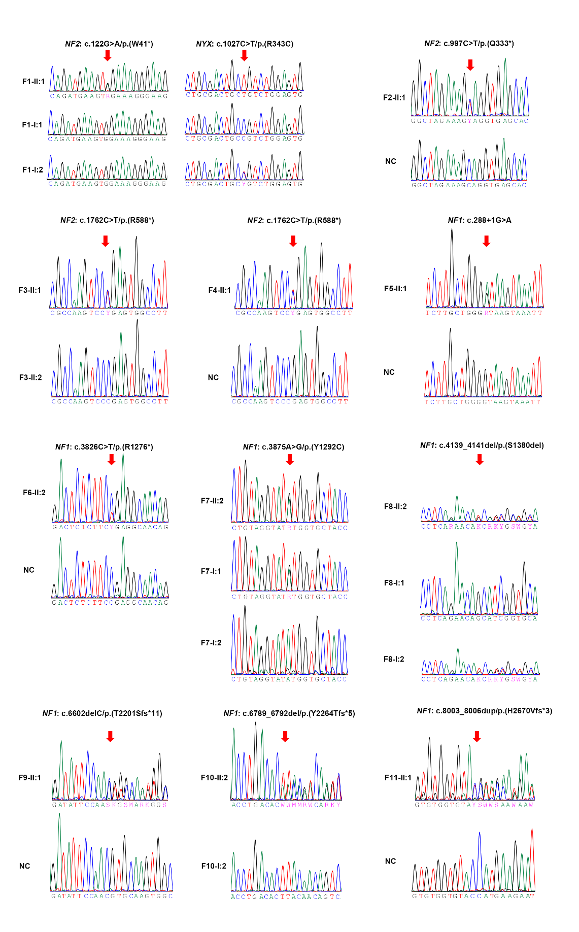
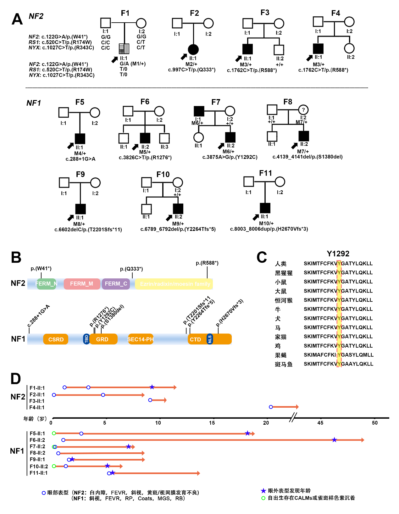
       基于课题组收集的高通量测序数据,共在1 1个不同眼病家系中发现10个NF1和NF2基因的致病/可能致病变异(表1和图1A、B),其中,3个为NF2基因变异(共4个家系),7个为NF1基因变异(共7个家系)。NF2基因的3个变异,即c.122G>A/p.(W41*)、c.997C>T/p.(Q333*)和c.1762C>T/p.(R588*),均为无义变异,且均已被既往研究报道(表1)[22-24]。而NF1基因的7个变异包括了5个功能缺失变异(loss-of-function, LoF变异)(1个无义变异、3个移码变异和1个影响剪接变异)、1个框内缺失和1个错义变异,其中4个为新发变异,即c.4139_4141del/p.(S1380del)、c.6602delC/p.(T2201Sfs*11)、c.6789_6792del/p.(Y2264Tfs*5)和c.8003_8006dup/p.(H2670Vfs*3)(表1)。根据BDGP、HSF、varSEAK、SpliceAI和Alamut预测,NF1基因c.288+1G>A变异影响剪接供体剪接位点;c.3875A>G/p.(Y1292C)变异则被5个工具预测为有害变异,且在不同物种中具有高度保守性(表1和图1C)。在gnomAD数据库中,除NF1基因变异c.3826C>T/p.(R1276*)频率为1/251202,以及变异c.3875A>G/p.(Y1292C)频率为1/251396外,其他变异均无人群频率报道。此外,上述变异已通过Sanger测序和家系共分离验证进一步证实(附图1)。基于ACMG/AMP标准,这10个变异中,除了NF1基因的c.3875A>G/p.(Y1292C)和c.4139_4141del/p.(S1380del)变异被评估为可能致病变异外,其余8个变异均被归类为致病变异(表1)。

图1 本研究中11个携带NF1和NF2致病变异的家系
Figure 1 The 11 pedigrees with variants in NF1 or NF2 in our cohort
(A)4个家系携带NF2致病变异以及7个家系携带NF1致病变异;(B)本研究中发现的NF1和NF2基因的致病或可能致病变异的分布;(C) Y1292在不同物种间氨基酸位置保守性预测(用黄色填充和红色虚线框突出显示);(D)携带NF2和NF1的致病或可能致病变异患者的眼部表型及眼外表型发生的时序。蓝色圆圈表示眼部表型发生的年龄,而绿色圆圈表示先证者出生时存在CALMs或雀斑样色素沉着。蓝色星星表示发现眼外病变的年龄,包括皮肤和神经系统病变。CALMs:牛奶咖啡斑。
(A) Four pedigrees had heterozygous variants in NF2, while seven pedigrees had heterozygous variants in NF1. (B) Distribution and frequency of P/LP variants in NF1 and NF2 in our cohort. (C) Conservation of mutant amino acid positions among different species, which were highlighted in yellow with red dashed box. (D) Age of ocular phenotypes occurred, and extraocular change identified. In patients with NF2, F1-II:1 and F2-II:1 had strabismus and cataract as the initial symptom, respectively, whereas they were both diagnosis as FEVR initially after a detailed eye examination. Other probands were with different ocular phenotypes initially. And all these 11 probands were revised as NF1 or NF2 after gene test and follow-up examination. The blue circle represents the age of ocular phenotypes occurred, while the green circle represents probands were with CALMs or freckling at birth. The blue star represents the age at identification of extraocular changes, including the skin and neurologic changes.

本研究在11个家系中发现10NF1NF2变异

Table 1. Ten variants in NF1 and NF2 identified in 11 families with different eye conditions in our cohort

变异编号

外显子

碱基改变

氨基酸改变

家系数

ACMG/AMP分类

ACMG/AMP证据

HGMD

首次报道

NF2 (NM_000268.3)


M1

2

c.122G>A

p.(W41*)

1

P

PVS1, PS2, PM2

DM

Evans DG, Trueman L, Wallace A, et al. Genotype/phenotype correlations in type 2 neurofibromatosis (NF2): evidence for more severe disease associated with truncating mutations[J]. J Med Genet, 1998, 35(6): 450-455. DOI: 10.1136/jmg.35.6.450

M2

10

c.997C>T

p.(Q333*)

1

P

PVS1, PM2, PM6

DM

Sestini R, Vivarelli R, Balestri P, et al. Neurofibromatosis type 2 attributable to gonosomal mosaicism in a clinically normal mother, and identification of seven novel mutations in the NF2 gene[J]. Hum Genet, 2000, 107(4): 366-371. DOI: 10.1007/s004390000378.

M3

16

c.1762C>T

p.(R588*)

2

P

PVS1_Moderate, PM2, PM6

DM

Van Hout CV, Tachmazidou I, Backman JD, et al. Exome sequencing and characterization of 49, 960 individuals in the UK Biobank[J]. Nature, 2020, 586: 749-756. DOI: 10.1038/s41586-020-2853-0.

NF1 (NM_000267.3)


M4

3

c.288+1G>A

/

1

P

PVS1, PM2, PM6

DM

A M John, M Ruggieri, R Ferner, M Upadhyaya.A search for evidence of somatic mutations in the NF1 gene[J].J Med Genet. 2000, 37(1):44-9. DOI: 10.1136/jmg.37.1.44.

M5

28

c.3826C>T

p.(R1276*)

1

LP

PVS1, PM6

DM

R A Heim, L N Kam-Morgan, C G Binnie, et al.Distribution of 13 truncating mutations in the neurofibromatosis 1 gene[J]. Hum Mol Genet. 1995, 4(6):975-81. DOI: 10.1093/hmg/4.6.975

M6

29

c.3875A>G

p.(Y1292C)

1

LP

PS1, PP1, PP3

DM

Ruen Yao, Tingting Yu, Yufei Xu, et al. Clinical Presentation and Novel Pathogenic Variants among 68 Chinese Neurofibromatosis 1 Children[J]. Genes (Basel). 2019, 10(11):847. DOI: 10.3390/genes10110847

M7

31

c.4139_4141del

p.(S1380del)

1

LP

PVS1, PM2, PM4

/

本研究

M8

43

c.6602delC

p.(T2201Sfs*11)

1

P

PVS1, PM2, PM6

/

本研究

M9

45

c.6789_6792del

p.(Y2264Tfs*5)

1

P

PVS1, PM2, PM6

/

本研究

M10

54

c.8003_8006dup

p.(H2670Vfs*3)

1

P

PVS1, PM2, PM6

/

本研究

注:DM:有害变异;P:致病;LP:可能致病。

Note: DM, deleterious mutation.

2.2 携带NF2致病变异患者表现复杂视网膜病变

       本研究中共有4个家系携带NF2基因致病变异,
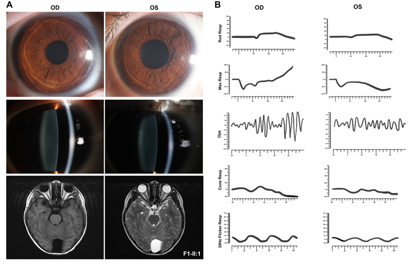
其临床表型各异,首诊表现均为复杂的眼部表型。其中,F1-Ⅱ:1为1例10岁男孩,该患儿3岁时因“出生后3个月起发现斜视”来诊,其右眼BCVA为0.6,左眼BCVA为指数眼前,屈光度数右眼为+0.50 DS/–1.00 DC ×80 °,左眼为–0.25 DS/–3.00 DC×10 °,眼前节未见明显异常(附图2A)。眼底检查显示右眼黄斑中央凹反光不明显;左眼视盘发育不全,黄斑区纤维增殖膜及血管从视盘区拖拽至颞侧中周部视网膜,与FEVR样改变相类似。OCT显示双眼黄斑区视网膜增厚,左眼黄斑前膜。FFA未观察到明显血管渗漏[25]该患儿自症状起曾多次于各地眼科门诊就诊,初步诊断均疑诊为FEVR。为进一步明确其致病原因,对患儿外周血DNA行TES[17]检测,结果发现在RS1基因中存在一个半合子致病变异c.520C>T/p.(R174W),但在FEVR相关基因中并未发现致病变异,因此考虑修改该患者诊断为X连锁视网膜劈裂症(X-linked retinoschisis, XLRS)[25]。然而,该基因变异尚未能解释患儿复杂眼部改变,因此,为了进一步探讨并发视网膜病变的可能原因,对其进行了WGS检测。结果,患者除了存在RS1致病变异外,还检测到另外2个基因的致病变异,包括NYX半合子变异c.1027C>T/p.(R343C)、NF2新发变异c.122G>A/p.(W41*)(表1)。根据上述结果,在患儿10岁时对其进行了回访,包括全身皮肤观察、CFP、SLO、FAF、OCT、ERG、颅脑和脊椎MRI等系统检查。结果发现,患儿全身皮肤并未发现任何CALMs、雀斑样色素沉着等,而双眼眼底改变(图2A)与7年前相似[25]。ERG结果显示患儿视杆反应明显下降,出现b波负波改变,且视锥振幅轻度降低(附图2B),提示患儿为完全型先天性静止性夜盲(complete congenital stationary night blindness, CSNB1)。完善颅脑和脊椎MRI检查,发现患儿双侧VS,延髓内存在一扩张性髓内强化信号(考虑星形细胞瘤可能性大),C1—2、T8—L5脊柱多发神经鞘瘤(图2B),眶周组织未见明显异常(附图2A),表明患儿表型与NF2基因变异相符。因此,基于基因检测结果以及眼科和神经学的系统检查,该患儿最终诊断为NF2相关神经鞘瘤病/XLRS/CSNB1。
       
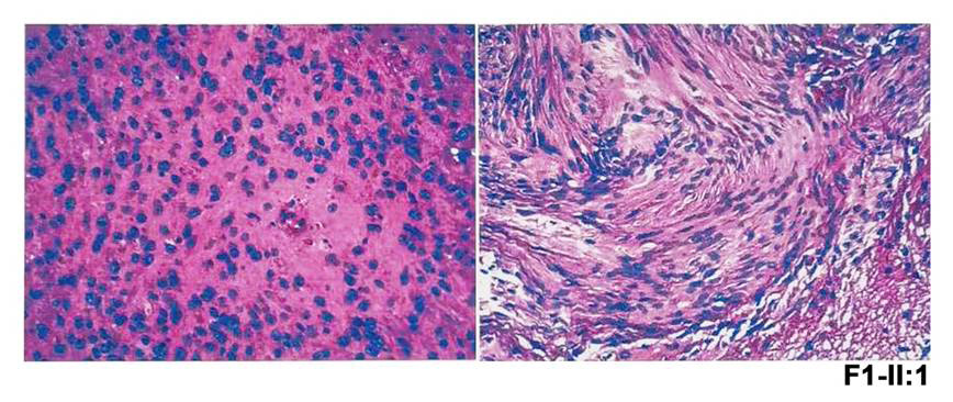
伴随眼部变化的NF患者常存在侵袭性的中枢神经系统肿瘤[26],且NF2无义变异与NF2相关神经鞘瘤病的严重程度相关[3, 27],本研究患儿与之相符。由于延髓中的肿瘤可能存在压迫呼吸中枢的风险而危及生命,因此,该患儿转诊至神经外科,行显微手术切除肿瘤,并康复出院。术后组织病理学检查显示该肿瘤为脊髓室管膜瘤(中枢神经细胞瘤WHO Ⅱ级),伴神经鞘瘤(附图3)。针对该患儿其他病变的处理,主要是神经科随访,以及针对NF2相关神经鞘瘤病的相关眼部改变和视网膜劈裂,每年进行眼科检查,密切监测眼部变化。
       此外,本研究在另外3例不同眼病且不相关的先证者中发现NF2另外两个致病变异(表1和表2)。F2-Ⅱ:1为5岁女孩,因双眼视力差首诊,检查发现双眼白内障,右眼视网膜脱离,FFA提示周边部视网膜血管毛刷样变及荧光渗漏,OCT提示左眼黄斑区视网膜前膜牵拉视网膜增厚,该患儿初诊为双眼先天性白内障,双眼FEVR,右眼视网膜脱离(图2C)。为进一步明确病因,完善基因检测,同样地,该患儿的基因检测结果并未发现FEVR相关致病基因变异,但发现NF2基因致病变异c.997C>T/p.(Q333*)。因此,结合基因检测结果(NF2基因致病变异)及OCT(左眼黄斑前膜伴视网膜错构瘤)(图2C),考虑F2-Ⅱ:1为NF2相关神经鞘瘤病。另外携带NF2基因c.1762C>T/p.(R588*)的2例先证者(F3-Ⅱ:1和F4-Ⅱ:1)则表现为黄斑萎缩和杯盘比增大(表2)。

图 2 携带NF2致病变异先证者的临床表型
Figure 2 Clinical phenotypes of individuals with variants in NF2
(A)F1-Ⅱ:1的CFP和OCT,CFP和FAF显示右眼黄斑中央凹反光不明显;左眼视盘发育不全,黄斑区纤维增殖膜及血管从视盘区拖拽至颞侧中周部视网膜。OCT显示双眼黄斑区视网膜增厚,左眼黄斑前膜。(B)F1-Ⅱ:1颅脑及脊椎MRI,颅脑MRI中红色箭头指向双侧前庭神经鞘瘤。颈椎MRI显示颈髓处存在一扩张性髓内强化病变,提示星形细胞瘤(黄色箭头),以及神经纤维瘤(红色箭头)。胸腰椎MRI显示T8—L5脊椎内多发神经纤维瘤(红色箭头)。(C) F2-Ⅱ:1的眼部检查结果。SLO显示右眼视网膜脱离,左眼黄斑区视网膜褶皱。FFA显示右眼周边视网膜血管渗漏。OCT显示左眼视网膜明显增厚,黄斑前膜伴视网膜褶皱。OD:右眼,OS:左眼。
(A) Fundus photograph and OCT of F1-II:1. Fundus photograph and autofluorescence showed dysplastic optic disc, retinal folds, macular retinal vasculature pulled towards temporally, and peripheral retinal degeneration in the left eye, and ERM in the right eye. OCT exhibited evident retinal thickening with an ERM in the left eye, and ERM extending to the vitreous in the right eye. MRI of the brain and spinal cord in F1-Ⅱ:1. (B) Brain imaging showed bilateral vestibular schwannomas (purple arrowhead). Cervical spine imaging demonstrated one expansive intramedullary enhancing lesion, suggestive of astrocytoma (yellow arrowhead), and one neurofibromas (red arrowhead). Thoracic and lumbar spine imaging showed multiple neurofibromas in T8-L5 spine (red arrowhead). (C) SLO showed retinal detachment of the right eye, while macular abnormity in the left eye in F2-II:1. FFA revealed vascular leakage of the peripheral retina of the right eye. OCT exhibited evident retinal thickening with an ERM accompanied with retinal folds in the left eye.


表2. 本研究11例携带NF1NF2致病/可能致病变异的不相关先证者的临床表型

Table 2. Clinical manifestations of 11 unrelated probands with P/LP variants in NF1 and NF2

先证者编号

变异编号

外显子

碱基改变

氨基酸改变

性别

发病年龄 (岁)#

眼科首诊

眼部表型

眼外表型

NF2 (NM_000268.3)


F1-II:1*

M1

2

c.122G>A

p.(W41*)

0.25

FEVR

黄斑中央凹反光不明显(右眼);视盘发育不全,黄斑区纤维增殖膜及血管从视盘区拖拽至颞侧中周部视网膜(左眼)

VS(双侧),脊髓室管膜瘤,多发神经鞘瘤 

F2-II:1

M2

10

c.997C>T

p.(Q333*)

5.6

FEVR

白内障和视网膜脱离(右眼),视网膜前膜及错构瘤(左眼) 

NA

F3-II:1

M3

16

c.1762C>T

p.(R588*)

9

RP

C/D=0.6

NA

F4-II:1

M3

16

c.1762C>T

p.(R588*)

33

RP

黄斑发育不良

NA

NF1 (NM_000267.3)


F5-II:1

M4

3

c.288+1G>A

/

4.5

FEVR

周边视网膜微血管异常

CALMs

F6-II:2

M5

28

c.3826C>T

p.(R1276*)

儿童期

RP

视网膜色素变性

雀斑样色素沉着

F7-II:2

M6

29

c.3875A>G

p.(Y1292C)

4

Coats

视网膜下渗出

雀斑样色素沉着

F8-II:2

M7

31

c.4139_4141del

p.(S1380del)

0.6

MGS

牵牛花样视盘改变

F9-II:1

M8

43

c.6602delC

p.(T2201Sfs*11)

1.3

OG

突眼

OG头部皮下肿块

F10-II:2

M9

45

c.6789_6792del

p.(Y2264Tfs*5)

1.4

斜视

斜视

CALMs,小脑神经纤维瘤浸润

F11-II:1

M10

54

c.8003_8006dup

p.(H2670Vfs*3)

6.4

OG

突眼

OG

备注:FEVR:家族性渗出性玻璃体视网膜病变;VS:前庭神经鞘瘤;RP:视网膜色素变性;MGS:牵牛花综合征;CALMs牛奶咖啡斑OG:神经胶质瘤;NA:未获得。
#:眼部表型发病年龄。

*F1-II:1基于基因检测及表型分析,最终诊断为NF2相关神经鞘瘤病/XLRS/CSNB1

Note: M, male; F, female; FEVR, Familial exudative vitreoretinopathy; ERM, epiretinal membrane; VS, vestibular schwannomas; CHRRPE, combined hamartoma of the retina with or without RPE; RD, retinal detachment; RP, retinitis pigmentosa; MD, macular dystrophy; CALMs, Café-au-lait macules; MGS, Morning glory disc; OG, optic glioma; NA, not available.   
#: age of onset of the ocular manifestations                                        
*F1-II:1 was finally diagnosis as NF2-related schwannomatosis/retinoschisis/CSNB1 due to pathogenic variants in three genes (NF2, RS1 and NYX).


图 3 携带NF1致病/可能致病变异先证者的临床表型
Figure 3 Clinical phenotypes of individuals with variants in NF1
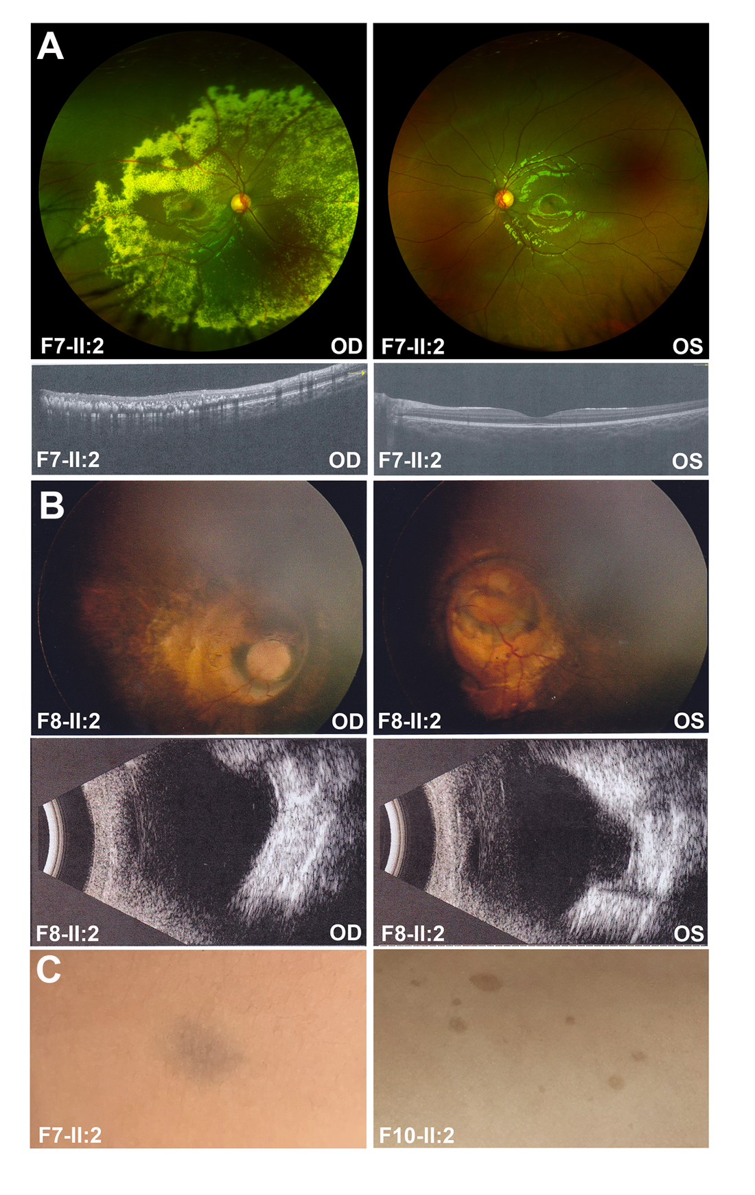
(A)F7-Ⅱ:2的OCT检查发现右眼视网膜下渗出和脉络膜高反射信号。(B)F8-Ⅱ:2眼底照相显示双眼牵牛花样视盘改变,B超同时显示双眼视盘凹陷。(C)F7-Ⅱ:2的肩胛骨上发现了雀斑样色素沉着,F10-Ⅱ:2全身皮肤发现存在6个以上CALMs。OD:右眼,OS:左眼。
(A) F7-Ⅱ:2 performed sub-retinal exudates from SLO and choroidal hyperreflective lesions from OCT of the right eye. (B) F8-Ⅱ:2 showed morning glory disc like fundus of both eyes, and B scan also showed depressed optic disc in both eyes. (C) A freckling on the scapula was also identified in F7-Ⅱ:2, and F10-Ⅱ:2 had more than six café-au-lait macules (CALMs) on the skin.

2.3 携带NF1致病/可能致病变异患者的临床表型

       本研究中7例携带NF1基因致病/可能致病变异的先证者中,2例以眼球突出和眼痛为首发症状(F9-Ⅱ:1右眼,F11-Ⅱ:1左眼),F9-Ⅱ:1眼球突出致眼睑闭合不全,且合并眶内占位性病变,及头部皮下肿块,而F11-Ⅱ:1的B超提示左眼内实性光团,头颅及眼眶MRI提示左眼球内占位性病变,未见明显眶内占位改变。2例患者均已行眼球摘除术,术后病理诊断为眼内神经胶质瘤。其余5例先证者的初始诊断则包括斜视、FEVR、RP、Coats病和MGS(图3A、B和表2),但在随访中,对其全身皮肤进行检查,发现这7例先证者中有4例存在皮肤改变,包括F5-Ⅱ:1和F10-Ⅱ:2全身多处CALMs,F6-Ⅱ:2、F7-Ⅱ:2在腋窝和肩胛骨处发现硬币样大小的雀斑样色素沉着(图3C和表2)。其中,F10-Ⅱ:2为斜视患者,随访行MRI检查发现右侧小脑半球异常信号,考虑神经纤维瘤浸润(表 2)。因此,结合临床表型及全身检查,上述眼部临床表现可能是NF1的非典型改变。

3 讨论

       本研究通过对不同眼部表型的先证者的高通量测序结果进行分析,共报告了11例携带NF1和NF2基因致病或可能致病变异先证者,这些先证者的临床表型包括FEVR、黄斑/视网膜发育不良、斜视、RP、Coats病和MGS等。其中1例初诊为FEVR的先证者携带了3个基因(NF2、RS1和NYX)的3个致病变异,而无FEVR相关致病基因变异,且其临床表型表现为复杂眼部表型(黄斑前膜合并视网膜错构瘤、视网膜劈裂和先天性静止性夜盲),以及MRI检查发现NF2致病变异相关神经系统改变,包括双侧前庭神经鞘瘤、脊髓室管膜瘤和多发性神经鞘瘤,最终诊断为NF2相关神经鞘瘤病/XLRS/CSNB1。
       NF2相关神经鞘瘤病(既往称为NF2)是一种常染色体显性遗传性多发性肿瘤综合征,以多发性中枢神经系统肿瘤为主要特征,包括VS、脑膜瘤、胶质瘤和脊髓室管膜瘤等[3, 11],而皮肤和眼部表型(包括白内障、视网膜错构瘤和视网膜前膜等)在儿童中较为常见[11]然而,NF2相关神经鞘瘤病的临床表型具有复杂性、进行性和高度可变性,给临床医生的诊断和治疗带来了巨大挑战,尤其是半数先证者无相关家族史。F1-Ⅱ:1眼底表现为单侧纤维增殖血管膜从视盘区伸向颞侧中周部视网膜,而F2-Ⅱ:1表现为周边部视网膜血管毛刷样变及荧光渗漏,均为FEVR(一种遗传性视网膜血管源性疾病,眼底改变多样)类似改变[28]。然而,视网膜病变局限于中周视网膜,而不累及外周视网膜血管是FEVR极为罕见的改变,尤其要指出的是,该患儿基因检测尚未提供支持FEVR诊断的基因证据。因此,上述结果表明该患者的眼底变化,包括单侧玻璃体视网膜牵拉和黄斑区拖拽的表现,可能是NF2相关神经鞘瘤病的一个非典型的临床表现。此外,尚未出现皮肤病变或神经系统症状,如肢体麻木、耳鸣、耳聋等,可能是导致该病例在3岁时无法被准确诊断的原因之一。与此同时,对于类似F1-Ⅱ:1的先证者而言,多系统的遗传性疾病的诊断也是一个挑战,且在携带3个基因致病变异患者中尤为困难。F1-Ⅱ:1携带NF2、RS1和NYX这3个基因致病变异,然而,F1-Ⅱ:1均未表现经典视网膜劈裂或夜盲,可能是由于这3个基因的3个致病变异同时出现,导致非典型眼底改变,即黄斑区NF2相关神经鞘瘤病眼底改变掩盖了视网膜裂的表征,且患儿生活在光线充足的环境中,夜盲症状常容易被忽视。因此,在尚未明确潜在基因变异的情况下识别上述疾病存在一定困难。
       NF1的典型表现为CALMs、雀斑样色素沉着、Lisch结节和OPG[29]。本研究中只有2例先证者初始表现为OPG,而其他携带NF1基因的致病或可能致病变异的先证者在初诊时主要表现为斜视、视网膜下渗出伴脉络膜高反射改变、视盘发育不良、FEVR和RP,其中,脉络膜高反射为既往报道NF1患者的眼部表现之[30]。如图1D所示,在7例携带NF1变异的先证者中,其中3例先证者在出生时即出现了CALMs或雀斑样色素沉着,然而直到眼部表型出现及明确基因检测结果后,这些皮肤改变才被临床医生注意到,提示本研究中发现的眼部改变可能是NF1的非典型表型。结合NF1基因的致病或可能致病变异的检出及对应眼部表现,说明需注意这类患者的皮肤改变,这可能对NF1的诊断有帮助,且建议对这些患者进行全身检查和长期随访观察。
       NF患者可能发展为良性或恶性肿瘤,存在危及生命的风险[31],迄今针对NF的治疗方法多种多样,包括药物和手术,目前为了进一步对其进行更有效的治疗,各类临床试验也有序开展[4, 31-33],因此,早发现、早诊断是治疗NF的关键。然而,其非典型的症状及眼部表现为诊治决策带来一定挑战性,但结合基因检测结果可发现潜在的NF,因此需根据基因检测结果完善神经系统检查,筛查潜在的神经系统肿瘤。结合本研究中F1-Ⅱ:1,初诊时表现为FEVR样眼底改变,但根据基因检测和随访结果,最终诊断为NF2相关神经鞘瘤病/XLRS/CSNB1,且该患儿及时行肿瘤摘除手术,解除了危及生命的相关风险。鉴于此,对于NF而言,基因检测和多学科合作至关重要,且对这类患者应每年进行神经科和眼科随访观察。然而,本研究中并非所有患者均完成回访以及完善全身皮肤情况、眼部表型、颅脑和脊髓MRI等检查,未进一步排除是否合并眼外其他器官异常,以及眼部表型是否进展。因此,希望后续能对这部分患者进行跟踪随访。此外,对于不明原因视网膜病变、疑似NF患者以及有NF家族史的人群应进行高通量测序分析鉴别复杂视网膜病变中的NF,以便早期诊断、早期治疗和进行遗传咨询。希望通过本研究拓展的NF的眼部表型,让更多临床医生更深入地了解NF,并结合高通量测序的研究结果,为NF的临床诊治提供依据。
       总而言之,本研究基于高通量测序分析,对不同视网膜病变进行鉴别,明确并扩展了NF1和NF2相关神经鞘瘤病的眼部表型谱,NF2相关神经鞘瘤病可表现为FEVR样眼底、视网膜或黄斑营养不良,而NF1可表现为Coats病、斜视、MGS、RP等。NF的诊断和治疗需要眼科学、遗传学、神经科学等多学科的合作,通过高通量测序鉴别视网膜病变中的NF,可为制定个性化的治疗方案提供依据。同时,需加强公众对NF的宣传和教育,提高其对该病的认识和了解,尤其是对于有特殊视网膜病变表现或家族史的人群,应及时进行基因检测和遗传咨询,早发现、早治疗,防范相关遗传风险。

利益冲突

所有作者均声明不存在利益冲突。

开放获取声明

本文适用于知识共享许可协议 ( Creative Commons),允许第三方用户按照署名(BY)-非商业性使用(NC)-禁止演绎(ND)(CC BY-NC-ND)的方式共享,即允许第三方对本刊发表的文章进行复制、发行、展览、表演、放映、广播或通过信息网络向公众传播,但在这些过程中必须保留作者署名、仅限于非商业性目的、不得进行演绎创作。

附图 1 本研究中携带NF1和NF2致病/可能致病变异的11个家系的Sanger测序图
Supplementary figure 1 Sanger sequencing chromatograph of 11 families with P/LP variants in NF1 and NF2 in this study
NF1和NF2的变异在测序图的上方显示,家系编号在左侧显示。
The variants in NF1 and NF2 were shown upward side of the sequence, and family numbers were shown in the left side.
附图 2 F1-Ⅱ:1的眼前段照相和ERG,以及MRI补充结果
Supplementary figure 2 Ocular anterior segment photograph and ERG, as well as additional MRI in F1-Ⅱ:1 
(A) F1-Ⅱ:1的眼前段未发现明显异常,眼眶MRI示眶组织未见异常。(B) ERG显示视杆细胞反应下降以及b波呈现负波形,视
锥细胞反应轻度下降。
(A) Ocular anterior segment photograph in F1-Ⅱ:1 was with normal limit, while the orbital MRI showed no abnormality in orbital tissue. 
(B) ERG showed significantly reduced rod responses and a negative waveform under bright white, with mildly reduced cone amplitude in F1-
Ⅱ:1.
附图 3 F1-Ⅱ:1术后病理检查结果
Supplementary figure 3 Histopathological examination of medulla oblongata and cervical cord from F1-Ⅱ:1
病理检查结果显示为脊髓室管膜瘤(中枢神经细胞瘤WHOⅡ级),伴神经鞘瘤。
spinal cord ependymoma (CNS WHO Level 2) accompanied with schwannomas was identified in medulla oblongata and cervical cord by histopathological examination.

1、Neurofibromatosis. Conference statement. national institutes of health consensus development conference[ J]. Arch Neurol, 1988, 45(5): 575- 578.Neurofibromatosis. Conference statement. national institutes of health consensus development conference[ J]. Arch Neurol, 1988, 45(5): 575- 578.
2、Evans%20DG.%C2%A0NF2-Related%20Schwannomatosis%5BM%5D%2F%2FAdam%20MP%2C%20Feldman%20%0AJ%2C%20Mirzaa%20GM%2C%20et%20al.%20Gene%20Reviews.%20Seattle%20(WA)%3A%20University%20of%20%0AWashington%2C%202023.Evans%20DG.%C2%A0NF2-Related%20Schwannomatosis%5BM%5D%2F%2FAdam%20MP%2C%20Feldman%20%0AJ%2C%20Mirzaa%20GM%2C%20et%20al.%20Gene%20Reviews.%20Seattle%20(WA)%3A%20University%20of%20%0AWashington%2C%202023.
3、Plotkin SR, Messiaen L, Legius E, et al. Updated diagnostic criteria and nomenclature for neurofibromatosis type 2 and schwannomatosis: an international consensus recommendation[ J]. Genet Med, 2022, 24(9): 1967-1977. DOI: 10.1016/j.gim.2022.05.007.Plotkin SR, Messiaen L, Legius E, et al. Updated diagnostic criteria and nomenclature for neurofibromatosis type 2 and schwannomatosis: an international consensus recommendation[ J]. Genet Med, 2022, 24(9): 1967-1977. DOI: 10.1016/j.gim.2022.05.007.
4、Tamura R. Current understanding of neurofibromatosis type 1, 2, and schwannomatosis[ J]. Int J Mol Sci, 2021, 22(11): 5850. DOI: 10.3390/ ijms22115850.Tamura R. Current understanding of neurofibromatosis type 1, 2, and schwannomatosis[ J]. Int J Mol Sci, 2021, 22(11): 5850. DOI: 10.3390/ ijms22115850.
5、Ferner RE, Huson SM, Thomas N, et al. Guidelines for the diagnosis and management of individuals with neurofibromatosis 1[ J]. J Med Genet, 2007, 44(2): 81-88. DOI: 10.1136/jmg.2006.045906.Ferner RE, Huson SM, Thomas N, et al. Guidelines for the diagnosis and management of individuals with neurofibromatosis 1[ J]. J Med Genet, 2007, 44(2): 81-88. DOI: 10.1136/jmg.2006.045906.
6、Evans DG, Sainio M, Baser ME. Neurofibromatosis type 2[ J]. J Med Genet, 2000, 37(12): 897-904. DOI: 10.1136/jmg.37.12.897.Evans DG, Sainio M, Baser ME. Neurofibromatosis type 2[ J]. J Med Genet, 2000, 37(12): 897-904. DOI: 10.1136/jmg.37.12.897.
7、Hirbe AC, Gutmann DH. Neurofibromatosis type 1: a multidisciplinary approach to care[ J]. Lancet Neurol, 2014, 13(8): 834-843. DOI: 10.1016/S1474-4422(14)70063-8.Hirbe AC, Gutmann DH. Neurofibromatosis type 1: a multidisciplinary approach to care[ J]. Lancet Neurol, 2014, 13(8): 834-843. DOI: 10.1016/S1474-4422(14)70063-8.
8、Ruggieri M, Huson SM. The clinical and diagnostic implications of mosaicism in the neurofibromatoses[ J]. Neurology, 2001, 56(11): 1433-1443. DOI: 10.1212/wnl.56.11.1433.Ruggieri M, Huson SM. The clinical and diagnostic implications of mosaicism in the neurofibromatoses[ J]. Neurology, 2001, 56(11): 1433-1443. DOI: 10.1212/wnl.56.11.1433.
9、Kehrer-Sawatzki H, Cooper DN. Challenges in the diagnosis of neurofibromatosis type 1 (NF1) in young children facilitated by means of revised diagnostic criteria including genetic testing for pathogenic NF1 gene variants[ J]. Hum Genet, 2022, 141(2): 177-191. DOI: 10.1007/s00439-021-02410-z.Kehrer-Sawatzki H, Cooper DN. Challenges in the diagnosis of neurofibromatosis type 1 (NF1) in young children facilitated by means of revised diagnostic criteria including genetic testing for pathogenic NF1 gene variants[ J]. Hum Genet, 2022, 141(2): 177-191. DOI: 10.1007/s00439-021-02410-z.
10、Monroe CL, Dahiya S, Gutmann DH. Dissecting clinical heterogeneity in neurofibromatosis type 1[ J]. Annu Rev Pathol, 2017, 12: 53-74. DOI: 10.1146/annurev-pathol-052016-100228.Monroe CL, Dahiya S, Gutmann DH. Dissecting clinical heterogeneity in neurofibromatosis type 1[ J]. Annu Rev Pathol, 2017, 12: 53-74. DOI: 10.1146/annurev-pathol-052016-100228.
11、Asthagiri AR, Parry DM, Butman JA, et al. Neurofibromatosis type 2[ J]. Lancet, 2009, 373(9679): 1974-1986. DOI: 10.1016/S0140- 6736(09)60259-2.Asthagiri AR, Parry DM, Butman JA, et al. Neurofibromatosis type 2[ J]. Lancet, 2009, 373(9679): 1974-1986. DOI: 10.1016/S0140- 6736(09)60259-2.
12、Coy S, Rashid R, Stemmer-Rachamimov A, et al. An update on the CNS manifestations of neurofibromatosis type 2[ J]. Acta Neuropathol, 2020, 139(4): 643-665. DOI: 10.1007/s00401-019-02029-5.Coy S, Rashid R, Stemmer-Rachamimov A, et al. An update on the CNS manifestations of neurofibromatosis type 2[ J]. Acta Neuropathol, 2020, 139(4): 643-665. DOI: 10.1007/s00401-019-02029-5.
13、Waisberg V, Rodrigues LO, Nehemy MB, et al. Spectral-domain optical coherence tomography findings in neurofibromatosis type 2[ J]. Invest Ophthalmol Vis Sci, 2016, 57(9): OCT262-OCT267. DOI: 10.1167/ iovs.15-18919.Waisberg V, Rodrigues LO, Nehemy MB, et al. Spectral-domain optical coherence tomography findings in neurofibromatosis type 2[ J]. Invest Ophthalmol Vis Sci, 2016, 57(9): OCT262-OCT267. DOI: 10.1167/ iovs.15-18919.
14、Emmanouil B, Wasik M, Charbel Issa P, et al. Structural abnormalities of the central retina in neurofibromatosis type 2[ J]. Ophthalmic Res, 2022, 65(1): 77-85. DOI: 10.1159/000519143.Emmanouil B, Wasik M, Charbel Issa P, et al. Structural abnormalities of the central retina in neurofibromatosis type 2[ J]. Ophthalmic Res, 2022, 65(1): 77-85. DOI: 10.1159/000519143.
15、Evans DG, Bowers N, Burkitt-Wright E, et al. Comprehensive RNA analysis of the NF1 gene in classically affected NF1 affected individuals meeting NIH criteria has high sensitivity and mutation negative testing is reassuring in isolated cases with pigmentary features only[ J]. EBioMedicine, 2016, 7: 212-220. DOI: 10.1016/j.ebiom.2016.04.005.Evans DG, Bowers N, Burkitt-Wright E, et al. Comprehensive RNA analysis of the NF1 gene in classically affected NF1 affected individuals meeting NIH criteria has high sensitivity and mutation negative testing is reassuring in isolated cases with pigmentary features only[ J]. EBioMedicine, 2016, 7: 212-220. DOI: 10.1016/j.ebiom.2016.04.005.
16、Jiang Y, Yi Z, Zheng Y, et al. The systemic genotype-phenotype characterization of PAX6-related eye disease in 164 Chinese families[ J]. Invest Ophthalmol Vis Sci, 2024, 65(10): 46. DOI: 10.1167/ iovs.65.10.46.Jiang Y, Yi Z, Zheng Y, et al. The systemic genotype-phenotype characterization of PAX6-related eye disease in 164 Chinese families[ J]. Invest Ophthalmol Vis Sci, 2024, 65(10): 46. DOI: 10.1167/ iovs.65.10.46.
17、Wang P, Li S, Sun W, et al. An ophthalmic targeted exome sequencing panel as a powerful tool to identify causative mutations in patients suspected of hereditary eye diseases[ J]. Transl Vis Sci Technol, 2019, 8(2): 21. DOI: 10.1167/tvst.8.2.21.Wang P, Li S, Sun W, et al. An ophthalmic targeted exome sequencing panel as a powerful tool to identify causative mutations in patients suspected of hereditary eye diseases[ J]. Transl Vis Sci Technol, 2019, 8(2): 21. DOI: 10.1167/tvst.8.2.21.
18、Li J, Jiang D, Xiao X, et al. Evaluation of 12 myopia-associated genes in Chinese patients with high myopia[ J]. Invest Ophthalmol Vis Sci, 2015, 56(2): 722-729. DOI: 10.1167/iovs.14-14880.Li J, Jiang D, Xiao X, et al. Evaluation of 12 myopia-associated genes in Chinese patients with high myopia[ J]. Invest Ophthalmol Vis Sci, 2015, 56(2): 722-729. DOI: 10.1167/iovs.14-14880.
19、Xiao X, Li S, Jia X, et al. X-linked heterozygous mutations in ARR3 cause female-limited early onset high myopia[ J]. Mol Vis, 2016, 22: 1257-1266.Xiao X, Li S, Jia X, et al. X-linked heterozygous mutations in ARR3 cause female-limited early onset high myopia[ J]. Mol Vis, 2016, 22: 1257-1266.
20、Wang Q, Wang P, Li S, et al. Mitochondrial DNA haplogroup distribution in Chaoshanese with and without myopia[ J]. Mol Vis, 2010, 16: 303-309.Wang Q, Wang P, Li S, et al. Mitochondrial DNA haplogroup distribution in Chaoshanese with and without myopia[ J]. Mol Vis, 2010, 16: 303-309.
21、Richards S, Aziz N, Bale S, et al. Standards and guidelines for the interpretation of sequence variants: a joint consensus recommendation of the American College of Medical Genetics and Genomics and the Association for Molecular Pathology[ J]. Genet Med, 2015, 17(5): 405- 424. DOI: 10.1038/gim.2015.30.Richards S, Aziz N, Bale S, et al. Standards and guidelines for the interpretation of sequence variants: a joint consensus recommendation of the American College of Medical Genetics and Genomics and the Association for Molecular Pathology[ J]. Genet Med, 2015, 17(5): 405- 424. DOI: 10.1038/gim.2015.30.
22、Evans DG, Trueman L, Wallace A, et al. Genotype/phenotype correlations in type 2 neurofibromatosis (NF2): evidence for more severe disease associated with truncating mutations[ J]. J Med Genet, 1998, 35(6): 450-455. DOI: 10.1136/jmg.35.6.450.Evans DG, Trueman L, Wallace A, et al. Genotype/phenotype correlations in type 2 neurofibromatosis (NF2): evidence for more severe disease associated with truncating mutations[ J]. J Med Genet, 1998, 35(6): 450-455. DOI: 10.1136/jmg.35.6.450.
23、Sestini R , Vivarelli R , Balestri P, et al. Neurofibromatosis type 2 attributable to gonosomal mosaicism in a clinically normal mother, and identification of seven novel mutations in the NF2 gene[ J]. Hum Genet, 2000, 107(4): 366-371. DOI: 10.1007/s004390000378.Sestini R , Vivarelli R , Balestri P, et al. Neurofibromatosis type 2 attributable to gonosomal mosaicism in a clinically normal mother, and identification of seven novel mutations in the NF2 gene[ J]. Hum Genet, 2000, 107(4): 366-371. DOI: 10.1007/s004390000378.
24、Van Hout CV, Tachmazidou I, Backman JD, et al. Exome sequencing and characterization of 49, 960 individuals in the UK Biobank[ J]. Nature, 2020, 586: 749-756. DOI: 10.1038/s41586-020-2853-0.Van Hout CV, Tachmazidou I, Backman JD, et al. Exome sequencing and characterization of 49, 960 individuals in the UK Biobank[ J]. Nature, 2020, 586: 749-756. DOI: 10.1038/s41586-020-2853-0.
25、Xiao S, Sun W, Xiao X , et al. Clinical and genetic features of retinoschisis in 120 families with RS1 mutations[ J]. Br J Ophthalmol, 2023, 107(3): 367-372. DOI: 10.1136/bjophthalmol-2021-319668.Xiao S, Sun W, Xiao X , et al. Clinical and genetic features of retinoschisis in 120 families with RS1 mutations[ J]. Br J Ophthalmol, 2023, 107(3): 367-372. DOI: 10.1136/bjophthalmol-2021-319668.
26、Sisk RA, Berrocal AM, Schefler AC, et al. Epiretinal membranes indicate a severe phenotype of neurofibromatosis type 2[ J]. Retina, 2010, 30(4 Suppl): S51-S58. DOI: 10.1097/IAE.0b013e3181dc58bf.Sisk RA, Berrocal AM, Schefler AC, et al. Epiretinal membranes indicate a severe phenotype of neurofibromatosis type 2[ J]. Retina, 2010, 30(4 Suppl): S51-S58. DOI: 10.1097/IAE.0b013e3181dc58bf.
27、Halliday D, Emmanouil B, Pretorius P, et al. Genetic Severity Score predicts clinical phenotype in NF2[ J]. J Med Genet, 2017, 54(10): 657-664. DOI: 10.1136/jmedgenet-2017-104519.Halliday D, Emmanouil B, Pretorius P, et al. Genetic Severity Score predicts clinical phenotype in NF2[ J]. J Med Genet, 2017, 54(10): 657-664. DOI: 10.1136/jmedgenet-2017-104519.
28、Ranchod TM, Ho LY, Drenser KA, et al. Clinical presentation of familial exudative vitreoretinopathy[ J]. Ophthalmology, 2011, 118(10): 2070-2075. DOI: 10.1016/j.ophtha.2011.06.020.Ranchod TM, Ho LY, Drenser KA, et al. Clinical presentation of familial exudative vitreoretinopathy[ J]. Ophthalmology, 2011, 118(10): 2070-2075. DOI: 10.1016/j.ophtha.2011.06.020.
29、Gutmann DH, Ferner RE, Listernick RH, et al. Neurofibromatosis type 1[ J]. Nat Rev Dis Primers, 2017, 3: 17004. DOI: 10.1038/nrdp.2017.4.Gutmann DH, Ferner RE, Listernick RH, et al. Neurofibromatosis type 1[ J]. Nat Rev Dis Primers, 2017, 3: 17004. DOI: 10.1038/nrdp.2017.4.
30、Godinho G, Esteves-Leandro J, Alves G, et al. Correlation between ophthalmologic and neuroradiologic findings in type 1 neurofibromatosis[ J]. J Neuroophthalmol, 2022, 42(1): 101-107. DOI: 10.1097/WNO.0000000000001241.Godinho G, Esteves-Leandro J, Alves G, et al. Correlation between ophthalmologic and neuroradiologic findings in type 1 neurofibromatosis[ J]. J Neuroophthalmol, 2022, 42(1): 101-107. DOI: 10.1097/WNO.0000000000001241.
31、Lin AL, Gutmann DH. Advances in the treatment of neurofibromatosisassociated tumours[ J]. Nat Rev Clin Oncol, 2013, 10(11): 616-624. DOI: 10.1038/nrclinonc.2013.144.Lin AL, Gutmann DH. Advances in the treatment of neurofibromatosisassociated tumours[ J]. Nat Rev Clin Oncol, 2013, 10(11): 616-624. DOI: 10.1038/nrclinonc.2013.144.
32、Wilson BN, John AM, Handler MZ, et al. Neurofibromatosis type 1: new developments in genetics and treatment[ J]. J Am Acad Dermatol, 2021, 84(6): 1667-1676. DOI: 10.1016/j.jaad.2020.07.105.Wilson BN, John AM, Handler MZ, et al. Neurofibromatosis type 1: new developments in genetics and treatment[ J]. J Am Acad Dermatol, 2021, 84(6): 1667-1676. DOI: 10.1016/j.jaad.2020.07.105.
33、Farschtschi S, Mautner VF, McLean ACL, et al. The neurofibromatoses[ J]. Dtsch Arztebl Int, 2020, 117(20): 354-360. DOI: 10.3238/ arztebl.2020.0354.Farschtschi S, Mautner VF, McLean ACL, et al. The neurofibromatoses[ J]. Dtsch Arztebl Int, 2020, 117(20): 354-360. DOI: 10.3238/ arztebl.2020.0354.
1、国家自然科学基金面上项目(82171056);广东省区域联合基金-青年基金项目 (2022A1515111060);广州市科技计划项目(SL2024A03J00525);眼科学国家重点实验室专项经费。
This work was supported by the National Natural Science Foundation of China (82171056);the Guangdong Basic and Applied Basic Research Foundation (2022A1515111060);the Science and Technology Planning Projects of Guangzhou (SL2024A03J00525);the Fundamental Research Funds of the State Key Laboratory of Ophthalmology()
上一篇
下一篇
其他期刊
  • 眼科学报

    主管:中华人民共和国教育部
    主办:中山大学
    承办:中山大学中山眼科中心
    主编:林浩添
    主管:中华人民共和国教育部
    主办:中山大学
    浏览
  • Eye Science

    主管:中华人民共和国教育部
    主办:中山大学
    承办:中山大学中山眼科中心
    主编:林浩添
    主管:中华人民共和国教育部
    主办:中山大学
    浏览
推荐阅读
出版者信息
目录