您的位置: 首页 > 2024年10月 第39卷 第10期 > 文字全文
2023年7月 第38卷 第7期11
目录

托珠单抗调控角膜碱烧伤后修复的研究

Study on the regulation of tocilizumab on corneal repair after alkali burns

来源期刊: 眼科学报 | 2024年10月 第39卷 第10期 489-500 发布时间:2024-10-28 收稿时间:2024/11/6 10:57:04 阅读量:238
作者:
关键词:
角膜碱烧伤白介素-6托珠单抗
corneal alkali burn interleukin-6 tocilizumab
DOI:
10.12419/24071007
收稿时间:
2024-07-10 
修订日期:
2024-08-22 
接收日期:
2024-10-18 
目的:评估外用白介素(Interleukin, IL)-6特异性抑制剂托珠单抗滴眼液在调控角膜碱烧伤后修复的安全性和有效性。方法:6只角膜假烧伤小鼠局部使用托珠单抗滴眼(2.5 mg/mL)和6只角膜假烧伤小鼠局部使用生理盐水滴眼,分别作为实验组和空白组以评估托珠单抗滴眼液的安全性。30只碱烧伤小鼠,按照1∶1随机分配到治疗组和对照组,治疗组使用托珠单抗滴眼液滴眼,对照组使用生理盐水,每日6次,连续用14 d。通过前段光学相干断层扫描观察虹膜前粘连、角膜后弹力层脱离及角膜水肿,在体视显微镜下检查角膜瘢痕形成及上皮伤口愈合。在角膜切片上评估IL-6定位、肌成纤维细胞、免疫细胞浸润和角膜上皮化生。在角膜铺片上评估角膜新生血管和新生淋巴管面积。通过实时荧光定量聚合酶链式反应(quantitative real-time polymerase chain reaction, qRT-PCR)方法检测小鼠角膜 IL-6的表达水平。果:对未进行碱烧伤的角膜使用托珠单抗治疗未观察到明显的角膜结构的损伤。角膜碱烧伤后可见角膜结构的破坏,角膜瘢痕形成并伴有角膜上皮伤口的延迟愈合。使用托珠单抗治疗后,虹膜前粘连的发生率从86.67%下降至20%(P <0.01),角膜后弹力层脱离的发生率从93.33%下降至53.33%(P <0.05),角膜厚度小于对照组[(100.03±15.73)μ m vs. (207.02±56.30)μ mP<0.001],角膜混浊评分从对照组的3.76±0.44下降到治疗组的1.94±0.83(<0.001),治疗组在第5天(<0.05)、第10天(P <0.001)和第14天(<0.001)的上皮愈合率高于对照组。角膜碱烧伤后可见IL-6大量分布于角膜全层,且可见大量肌成纤维细胞形成及免疫细胞浸润,托珠单抗治疗后抑制了IL-6的表达(下降77.5%,P <0.05),肌成纤维细胞数量从每视野(91.44±65.60)个减少至(12.89±10.51)个(P <0.01),免疫细胞的数量从每视野(60.30±28.71)个细胞减少至每视野(6.80±3.82)个细胞(P <0.001)。此外,托珠单抗还减少角膜切片中每视野的杯状细胞数目由(11.3±5.29)个减少至(2.0±1.90个)(P <0.01),并减少角膜新生血管和新生淋巴管的形成(分别减少了76.86%和71.16%,均P <0.001)。结论:局部使用托珠单抗抑制IL-6未见明显角膜毒性,且可以调控角膜碱烧伤后的修复。
Objective: To evaluate the safety and effect of topical IL-6 inhibitor tocilizumab eye drops in regulating corneal alkali burn repair. Methods: Six mice without corneal burns were locally treated with tocilizumab eye drops (2.5 mg/mL) and six mice with corneal pseudo burn were treated with saline, respectively, as experimental and blank groups to evaluate the safety of tocilizumab eye drops. 30 alkali burned mice were randomly divided into a treatment group and a control group in a 1:1 ratio. The treatment group received tocilizumab eye drops, while the control group received physiological saline solution 6 times per day for 14 days. Observe the anterior adhesion of the iris, detachment of the Descemet membrane, and corneal edema through anterior segment optical coherence tomography (AS-OCT), and examine corneal scarring and epithelial wound healing under a stereomicroscope. Evaluate IL-6 localization, myofibroblasts, immune cell infiltration, and corneal epithelial metaplasia on corneal sections. Evaluate corneal neovascularization and neovascularization area by whole-mount cornea staining. Detect the expression level of IL-6 in mouse cornea by qRT-PCR. Results: No significant damage to the corneal structure was observed in the treatment of unburned corneas with tocilizumab. After corneal alkali burns, the corneal structure was damaged, corneal scarring was formed, and delayed healing of corneal epithelial wounds was observed.After treatment with tocilizumab, the incidence of anterior synechia of the iris significantly decreased from 86.67% to 20% (P <0.01), the incidence of Descemet membrane detachment decreased from 93.33% to 53.33% (P <0.05), the corneal thickness was significantly less than that of the control group (100.03±15.73) μ m vs. (207.02±56.30)μ m (P <0.001), the corneal opacity score decreased from 3.76±0.44 in the control group to 1.94±0.83 in the treatment group (P <0.001), and the epithelial healing rate in the treatment group was significantly higher than that in the control group on day 5 (P <0.05), day 10 (P <0.001), and day 14 (P <0.001).After corneal alkali burns, IL-6 was distributed throughout the corneal layer, and a large number of myofibroblasts and immune cells were observed. After treatment with tocilizumab, the expression of IL-6 was inhibited (decreased by 77.5%, P <0.05), the number of myofibroblasts decreased from (91.44±65.60) per field to (12.89±10.51) per field (P <0.01), and the number of immune cells decreased from (60.30±28.71) cells per field to (6.80±3.82) cells per field (P <0.001). In addition, tocilizumab also reduced the number of goblet cells per field in corneal sections (from 11.3±5.29 to 2.0±1.90) (P <0.01), and reduced the formation of corneal neovascularization and neovascular lymphatic vessels (by 76.86% and 71.16%, respectively, both P <0.001). Conclusion: Topical use of tocilizumab to inhibit IL-6 showed no significant corneal toxicity and can regulate the repair of cornea after alkali burns.

文章亮点

1. 关键发现

 • 局部应用托珠单抗不会对正常角膜产生明显损伤,且可以抑制碱烧伤后角膜的炎症反应,调控角膜修复。

2. 已知与发现

 • 免疫细胞浸润是角膜碱烧伤后的重要病理生理过程,而抗炎治疗是角膜碱烧伤后最常见的治疗方法,然而常用的糖皮质激素会产生许多并发症。白介素 -6 被发现具有强烈的刺激免疫细胞浸润的作用,而其特异性抑制剂托珠单抗被发现在多种炎症相关疾病中具有良好的治疗效果。
 • 角膜碱烧伤后局部使用托珠单抗可以抑制角膜结构被破坏,促进角膜上皮的生长,抑制角膜上皮化生、瘢痕形成、新生血管和新生淋巴管形成等角膜病理性修复,且不会对正常角膜产生明显损伤。

3. 意义与改变

 • 角膜碱烧伤后局部使用托珠单抗可以促进角膜上皮生长,抑制角膜的病理性修复,可能可以为角膜碱烧伤患者提供一种安全且能有效改善预后的非手术治疗手段。

       由各种化学物质或热源导致的角膜烧伤是常见急症,在全院急诊患者中占比约为7%[1-2],其中碱性物质导致的角膜碱烧伤患者在各种原因的眼外伤患者中的视力预后最差[3]。角膜碱烧伤后,角膜缘干细胞坏死及眼表微环境被破坏,角膜发生病理性的修复,可导致角膜瘢痕形成、角膜上皮化生、新生血管和淋巴管形成,严重影响患者外观及视力。手术是唯一有效的复明方法,然而由于供体短缺、价格高昂等因素,仅有少部分患者可以恢复视力[4]。角膜碱烧伤患者易发生供体排斥[5],排斥率超过 50%[5],此外有三分之一的患者需反复进行角膜移植术[3]。因此需要研究调控角膜碱烧伤后修复的机制及探索可用于治疗角膜碱烧伤患者的药物。
       角膜碱烧伤后,大量免疫细胞浸润于角膜中,分泌大量的细胞因子和蛋白酶,促使角膜基质层中的角膜基质细胞转化为角膜肌成纤维细胞,导致角膜瘢痕及新生血管形成[6]。抗炎治疗,即通过药物抑制免疫细胞浸润是角膜碱烧伤后最常见的治疗方法[7]。糖皮质激素(激素)用于治疗角膜碱烧伤,可减少炎性细胞浸润,稳定多形核白细胞的细胞膜,减少炎性物质的释放[7]。然而激素也会带来一些棘手的并发症,如青光眼、角膜溶解等[8- 9];激素类药物也会抑制角膜上皮的修复[10];非甾体类药物也可用于抗炎治疗,然而其会增加上皮缺损情况下角膜溶解的发生率[11];因此有必要寻找更安全、更有效的药物。
       白介素(Interleukin, IL)-6是趋化因子家族的一种细胞因子。可由多种细胞如B细胞、巨噬细胞、树突细胞、神经胶质细胞、上皮细胞和某些肿瘤细胞分泌。可通过与IL-6特异性受体(IL-6 receptor, IL-6R)结合而发挥作用。IL-6被发现参与了多种眼部疾病的发生和发展,如干眼症[12]、翼状胬肉[13]和后发性白内障[14]等,作为调控免疫细胞浸润的重要因子,IL-6已被发现参与角膜炎[15-16]、角膜新生血管形成[17]的病理过程,且IL-6也参与了促进角膜上皮生长[18],以上均体现了IL-6功能的多样性。
       托珠单抗是一种IL-6抑制剂,可阻断IL-6和IL-6受体之间的相互作用,因而阻断IL-6通路的信号传导。目前托珠单抗被用于部分炎症性疾病,包括类风湿关节炎、青少年系统性特发性关节炎和新型冠状病毒感染的抗炎治疗[19-21]。此外,也有研究人员使用托珠单抗治疗部分纤维化疾病,如嗜酸性筋膜炎和系统性硬化症[22- 23]
       然而,目前尚未有报道在正常眼表使用托珠单抗滴眼液以反映其安全性,也无报道在碱烧伤后局部使用托珠单抗调控角膜修复的研究。本研究的目的是观察托珠单抗滴眼液局部阻断IL-6是否有明显角膜毒性,以及其是否可以抑制小鼠眼碱烧伤模型中的炎症和瘢痕形成。

1 材料与方法

1.1 动物模型的构建

       所有动物实验均经中山眼科中心动物伦理委员会批准(伦理批件号:O2022065),并按照视觉与眼科研究协会制定的《动物在眼科与视觉研究中的应用声明》指南进行。在所有实验中使用6~8周龄的雌性C57BL/6小鼠(体质量约20 g)。在实验之前,对动物进行适应性饲养1周。
       实验当天小鼠禁食、禁水。首先检测托珠单抗是否会对角膜造成损伤,将小鼠麻醉后由同一名研究人员对小鼠进行右眼角膜假烧伤:直径2 mm的滤纸圆盘在生理盐水中浸泡10 s以充分浸湿圆盘,迅速贴附于每只小鼠的右眼角膜中央进行假烧灼10 s,随后立即用15 mL生理盐水冲洗眼球表面。然后将动物随机分为2组:实验组(n=6)接受10 μ L托珠单抗溶液(2.5 mg/mL稀释于生理盐水;MedChemExpress,美国)滴眼,每天6次,持续14 d,而空白组(=6)左眼也进行假烧伤,然后双眼同时予10 μ L生理盐水滴眼。空白组共使用12颗眼球,其中6颗眼球用于制备角膜切片,6颗眼球用于实时荧光定量聚合酶链式反应(quantitative real-time polymerase chain reaction, qRT-PCR)实验。
       随后检测托珠单抗对角膜碱烧伤后修复的作用:将小鼠麻醉后由同一名研究人员对小鼠进行右眼角膜碱烧伤:直径2 mm的滤纸圆盘在1 mL的1 mol/L NaOH溶液中浸泡10 s以充分浸湿圆盘,迅速贴附于每只小鼠的右眼角膜中央进行烧灼10 s,随后立即用15 mL生理盐水冲洗眼球表面以冲去残留的NaOH溶液。然后将动物随机分为2组:治疗组(n =15)接受10 μ L托珠单抗溶液(2.5 mg/mL稀释于生理盐水;MedChemExpress,美国)滴眼,每天6次,持续14 d,而对照组(=15)接受10 μ L生理盐水滴眼。
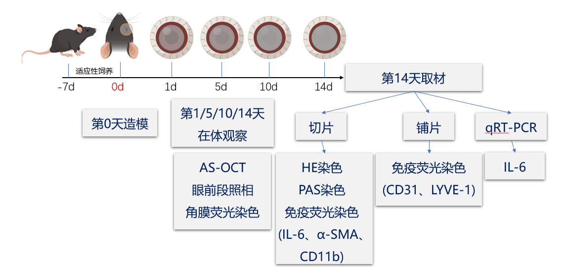
       术后第14天通过吸入二氧化碳和颈椎脱臼将小鼠安乐死,立即取出小鼠眼球以进行下一步实验。实验流程见图1。

图 1 角膜碱烧伤实验流程图
Figure 1 Experiment flow chart of corneal alkali burn

1.2 临床指标

       手术后第14天,使用连接到立体显微镜(SZM-7045T2, Phenix, 中国)的数码相机(MC-1400UK, Phenis, 中国)拍摄角膜照片并进行角膜混浊评分(0~4分):其中0分为完全透明;1分为稍不透明,虹膜和瞳孔很容易看到;2分为较不透明,虹膜和瞳孔仍可检测到;3=不透明,瞳孔几乎分为无法检测;4分为完全不透明,看不到瞳孔。角膜表面滴加应用0.1%荧光素钠滴眼液10 μ L在钴蓝灯下拍照用于评价角膜上皮伤口修复情况。使用iVue仪的“角膜厚度扫描模式”对准小鼠瞳孔中央进行前段光学相干断层扫描(anterior segment optical coherence tomography, AS-OCT)观察前房渗出、眼前段结构损伤及角膜厚度变化。

1.3 组织切片苏木精-伊红染色和过碘酸-雪夫染色

       将眼球于4%多聚甲醛(paraformaldehyde, PFA)中4℃过夜固定后,通过梯度浓度乙醇脱水并使用二甲苯使组织透明化,制备为石蜡组织块后通过切片机获得5 μ m组织切片。组织切片通过梯度浓度乙醇水化后按照以往的方法进行苏木精-伊红染色和过碘酸雪夫(periodic acid-schiff, PAS)染色[24]。通过Axio Imager Z2正置荧光显微镜获得图像。每个样本选取2~4个视野进行分析。新生血管内皮细胞被认为是围成血管的单层扁平上皮,杯状细胞被认为是PAS染色阳性的细胞。

1.4 组织切片免疫荧光

       将石蜡切片水化后使用柠檬酸修复液在微波炉中进行抗原修复,然后在室温下使用10%牛血清白蛋白(bovine serum albumin, BSA)溶液进行封闭,随后4 ℃孵育一抗过夜并使用磷酸盐缓冲液洗5 min共3次;荧光二抗室温避光孵育1 h,磷酸盐缓冲液冲洗5 min共4次;使用含4', 6-二脒基-2-苯基吲哚(4', 6-diamidino-2-phenylindole, DAPI)的抗荧光淬灭封片液封片,通过Axio Imager Z2正置荧光显微镜获得图像。每个样本选取2~4个视野进行分析。
       所用抗体及稀释比例如下所示:IL-6 (1:200, 21865-1-AP, Proteintech), α-SMA (1:200, A2547, MilliporeSigma), CD11b (1:200, 14 - 0112 - 82, ThermoFisher),山羊抗鼠荧光二抗 (Alexa Fluor® 555) (1:1 000, ab150114, Abcam) 和山羊抗兔荧光二抗(Alexa Fluor® 488) (1:1 000, ab150077, Abcam)。

1.5 角膜铺片免疫荧光

       碱烧伤后第14天,通过解剖获得小鼠完整眼球,在100%甲醇溶液中固定1 h,然后在角膜缘区域周围小心解剖获得整个角膜,并在37 °C下在0.02 mmol/LEDTA溶液中孵育过夜,使用有齿镊去除上皮。随后用5%BSA+0.1%Triton溶液在室温下封闭角膜1 h。然后将角膜与抗CD31兔单克隆抗体(1:100,ab28364, Abcam)和抗淋巴管内皮透明质酸受体(lymphatic vessel endothelial hyaluronic acid receptor, LYVE)-1小鼠单克隆抗体(1:100,14-0443-82, ThermoFisher)使用相同的封闭液稀释并在4°C下孵育过夜和37°C下孵育1 h。洗涤后,将角膜与山羊抗鼠IgG H&L(Alexa Fluor®555)(1:1 000,ab150114, Abcam)和山羊抗兔IgG H&L (1:1 000,ab150077, Abcam)在室温下孵育1 h,然后将角膜使用剪刀剪开呈“四叶草状”后封片。通过Axio Imager Z2拍摄荧光图像。

1.6 qRT-PCR

       参考以往的研究方法,但以2个同组小鼠角膜作为一个样本进行qRT-PCR实验[25]。使用TRIzol溶液提取样本总RNA,使用260 nm和280 nm处的吸光度比值评估总RNA样品的纯度,后使用逆转录试剂盒配置反应体系进行逆转录:Oligo(dT) 0.5 µ L + 随机引物 0.5 µ L + 2倍稀释 ES Reaction Mix 5 µ L+ Enzyme Mix 0.5 µ L + RNA样品 1.0 µ g,加入无酶水将体积补至10 µ L,然后配置qRT-PCR的反应体系:cDNA(1∶20稀释)5.0 µ L+ 上游引物0.5 µ L+ 下游引物 0.5 µ L + 2倍稀释SYBR Green qRT-PCR SuperMix 10 µ L,加入ddH2O将体积补至20 µ L。设定反应程序为95℃ 5 min,95℃ 15 s、60℃ 32 s共 40次循环并进行熔解曲线分析。使用ΔΔCT方法进行半定量分析。引物设计:GAPDH正向引物为5'- GGCCTCAAGGATAA-3',逆向引物为5'- GCCCCTCTCGTTATGG-3'),IL-6正向引物为5'- GTTGCCTCTTGGGACTGAT -3',逆向引物为5'- TTTCCACGATTTCCCAGAGA -3'。

1.7 统计学分析

       在R(4.2.1)中通过Fisher确切概率法或χ 2检验比较组间虹膜前粘连和角膜后弹力层脱离发生率的差异。在进行Shapiro-Wilk正态性检验和Levene方差齐性检验后,结果以x±或中位数[四分位数间距(interquartile range, IQR)]表示。P <0.05为差异有统计学意义。过Mann-Whitney 检验比较不同组间角膜混浊评分的差异,Student-检验和Welch-检验比较不同组间同一时间段内的角膜厚度、角膜上皮缺损面积、每视野内各类细胞数量、qRT-PCR数据、新生血管和淋巴管面积的差异,配对样本检验比较同一组内不同时间段的角膜厚度的差异。

2 结果

2.1 应用托珠单抗未见明显角膜毒性

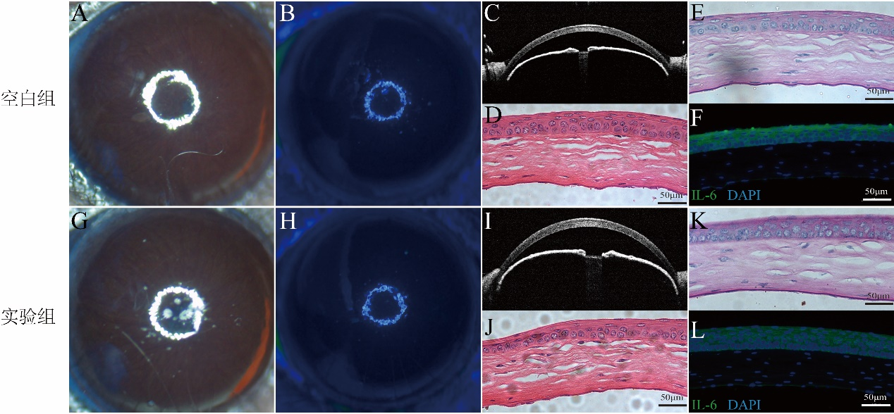
       对小鼠角膜局部使用托珠单抗后,第14天的眼前段照相显示空白组(图2A&图2B)和实验组(图2G&图2H)的角膜均透明,可见瞳孔和虹膜,且未见角膜上皮缺损。AS-OCT图像提示托珠单抗未导致炎性渗出及结构损伤(图2I),苏木精-伊红染色显示使用托珠单抗后,角膜上皮层完整,基质层中胶原排列整齐(图2J),PAS染色未见杯状细胞(图2K)。免疫荧光实验显示IL-6仍仅分布于角膜上皮层,基质层中未见IL-6表达(图2L),这提示局部使用托珠单抗对正常眼无明显损伤。

图 2 局部使用托珠单抗治疗未见明显角膜毒性
Figure 2 Topical tocilizumab treatment showed no significant corneal toxicity
(A、B)空白组第14天的眼前段照相的代表性图像,角膜透明,可见瞳孔和虹膜,角膜上皮未见缺损。(C)空白组第14天的AS-OCT的代表性图像,前房未见渗出,无眼前段结构破坏。(D)空白组第14天的苏木精-伊红染色(×200)显示:角膜上皮完整,角膜基质层中胶原纤维排列整齐。(E)空白组第14天的PAS染色(×200)显示:角膜上皮层中未见杯状细胞。(F)空白组第14天的免疫荧光染色(×200)显示:IL-6仅分布于上皮层,角膜基质层中IL-6几乎不表达。(G、H)使用14 d托珠单抗后,实验组角膜仍透明,可见瞳孔和虹膜,角膜上皮未见明显缺损。(I)实验组前房未见渗出物,眼前段结构未见明显破坏。(J)苏木精-伊红染色(×200)显示:实验组角膜上皮未见明显破坏,角膜基质中胶原纤维排列未见明显紊乱。(K)PAS染色(×200)显示:实验组使用托珠单抗后,角膜上皮层未检出杯状细胞。(L)免疫荧光染色(×200)显示:实验组角膜中的IL-6仍仅分布于上皮层中,角膜基质层中未见明显IL-6的信号。
(A, B) Representative images of the anterior segment of the eye taken on day 14 in the blank group, show transparent cornea, visible pupil and iris, and no defects in corneal epithelium. (C) The representative image of AS-OCT on the 14th day of the blank group showed no exudation in the anterior chamber and no damage to the anterior segment structure. (D) On the 14th day of the blank group, hematoxylin eosin staining (×200) showed that the corneal epithelium was intact and the collagen fibers in the corneal stromal layer were arranged neatly. (E) PAS staining (×200) on the 14th day of the blank group showed that no goblet cells were found in the corneal epithelium. (F) On the 14th day of the blank group, immunofluorescence staining (×200) showed that IL-6 was only distributed in the epithelial layer, and IL-6 was almost not expressed in the corneal stromal layer. (G, H) After 14 days of treating by tocilizumab, the cornea of the experimental group remained transparent, with visible pupils and irises, and no significant defects in the corneal epithelium. (I) No exudate was found in the anterior chamber of the experimental group, and no significant damage was observed in the anterior segment structure. (J) Hematoxylin eosin staining (×200) showed that there was no significant damage to the corneal epithelium in the experimental group, and there was no obvious disorder in the arrangement of collagen fibers in the corneal stroma. (K) PAS staining (×200) showed that no goblet cells were detected in the corneal epithelial layer of the experimental group after using tocilizumab. (L) Immunofluorescence staining (×200) showed that IL-6 in the experimental group's cornea was only distributed in the upper cortex, and no obvious IL-6 signal was observed in the corneal stromal layer.

2.2 使用托珠单抗局部滴眼调控角膜碱烧伤后的修复过程

       2.2.1 托珠单抗可抑制碱烧伤对角膜结构的破坏
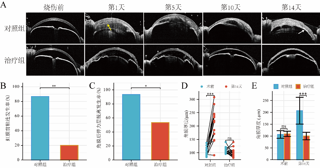
       根据AS-OCT图像,碱烧伤后的前房中可以检测到大量炎症渗出物,并伴有前粘连、后弹力膜脱落和角膜明显水肿(图3A中的对照组)。对照组和治疗组烧伤前的角膜厚度基本一致(=0.456, P >0.05),见图3E。托珠单抗治疗后,虹膜前粘连的发生率从86.67%显著下降至20%(χ 2=26.000, P <0.01),见图3B,角膜后弹力层脱离的发生率从93.33%下降至53.33%(χ 2=12.250, P <0.05),见图3C。对照组烧伤后第14天角膜明显水肿,角膜厚度从(106.89±16.17)μ m增加到(207.02±56.30)μ m(=7.456, <0.001),见图3D。治疗组烧伤后角膜未发生明显水肿,厚度未见明显变化[(100.03±15.73)μ m vs. (109.19±11.03)μ m,t=−1.955, P >0.05],见图3D,且治疗组的角膜厚度明显小于对照组[(100.03±15.73)μ m vs. (207.02±56.30)μ m,−7.089, P<0.001],见图3E。

图 3 托珠单抗抑制角膜结构被破坏
Figure 3 Tocilizumab inhibits the destruction of corneal structure
(A)治疗组与对照组相比,前房炎症渗出都较轻,且在对照组中可观察到眼前段结构损伤,如后弹力膜脱落(黄色箭头指示)、虹膜前粘连(白色箭头指示)。(B)托珠单抗治疗后,虹膜前粘连的发生率明显下降。(C)托珠单抗治疗后,角膜后弹力层脱离的发生率明显下降。(D)在烧伤后第14天,对照组角膜明显水肿,角膜厚度明显增加,而治疗组的角膜厚度没有明显变化。(E)对照组和治疗组在造模前角膜厚度基本一致,而托珠单抗减轻了角膜水肿。nsP >0.05,*P<0.05,**P<0.01,***P<0.001。
(A) Compared with the control group, the treatment group had milder anterior chamber inflammation and exudation, while structural damage to the anterior segment of the eye could be observed in the control group, such as posterior elastic membrane detachment (indicated by the yellow arrow) and anterior iris adhesion (indicated by the white arrow). (B) After treatment with tocilizumab, the incidence of anterior iris adhesion significantly decreased. (C) After treatment with tocilizumab, the incidence of Descemet membrane detachment significantly decreased. (D) On the 14th day after burn, the control group showed significant corneal edema and increased corneal thickness, while the treatment group showed no significant change in corneal thickness. (E) The control group and the treatment group had similar preoperative corneal thickness, and tocilizumab reduced corneal edema. nsP >0.05,*P<0.05,**P<0.01,***P<0.001.
       2.2.2 托珠单抗可抑制角膜碱烧伤后的瘢痕形成,并促进上皮生长
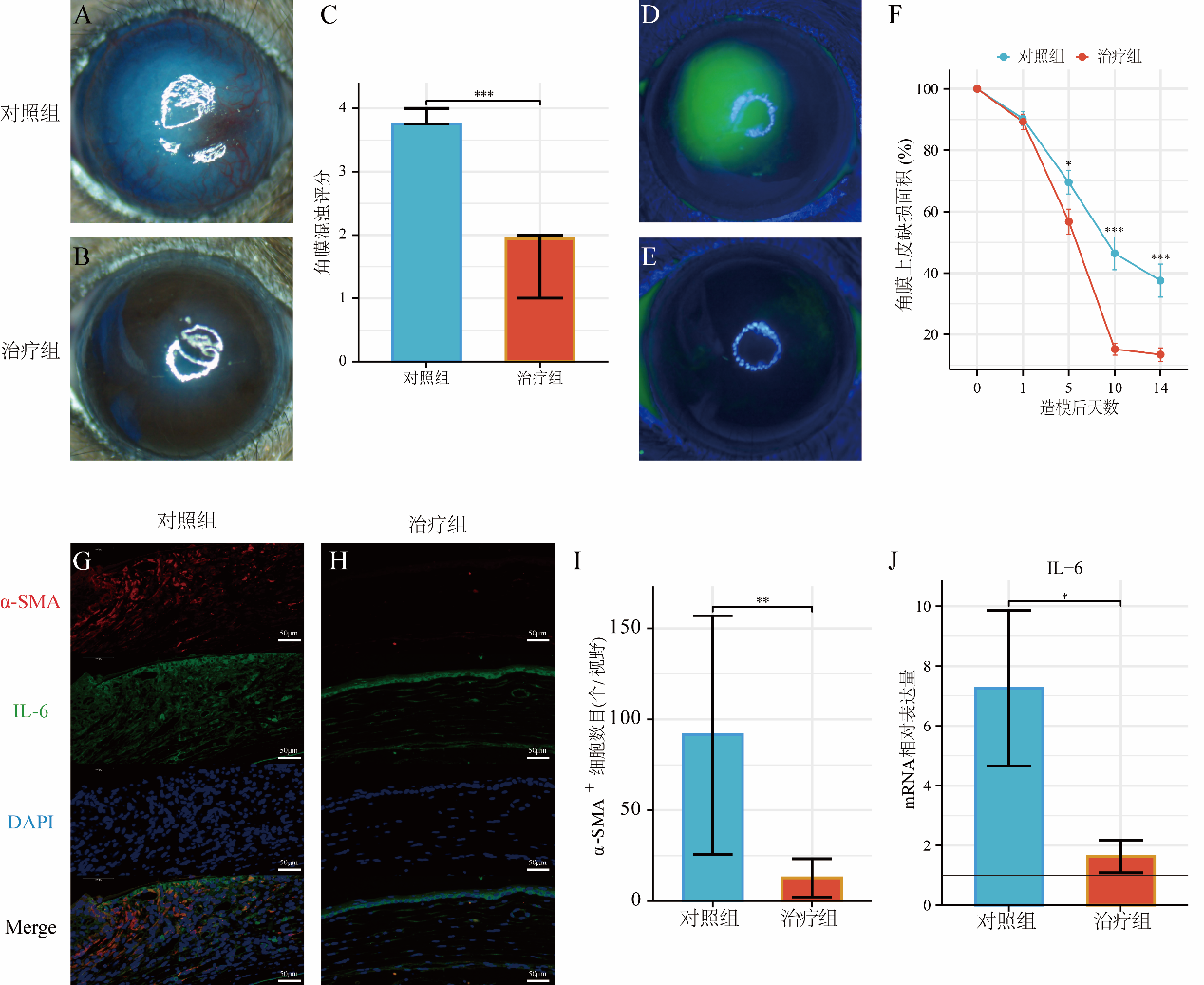
       角膜碱烧伤后,对照组角膜瘢痕形成,角膜明显混浊(角膜混浊评分为3.76±0.44),见图4A;且角膜上皮延迟愈合(碱烧伤后第14天角膜上皮缺损面积仍可达37.53%,见图4D)。在免疫荧光染色实验中,可观察到碱烧伤后大量肌成纤维细胞形成,IL-6分布于角膜全层中,见图4G。通过对碱烧伤角膜使用托珠单抗治疗后,抑制角膜碱烧伤后的瘢痕形成,见图4B,角膜混浊评分从对照组的4(IQR=0.25)下降到治疗组的2(IQR=1)(U=242, <0.001),见图4C,并促进了角膜上皮的愈合:治疗组的上皮缺损面积小于对照组,在第5天(=−2.295, P <0.05)、第10天(=−5.500, P <0.001)和第14天(t =−4.126, P <0.001)具有统计学意义,见图4D~4F。托珠单抗治疗后,每个视野的平均肌成纤维细胞数量从91.44±65.60减少到12.89±10.51(=−3.547, P <0.01),且IL-6的荧光强度也降低,qRT-PCR实验显示IL-6在转录水平的表达减少了约77.5%(t =−3.6658, P <0.05),见图4G~4J。

图 4 托珠单抗治疗抑制角膜瘢痕形成,促进上皮再生,并抑制IL-6的表达
Figure 4 Tocilizumab treatment reduced corneal scarring, promoted epithelial regeneration, and suppressed IL-6 expressionlevel
(A、B)分别为碱烧伤后第14天对照组和治疗组的代表性眼前段照相;对照组中角膜不透明,瞳孔和虹膜不可见,角膜混浊评分为4;治疗组中角膜较不透明,但虹膜和瞳孔仍可检测到,角膜混浊评分为2。(C)托珠单抗治疗后角膜混浊评分显著降低。(D、E)分别为碱烧伤后对照组和治疗组的代表性钴蓝灯下眼前段照相,均取自第14天,绿色部分代表角膜上皮缺损。(F)托珠单抗治疗后角膜上皮更快愈合。(G~I)免疫荧光分析(×200放大倍数)显示,与对照组相比,治疗组的肌成纤维细胞(以α-SMA为标记)数量减少,IL-6荧光强度降低。(J)与对照组相比,治疗组角膜中IL-6的mRNA表达量降低。黑线代表空白组IL-6mRNA的相对表达量,被设定为1。*P<0.05,**P<0.01,***P<0.001。
(A, B) Representative anterior segment photographs of the control group and treatment group on the 14th day after alkali burn injury, respectively; In the control group, the cornea was opaque, with invisible pupils and irises, whose corneal opacity score was 4; In the treatment group, the cornea was relatively opaque, but the iris and pupil can be detected, with a corneal opacity score of 2. (C) After treatment with tocilizumab, the corneal opacity score was significantly decreased. (D, E) Representative cobalt blue lamp images of the anterior segment of the eye in the control group and treatment group after alkali burns, both taken on day 14. The green area represents corneal epithelial defects. (F) After treatment with tocilizumab, the corneal epithelium healed faster. (G~I) Immunofluorescence analysis (x 200 magnification) showed that compared with the control group, the treatment group had a decrease in the number of myofibroblasts (labeled with α- SMA) and a decrease in IL-6 fluorescence intensity. (J) Compared with the control group, the mRNA expression of IL-6 in the cornea of the treatment group decreased. The black line represents the relative expression level of IL-6 mRNA in the blank group, which is set to 1. *P<0.05,**P<0.01,***P<0.001.
       2.2.3 托珠单抗可抑制角膜上皮化生、角膜新生血管和新生淋巴管生成
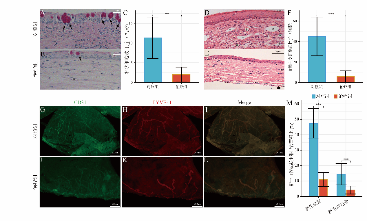
       角膜切片PAS染色显示,托珠单抗治疗后抑制了角膜上皮化生,即治疗组每视野角膜上皮中检测到的杯状细胞减少(2.0±1.90 vs. 11.3±5.29,=−4.100, <0.01),见图5A~5C。此外,角膜碱烧伤后还可以观察到大量新生血管,见图4A。烧伤的角膜的苏木精-伊红染色切片中,可以在基质层中观察到许多新生血管,每个视野的血管内皮细胞数量为44.92±18.98,见图5D。经过治疗后,基质中仅可见少量血管,见图5E;血管内皮细胞数量显著减少至每视野5.60±5.59(t =−6.528 6, P <0.001),见图5F。角膜铺片的免疫荧光显示,新生血管面积(以CD31为标志)从对照组的(47.33±9.52)%降至治疗组的(10.95±4.59)% (t =−12.559, P <0.001),淋巴管面积(以LYVE-1为标志)由对照组的(14.39±6.92)%降至治疗组的(4.15±2.58)% (t =−5.1066, P <0.001),见图5G~5M。

图 5 托珠单抗治疗抑制角膜上皮化生、角膜新生血管和淋巴血管生成
Figure 5 Tocilizumab treatment inhibited corneal conjunctivalization,neovascularization, and lymphangiogenesis
(A、B)分别为碱烧伤后第14天对照组和治疗组的代表性角膜PAS染色切片图像(× 400) “*”指示新生血管管腔,箭头(↑)指示杯状细胞。(C)角膜苏木精-伊红染色切片中的血管内皮细胞数量在治疗后减少。(D、E)分别为碱烧伤后第14天对照组和治疗组的代表性角膜苏木精-伊红染色切片图像(×200),“*”指示新生血管管腔。(F)治疗组角膜上皮化生减轻,以角膜上皮中杯状细胞的数量为标准。(G~M)CD31标记的角膜新生血管和LYVE-1标记的角膜新生淋巴管覆盖的面积(G~L,×50)在托珠单抗治疗后明显减少。**P<0.01,***P<0.001。
(A, B) Representative corneal PAS stained section images (× 400) of the control group and treatment group on the 14th day after alkali burn, respectively. “*” indicates the lumen of neovascularization, and the arrow (↑) indicates goblet cells. (C) The number of endothelial cells in corneal hematoxylin-eosin slices decreased after treatment. (D, E) Representative corneal hematoxylin-eosin stained section images (×200) of the control group and treatment group on the 14th day after alkali burn, respectively, “*” indicating the lumen of neovascularization. (F) The treatment group showed a reduction in corneal epithelial metaplasia, based on the number of goblet cells in the corneal epithelium. The coverage area (G~L, ×50) of CD31 labeled corneal neovascularization and LYVE-1 labeled corneal lymphangiogenesis was significantly reduced after treatment with tocilizumab. **P<0.01,***P<0.001.
       2.2.4 托珠单抗可抑制角膜免疫细胞浸润
       组织切片免疫荧光实验显示碱烧伤后角膜基质层中有大量CD11b标记的免疫细胞,每视野为(60.30±28.71)个细胞,而治疗组减轻了免疫细胞浸润,每视野为(6.80±3.82)个细胞(t =-5.8421, P <0.001),图6A&6B。

图 6 托珠单抗治疗减轻角膜免疫细胞浸润
Figure 6 Tocilizumab treatment alleviated corneal immune cell infiltration
(A)对照组和治疗组免疫荧光染色不同通道的代表性图像(×200);(B)托珠单抗通过抑制IL-6减轻角膜基质中的免疫细胞(以CD11b为标志)浸润。 ***P<0.001。
(A) Representative images of different channels in immunofluorescence staining of control group and treatment group (×200); (B) Tocilizumab reduced immune cell infiltration (marked by CD11b) in the corneal stroma by inhibiting IL-6. ***P<0.001.

3 讨论

       角膜碱烧伤是目前棘手的眼科急症,通常会遗留影响视力的并发症,如角膜穿孔、瘢痕形成和新生血管形成。目前的治疗旨在通过合理地使用激素以控制炎症,但激素会产生许多并发症,影响患者的视力。因此有必要寻找新的治疗靶点以及更安全且有效的药物。
       IL-6参与免疫细胞浸润及多种组织的病理性修复过程[26],其仅能通过与其特异性受体 结合发挥其生物活性[15]。IL-6通过两种途径发出信号:通过膜结合IL-6受体(membrane-bound form of interleukin-6 receptor, mIL-6R)的经典信号传导通路和通过可溶性IL-6受体(soluble form of interleukin-6 receptor, sIL-6R)的反式信号传导通[15, 27]。角膜上皮细胞被认为具有产生sIL-6R的能力,在体外实验中可激活角膜成纤维细胞内的信号转导及转录活化因子3(signal transducer and activator of transcription 3, STAT-3)的表达[28],而STAT-3被发现与角膜新生血管形成有关[29]。抑制IL-6受体可以有效地阻止中性粒细胞在角膜中的浸润[27, 30]
       本研究中首先评估了通过托珠单抗抑制IL-6对于正常眼的影响,对实验组的小鼠局部使用托珠单抗后未见明显的角膜毒性,在一定程度上说明了其安全性。
       在角膜碱烧伤模型中,托珠单抗抑制IL-6可以有效缓解免疫细胞的浸润。抑制免疫细胞浸润后,角膜基质细胞较少转化为肌成纤维细胞,抑制了角膜损伤后的病理性修复,表现在抑制角膜厚度的增加及角膜瘢痕形成。此外,由于抑制免疫细胞浸润,减少炎性反应,表现在减少前房渗出及减轻眼前段结构的破坏。
       在角膜损伤愈合过程中,角膜上皮伤口快速闭合可以减少上皮细胞和泪液中的促炎蛋白进入基质,可阻止角膜基质层的病理性修复,而上皮修复过程越快,形成的角膜基质成分越少,角膜透明度越[31]。既往研究指出在体外培养的角膜上皮细胞中加入IL-6可以促进角膜上皮的再生[18],使用刀片划伤角膜后IL-6的表达增加也促进了角膜上皮的修复[27]。然而本研究显示,托珠单抗抑制IL-6后反而促进了上皮的再生,这可能是因为在角膜损伤后,修复过程中上皮细胞会被免疫细胞产生的蛋白酶溶解,导致上皮伤口难以愈合[32],抑制IL-6后免疫细胞浸润被抑制,使得上皮细胞得以顺利再生。此外,既往的体外实验无法完全模拟角膜碱烧伤后的严重炎症反应,使用刀片划伤角膜产生的炎症程度也与角膜碱烧伤不同,较强与较弱的炎症程度也可能决定IL-6分别发挥着抑制和促进角膜上皮修复的作用,这仍有待进一步的实验阐述IL-6在角膜上皮修复中发挥的作用。此外,由于托珠单抗加速了角膜上皮的生长,因而减少了结膜上皮侵入角膜导致的角膜上皮化生。
       新生血管和新生淋巴管的形成可能通过抑制IL-6诱导的血管内皮生长因子(vascular endothelial growth factor, VEGF)表达实现的[17]。考虑到免疫细胞通过血管和淋巴管渗入角膜[5],因此可能降低角膜移植后排斥反应的概率。以上结果除了说明IL-6作为潜在的治疗靶点在调控角膜损伤修复的发挥重要作用外,还表明托珠单抗可作为抑制碱烧伤后角膜瘢痕和新生血管形成的药物。
       本研究仍存在局限。首先,目前已知sIL-6R广泛表达,但mIL-6R的表达仅限于特定细胞。mIL-6R是否存在于角膜基质细胞和角膜肌成纤维细胞尚未得到证实,尚不能明确抑制IL-6能够直接抑制角膜基质细胞向肌成纤维体转化。第二,本研究中缺少IL-6高表达的细胞定位的直接证据,后续可通过免疫荧光特异性地标记角膜上皮细胞、角膜肌成纤维细胞等,以更清晰地显示IL-6在角膜中的分布。第三,本研究缺少IL-6在翻译水平的直接证据,仅通过免疫荧光实验作为定性分析。第四,CD11b是单核细胞、巨噬细胞和粒细胞的黏附相互作用蛋白,尽管被Zhou等[33]将CD11b作为实验中免疫细胞的标志物,但CD11b并不能完全反映角膜中的免疫细胞,因此可能会存在一定误差,后续可通过使用CD45、CD3、CD4、CD8等不同类型的抗体组合进行流式细胞分析,以减少误差并明确托珠单抗是否对不同的免疫细胞有不同的作用,并进一步探究不同类型的免疫细胞在角膜碱烧伤后的修复过程中所起的作用。
       总而言之,角膜碱烧伤是最严重的眼部损伤之一,目前治疗方法有限。本研究显示,局部应用托珠单抗可能是用于角膜碱烧伤治疗的安全且有效的方法,其可以抑制碱烧伤后角膜结构被破坏,抑制角膜中的免疫细胞浸润,抑制瘢痕及新生血管形成,此外,托珠单抗还可促进角膜上皮生长。但仍需要进行大量研究,以期将这些发现转化为临床应用。

利益冲突

所有作者均声明不存在利益冲突。

开放获取声明

本文适用于知识共享许可协议 (Creative Commons),允许第三方用户按照署名(BY)-非商业性使用(NC)-禁止演绎(ND)(CC BY-NC-ND)的方式共享,即允许第三方对本刊发表的文章进行复制、发行、展览、表演、放映、广播或通过信息网络向公众传播,但在这些过程中必须保留作者署名、仅限于非商业性目的、不得进行演绎创作。


1、Wagoner MD. Chemical injuries of the eye: current concepts in pathophysiology and therapy[ J]. Surv Ophthalmol, 1997, 41(4): 275- 313. DOI: 10.1016/s0039-6257(96)00007-0.Wagoner MD. Chemical injuries of the eye: current concepts in pathophysiology and therapy[ J]. Surv Ophthalmol, 1997, 41(4): 275- 313. DOI: 10.1016/s0039-6257(96)00007-0.
2、Kate A, Sharma S, Yathish S, et al. Demographic profile and clinical characteristics of patients presenting with acute ocular burns[ J]. Indian J Ophthalmol, 2023, 71(7): 2694-2703. DOI: 10.4103/IJO. IJO_3330_22.Kate A, Sharma S, Yathish S, et al. Demographic profile and clinical characteristics of patients presenting with acute ocular burns[ J]. Indian J Ophthalmol, 2023, 71(7): 2694-2703. DOI: 10.4103/IJO. IJO_3330_22.
3、Kuckelkorn R, Schrage N, Keller G, et al. Emergency treatment of chemical and thermal eye burns[ J]. Acta Ophthalmol Scand, 2002, 80(1): 4-10. DOI: 10.1034/j.1600-0420.2002.800102.x.Kuckelkorn R, Schrage N, Keller G, et al. Emergency treatment of chemical and thermal eye burns[ J]. Acta Ophthalmol Scand, 2002, 80(1): 4-10. DOI: 10.1034/j.1600-0420.2002.800102.x.
4、Tran TM, Duong H, Bonnet C, et al. Corneal blindness in Asia: a systematic review and meta-analysis to identify challenges and opportunities[ J]. Cornea, 2020, 39(9): 1196-1205. DOI: 10.1097/ ICO.0000000000002374.Tran TM, Duong H, Bonnet C, et al. Corneal blindness in Asia: a systematic review and meta-analysis to identify challenges and opportunities[ J]. Cornea, 2020, 39(9): 1196-1205. DOI: 10.1097/ ICO.0000000000002374.
5、Williams%20KA%2C%20Esterman%20AJ%2C%20Bartlett%20C%2C%20et%20al.%20How%20effective%20is%20penetrating%20%0Acorneal%20transplantation%3F%20Factors%20influencing%20long-term%20outcome%20in%20%0Amultivariate%20analysis%5B%20J%5D.%20Transplantation%2C%202006%2C%2081(6)%3A%20896-901.%20DOI%3A%20%0A10.1097%2F01.tp.0000185197.37824.35.Williams%20KA%2C%20Esterman%20AJ%2C%20Bartlett%20C%2C%20et%20al.%20How%20effective%20is%20penetrating%20%0Acorneal%20transplantation%3F%20Factors%20influencing%20long-term%20outcome%20in%20%0Amultivariate%20analysis%5B%20J%5D.%20Transplantation%2C%202006%2C%2081(6)%3A%20896-901.%20DOI%3A%20%0A10.1097%2F01.tp.0000185197.37824.35.
6、Mohan RR, Kempuraj D, D'Souza S, et al. Corneal stromal repair and regeneration[ J]. Prog Retin Eye Res, 2022, 91: 101090. DOI: 10.1016/ j.preteyeres.2022.101090.Mohan RR, Kempuraj D, D'Souza S, et al. Corneal stromal repair and regeneration[ J]. Prog Retin Eye Res, 2022, 91: 101090. DOI: 10.1016/ j.preteyeres.2022.101090.
7、Sharma N, Kaur M, Agarwal T, et al. Treatment of acute ocular chemical burns[ J]. Surv Ophthalmol, 2018, 63(2): 214-235. DOI: 10.1016/ j.survophthal.2017.09.005.Sharma N, Kaur M, Agarwal T, et al. Treatment of acute ocular chemical burns[ J]. Surv Ophthalmol, 2018, 63(2): 214-235. DOI: 10.1016/ j.survophthal.2017.09.005.
8、Thathapudi NC, Groleau M, Degué DS, et al. Novel micellar CB2 receptor agonist with anti-inflammatory action for treating corneal alkali burns in a mouse model[ J]. Front Pharmacol, 2023, 14: 1270699. DOI: 10.3389/fphar.2023.1270699.Thathapudi NC, Groleau M, Degué DS, et al. Novel micellar CB2 receptor agonist with anti-inflammatory action for treating corneal alkali burns in a mouse model[ J]. Front Pharmacol, 2023, 14: 1270699. DOI: 10.3389/fphar.2023.1270699.
9、Donshik PC, Berman MB, Dohlman CH, et al. Effect of topical corticosteroids on ulceration in alkali-burned corneas[ J]. Arch Ophthalmol, 1978, 96(11): 2117-2120. DOI: 10.1001/archopht.1978. 03910060497024.Donshik PC, Berman MB, Dohlman CH, et al. Effect of topical corticosteroids on ulceration in alkali-burned corneas[ J]. Arch Ophthalmol, 1978, 96(11): 2117-2120. DOI: 10.1001/archopht.1978. 03910060497024.
10、Urru M, Ranieri G, Mencucci R , et al. Comparison of the antiinflammatory and cytotoxic potential of different corticosteroid eye drop preparations[ J]. Ocul Immunol Inflamm, 2020, 28(5): 839-845. DOI: 10.1080/09273948.2019.1634214.Urru M, Ranieri G, Mencucci R , et al. Comparison of the antiinflammatory and cytotoxic potential of different corticosteroid eye drop preparations[ J]. Ocul Immunol Inflamm, 2020, 28(5): 839-845. DOI: 10.1080/09273948.2019.1634214.
11、Hoffman RS, Braga-Mele R, Donaldson K, et al. Cataract surgery and nonsteroidal antiinflammatory drugs[ J]. J Cataract Refract Surg, 2016, 42(9): 1368-1379. DOI: 10.1016/j.jcrs.2016.06.006.Hoffman RS, Braga-Mele R, Donaldson K, et al. Cataract surgery and nonsteroidal antiinflammatory drugs[ J]. J Cataract Refract Surg, 2016, 42(9): 1368-1379. DOI: 10.1016/j.jcrs.2016.06.006.
12、Enriquez-de-Salamanca A, Castellanos E, Stern ME, et al. Tear cytokine and chemokine analysis and clinical correlations in evaporative-type dry eye disease [ J]. Mol Vis. 2010;16:862-73. https://www.ncbi.nlm. nih.gov/pubmed/20508732.Enriquez-de-Salamanca A, Castellanos E, Stern ME, et al. Tear cytokine and chemokine analysis and clinical correlations in evaporative-type dry eye disease [ J]. Mol Vis. 2010;16:862-73. https://www.ncbi.nlm. nih.gov/pubmed/20508732.
13、di Girolamo N, Kumar RK , Coroneo MT, et al. UVB-mediated induction of interleukin-6 and-8 in pterygia and cultured human pterygium epithelial cells[ J]. Invest Ophthalmol Vis Sci, 2002, 43(11): 3430-3437.di Girolamo N, Kumar RK , Coroneo MT, et al. UVB-mediated induction of interleukin-6 and-8 in pterygia and cultured human pterygium epithelial cells[ J]. Invest Ophthalmol Vis Sci, 2002, 43(11): 3430-3437.
14、Lewis AC. Interleukin-6 in the pathogenesis of posterior capsule opacification and the potential role for interleukin-6 inhibition in the future of cataract surgery[ J]. Med Hypotheses, 2013, 80(4): 466-474. DOI: 10.1016/j.mehy.2012.12.042.Lewis AC. Interleukin-6 in the pathogenesis of posterior capsule opacification and the potential role for interleukin-6 inhibition in the future of cataract surgery[ J]. Med Hypotheses, 2013, 80(4): 466-474. DOI: 10.1016/j.mehy.2012.12.042.
15、G h a s e m i H . R o l e s o f I L - 6 i n o c u l a r i n f l a m m a t i o n : a review[ J]. Ocul Immunol Inflamm, 2018, 26(1): 37-50. DOI: 10.1080/09273948.2016.1277247.G h a s e m i H . R o l e s o f I L - 6 i n o c u l a r i n f l a m m a t i o n : a review[ J]. Ocul Immunol Inflamm, 2018, 26(1): 37-50. DOI: 10.1080/09273948.2016.1277247.
16、Chucair-Elliott AJ, Jinkins J, Carr MM, et al. IL -6 contributes to corneal nerve degeneration after herpes simplex virus type I infection[ J]. Am J Pathol, 2016, 186(10): 2665-2678. DOI: 10.1016/ j.ajpath.2016.06.007.Chucair-Elliott AJ, Jinkins J, Carr MM, et al. IL -6 contributes to corneal nerve degeneration after herpes simplex virus type I infection[ J]. Am J Pathol, 2016, 186(10): 2665-2678. DOI: 10.1016/ j.ajpath.2016.06.007.
17、Sakimoto T, Sugaya S, Ishimori A, et al. Anti-inflammatory effect of IL-6 receptor blockade in corneal alkali burn[ J]. Exp Eye Res, 2012, 97(1): 98-104. DOI: 10.1016/j.exer.2012.02.015.Sakimoto T, Sugaya S, Ishimori A, et al. Anti-inflammatory effect of IL-6 receptor blockade in corneal alkali burn[ J]. Exp Eye Res, 2012, 97(1): 98-104. DOI: 10.1016/j.exer.2012.02.015.
18、Arranz-Valsero I, Soriano-Romaní L, García-Posadas L, et al. IL-6 as a corneal wound healing mediator in an in vitro scratch assay[ J]. Exp Eye Res, 2014, 125: 183-192. DOI: 10.1016/j.exer.2014.06.012.Arranz-Valsero I, Soriano-Romaní L, García-Posadas L, et al. IL-6 as a corneal wound healing mediator in an in vitro scratch assay[ J]. Exp Eye Res, 2014, 125: 183-192. DOI: 10.1016/j.exer.2014.06.012.
19、Vílchez-Oya F, Sánchez-Schmidt JM, Agustí A, et al. The use of tocilizumab in the treatment of refractory eosinophilic fasciitis: a casebased review[ J]. Clin Rheumatol, 2020, 39(5): 1693-1698. DOI: 10.1007/s10067-020-04952-5.Vílchez-Oya F, Sánchez-Schmidt JM, Agustí A, et al. The use of tocilizumab in the treatment of refractory eosinophilic fasciitis: a casebased review[ J]. Clin Rheumatol, 2020, 39(5): 1693-1698. DOI: 10.1007/s10067-020-04952-5.
20、Avci%20AB%2C%20Feist%20E%2C%20Burmester%20GR.%20Targeting%20IL-6%20or%20IL-6%20receptor%20in%20%0Arheumatoid%20arthritis%3A%20what%20have%20we%20learned%3F%5B%20J%5D.%20BioDrugs%2C%202024%2C%20%0A38(1)%3A%2061-71.%20DOI%3A%2010.1007%2Fs40259-023-00634-1.Avci%20AB%2C%20Feist%20E%2C%20Burmester%20GR.%20Targeting%20IL-6%20or%20IL-6%20receptor%20in%20%0Arheumatoid%20arthritis%3A%20what%20have%20we%20learned%3F%5B%20J%5D.%20BioDrugs%2C%202024%2C%20%0A38(1)%3A%2061-71.%20DOI%3A%2010.1007%2Fs40259-023-00634-1.
21、Geng C, Zhao W, Wang Z, et al. Acute necrotizing encephalopathy associated with COVID-19: case series and systematic review[ J]. J Neurol, 2023, 270(11): 5171-5181. DOI: 10.1007/s00415-023- 11915-8.Geng C, Zhao W, Wang Z, et al. Acute necrotizing encephalopathy associated with COVID-19: case series and systematic review[ J]. J Neurol, 2023, 270(11): 5171-5181. DOI: 10.1007/s00415-023- 11915-8.
22、Mendoza FA, Allawh T, Jimenez SA. Pharmacological treatment of systemic sclerosis-associated interstitial lung disease: an updated review and current approach to patient care[ J]. Clin Exp Rheumatol, 2023, 41(8): 1704-1712. DOI: 10.55563/clinexprheumatol/am4nmv.Mendoza FA, Allawh T, Jimenez SA. Pharmacological treatment of systemic sclerosis-associated interstitial lung disease: an updated review and current approach to patient care[ J]. Clin Exp Rheumatol, 2023, 41(8): 1704-1712. DOI: 10.55563/clinexprheumatol/am4nmv.
23、ElIskandarani S, Khasho M, MerashliM. Tocilizumab in the treatment of eosinophilic fasciitis: acase study and review of literature[ J]. MediterrJRheumatol, 2023, 34(1): 78-85. DOI: 10.31138/mjr.34.1.78.ElIskandarani S, Khasho M, MerashliM. Tocilizumab in the treatment of eosinophilic fasciitis: acase study and review of literature[ J]. MediterrJRheumatol, 2023, 34(1): 78-85. DOI: 10.31138/mjr.34.1.78.
24、Jeong MK, Kim BH. Grading criteria of histopathological evaluation in BCOP assay by various staining methods[ J]. Toxicol Res,2021, 38(1): 9-17. DOI: 10.1007/s43188-021-00099-w.Jeong MK, Kim BH. Grading criteria of histopathological evaluation in BCOP assay by various staining methods[ J]. Toxicol Res,2021, 38(1): 9-17. DOI: 10.1007/s43188-021-00099-w.
25、Ong HS, Riau AK, Yam GH, et al. Mesenchymal stem cell exosomes as immunomodulatory therapy for corneal scarring[ J]. Int J Mol Sci, 2023, 24(8): 7456. DOI: 10.3390/ijms24087456.Ong HS, Riau AK, Yam GH, et al. Mesenchymal stem cell exosomes as immunomodulatory therapy for corneal scarring[ J]. Int J Mol Sci, 2023, 24(8): 7456. DOI: 10.3390/ijms24087456.
26、Tanaka T, Narazaki M, Kishimoto T. IL-6 in inflammation, immunity, and disease[ J]. Cold Spring Harb Perspect Biol, 2014, 6(10): a016295. DOI: 10.1101/cshperspect.a016295.Tanaka T, Narazaki M, Kishimoto T. IL-6 in inflammation, immunity, and disease[ J]. Cold Spring Harb Perspect Biol, 2014, 6(10): a016295. DOI: 10.1101/cshperspect.a016295.
27、Ebihara N, Matsuda A, Nakamura S, et al. Role of the IL-6 classic- and trans-signaling pathways in corneal sterile inflammation and wound healing[ J]. Invest Ophthalmol Vis Sci, 2011, 52(12): 8549-8557. DOI: 10.1167/iovs.11-7956.Ebihara N, Matsuda A, Nakamura S, et al. Role of the IL-6 classic- and trans-signaling pathways in corneal sterile inflammation and wound healing[ J]. Invest Ophthalmol Vis Sci, 2011, 52(12): 8549-8557. DOI: 10.1167/iovs.11-7956.
28、Sugaya S, Sakimoto T, Shoji J, et al. Regulation of soluble interleukin-6 (IL-6) receptor release from corneal epithelial cells and its role in the ocular surface[ J]. Jpn J Ophthalmol, 2011, 55(3): 277-282. DOI: 10.1007/s10384-011-0002-x.Sugaya S, Sakimoto T, Shoji J, et al. Regulation of soluble interleukin-6 (IL-6) receptor release from corneal epithelial cells and its role in the ocular surface[ J]. Jpn J Ophthalmol, 2011, 55(3): 277-282. DOI: 10.1007/s10384-011-0002-x.
29、Yu B, Yang L, Song S, et al. LRG1 facilitates corneal fibrotic response by inducing neutrophil chemotaxis via Stat3 signaling in alkaliburned mouse corneas[ J]. Am J Physiol Cell Physiol, 2021, 321(3): C415-C428. DOI: 10.1152/ajpcell.00517.2020.Yu B, Yang L, Song S, et al. LRG1 facilitates corneal fibrotic response by inducing neutrophil chemotaxis via Stat3 signaling in alkaliburned mouse corneas[ J]. Am J Physiol Cell Physiol, 2021, 321(3): C415-C428. DOI: 10.1152/ajpcell.00517.2020.
30、McFarland-Mancini MM, Funk HM, Paluch AM, et al. Differences in wound healing in mice with deficiency of IL -6 versus IL -6 receptor[ J]. J Immunol, 2010, 184(12): 7219-7228. DOI: 10.4049/ jimmunol.0901929.McFarland-Mancini MM, Funk HM, Paluch AM, et al. Differences in wound healing in mice with deficiency of IL -6 versus IL -6 receptor[ J]. J Immunol, 2010, 184(12): 7219-7228. DOI: 10.4049/ jimmunol.0901929.
31、Geremicca W, Fonte C, Vecchio S. Blood components for topical use in tissue regeneration: evaluation of corneal lesions treated with platelet lysate and considerations on repair mechanisms[ J]. Blood Transfus, 2010, 8(2): 107-112. DOI: 10.2450/2009.0091-09.Geremicca W, Fonte C, Vecchio S. Blood components for topical use in tissue regeneration: evaluation of corneal lesions treated with platelet lysate and considerations on repair mechanisms[ J]. Blood Transfus, 2010, 8(2): 107-112. DOI: 10.2450/2009.0091-09.
32、Park SB, Jung SH, Jin H, et al. Bioluminescence imaging of matrix metalloproteinases-2 and-9 activities in ethanol-injured cornea of mice[ J]. In Vivo, 2021, 35(3): 1521-1528. DOI: 10.21873/ invivo.12405.Park SB, Jung SH, Jin H, et al. Bioluminescence imaging of matrix metalloproteinases-2 and-9 activities in ethanol-injured cornea of mice[ J]. In Vivo, 2021, 35(3): 1521-1528. DOI: 10.21873/ invivo.12405.
33、Zhou C, Singh A, Qian G, et al. Microporous drug delivery system for sustained anti-VEGF delivery to the eye[ J]. Transl Vis Sci Technol, 2020, 9(8): 5. DOI: 10.1167/tvst.9.8.5.Zhou C, Singh A, Qian G, et al. Microporous drug delivery system for sustained anti-VEGF delivery to the eye[ J]. Transl Vis Sci Technol, 2020, 9(8): 5. DOI: 10.1167/tvst.9.8.5.
1、广州市科技项目(202102080039)。
This work was supported by grants from the Science and Technology Project of Guangzhou Municipal (202102080039).()
上一篇
下一篇
其他期刊
  • 眼科学报

    主管:中华人民共和国教育部
    主办:中山大学
    承办:中山大学中山眼科中心
    主编:林浩添
    主管:中华人民共和国教育部
    主办:中山大学
    浏览
  • Eye Science

    主管:中华人民共和国教育部
    主办:中山大学
    承办:中山大学中山眼科中心
    主编:林浩添
    主管:中华人民共和国教育部
    主办:中山大学
    浏览
推荐阅读
出版者信息
目录