您的位置: 首页 > 2016年6月 第31卷 第2期 > 文字全文
2023年7月 第38卷 第7期11
目录

Curative effects of probing alone and probing combined with nasolacrimal injection of levofloxacin ophthalmic gel on congenital duct obstruction of children from 3–12 months of age

Curative effects of probing alone and probing combined with nasolacrimal injection of levofloxacin ophthalmic gel on congenital duct obstruction of children from 3–12 months of age

来源期刊: 眼科学报 | 2016年6月 第31卷 第2期 78-85 发布时间: 收稿时间:2024/11/29 21:51:04 阅读量:317
作者:
关键词:
Probing congenital nasolacrimal duct obstruction (CNLDO) bacterial culture levofloxacin ophthalmic gel susceptibility testing
DOI:
doi: 10.3978/j.issn.1000-4432.2016.06.04
收稿时间:
2016-04-01 
修订日期:
 
接收日期:
2016-06-12 
Background: To investigate the 1-time success rate of probing alone and nasolacrimal duct probing combined with nasolacrimal injection of levofloxacin ophthalmic gel on congenital nasolacrimal duct obstruction (CNLDO) in young children.
Methods: A retrospective case series was performed on 494 cases (647 eyes) of 3–12 month-old children with CNLDO between July 2014 and July 2015. Material obtained from the lacrimal sac was cultured to isolate infectious agents. Susceptibility testing was done. Children from 3–12 months of age who were found to be sensitive to Levofloxacin (n=493 eyes) were separated into two groups: 3–6 months of age (276 eyes) and 7–12 months of age (217 eyes). Each of the groups were then randomized into group A (138 eyes of 3–6 months of age; 102 eyes of 7–12 months of age) and group B (138 eyes of 3–6 months of age; 115 eyes of 7–12 months of age). Children in group A underwent nasolacrimal duct probing alone; those in group B underwent nasolacrimal duct probing plus nasolacrimal duct injection of levofloxacin and the efficacy of probing was evaluated.
Results: The average detection rate of pathogenic bacteria in dacryocystitis was 75.1%, and Staphylococcus aureus was found to be the main pathogenic bacteria (42.59%, 106 cases). Among children from 7–12 months of age, the 1-time success rate of nasolacrimal duct probing alone was 88.24% and the 1-time success rate of probing combined with nasolacrimal duct injection of levofloxacin ophthalmic gel was 96.52% (statistical significance, P=0.02<0.05).
Conclusions: Most pathogenic bacteria (96.81%) were sensitive to levofloxacin. Nasolacrimal duct probing combined with nasolacrimal duct injection of levofloxacin may improve the success rate of probing in children older than 6 months of age.
Background: To investigate the 1-time success rate of probing alone and nasolacrimal duct probing combined with nasolacrimal injection of levofloxacin ophthalmic gel on congenital nasolacrimal duct obstruction (CNLDO) in young children.
Methods: A retrospective case series was performed on 494 cases (647 eyes) of 3–12 month-old children with CNLDO between July 2014 and July 2015. Material obtained from the lacrimal sac was cultured to isolate infectious agents. Susceptibility testing was done. Children from 3–12 months of age who were found to be sensitive to Levofloxacin (n=493 eyes) were separated into two groups: 3–6 months of age (276 eyes) and 7–12 months of age (217 eyes). Each of the groups were then randomized into group A (138 eyes of 3–6 months of age; 102 eyes of 7–12 months of age) and group B (138 eyes of 3–6 months of age; 115 eyes of 7–12 months of age). Children in group A underwent nasolacrimal duct probing alone; those in group B underwent nasolacrimal duct probing plus nasolacrimal duct injection of levofloxacin and the efficacy of probing was evaluated.
Results: The average detection rate of pathogenic bacteria in dacryocystitis was 75.1%, and Staphylococcus aureus was found to be the main pathogenic bacteria (42.59%, 106 cases). Among children from 7–12 months of age, the 1-time success rate of nasolacrimal duct probing alone was 88.24% and the 1-time success rate of probing combined with nasolacrimal duct injection of levofloxacin ophthalmic gel was 96.52% (statistical significance, P=0.02<0.05).
Conclusions: Most pathogenic bacteria (96.81%) were sensitive to levofloxacin. Nasolacrimal duct probing combined with nasolacrimal duct injection of levofloxacin may improve the success rate of probing in children older than 6 months of age.

Introduction

Congenital nasolacrimal duct obstruction (CNLDO) is commonly caused by obstruction of the valve of Hasner at the distal end of the nasolacrimal duct. The cardinal symptoms are epiphora and increased secretions. In children, the immune system is undeveloped. In particular, their tears (1,2) are lacking in IgA and IgM, allowing pathogenic bacteria to proliferate and leading to copious secretions, which may in turn cause dacryocystitis. Secretions cover the whole eyelid and conjunctiva, which makes the eyes closed. Studies by Eshraghi et al. (3), Kim et al. (4), Ozgur et al. (5), and Piotrowski et al. (6) implicated dacryocystitis as the possible cause of amblyopia and anisometropia.
Antibiotic eye drops are important in the treatment of dacryocystitis secondary to nasolacrimal duct obstruction. However, because the immune system (7,8) in younger children is immature, long-term and irregular application of antibiotics may lead to the increase of drug-resistant bacteria. Thus, reliable bacterial culture and susceptibility testing are essential (9).

Traditional nasolacrimal duct probing (10) is the firstline treatment for CNLDO; however, the time for primary probing is controversial. Although early probing performed before the age of 1 year was traditionally recommended, many reports (11-13) have since confirmed high frequencies of spontaneous resolution during the first year of life. Accordingly, a ‘wait-and-see’ approach, combined with conservative therapies, is judged to be the best option in infants aged <1 year. However, this must be balanced against the decrease in success rates for probing that accompanies advancing age (14-16). If conservative management fails, persistent CNLDO beyond 1 year of age should be managed by primary probing or even by advanced treatment such as balloon catheter dilation, silicone tube intubation or dacryocystorhinostomy. Katowitz (17) found that the 1-time success rate of nasolacrimal duct probing was 97% in children before 13 months of age and decreased to 54.7% after 13 months. Moreover, children (18) younger than 12 months of age may be well fixed in local anesthesia undergoing probing. Thus, many scholars (19) have suggested that primary probing is best performed within the 12 months of age.

As the duration of the obstruction increases, serious infective complications and proliferation will occurs, traditional nasolacrimal duct probing may inevitably cause mucosa edema of the lacrimal duct and even injury to mucosal epithelial cells. If inflammation lasts for a long time and postoperative tissue edema and reaction are severe, re-adhesion and obstruction may occur. Thus, the 1-time success rate decreased. Repeated probing may increase the pain index, affect physiological and psychological development, and increase the difficulty of subsequent probing. The present study aimed to how to increase its 1-time success rate by using levofloxacin gel.

Methods

Study subjects criteria

A prospective clinical study was conducted. We selected 494 children (237 boys, 257 girls, total 647 eyes; age range, 3 to 12 months) with CNLDO between July 2014 and July 2015. Three hundred and forty one of who had unilateral dacryocystitis and 153 with bilateral dacryocystitis. The children had been referred to the Department of Ophthalmology of Guangdong Women and Children Hospital due to chronic infections of the lacrimal sac. The clinical diagnosis of CNLDO was defined as an infant who presented with typical epiphora and/or eye discharge in the first few months of life. The irrigation of lacrimal passage visible purulent or sticky purulent sex secretion overflow, no liquid swallowing. All cases of pseudoepiphora and epiphora caused by diagnoses other than CNLDO (such as neonatal conjunctivitis, congenital glaucoma, congenital, entropion trichiasis, keratitis) were excluded from the study. Children with a recent history (within the last 4 weeks) of topical antibiotic use in the affected eye/eyes were excluded, on the assumption that the use of antibiotics might affect the species of organisms isolated from the eye. Informed consent was obtained from the parents or guardians of all patients enrolled in the study. Approval for this study was obtained from the University Institutional Review Board (IRB) (Guangdong Women and Children Hospital 201601028).

Bacterial culture and susceptibility testing

Secretions were taken from the lacrimal sac of study participants. Samples were collected with sterile cotton wool swabs, ensuring that the lid margin or the conjunctiva was not touched. We applied pressure over the lacrimal sac and allowed the discharge to reflux through the nasolacrimal punctum. 

Discharge samples were respectively inoculated onto blood agar plate, chocolate plate, MacConkey plate, and fungi medium. Samples with suspected bacteria were subjected to gram staining and the bacterial morphology and staining properties were observed under optical microscope. Next, bacterial culture and strain identification were carried out. All work was performed under sterile conditions. A VITEK 2 full automatic microbial analysis system and susceptibility test kit (both from BioMerieux, France) were used. Bacteria identification was performed according to the procedure described in the National Guide to Clinical Laboratory Procedures (20). Assessment of the susceptibility testing results was referred to the National Committee for Clinical Laboratory Standards (21). The diameter of inhibition zone was used as the judgment standard and the units were measured in millimeters.

Grouping of cases

Children ranging from 3–12 months of age (n=493 eyes) whose susceptibility tests showed sensitivity to levofloxacin were divided the patients into two groups by stratified random method: divided into two groups: 3–6 months of age (276 eyes) and 7–12 months of age (217 eyes). A single blind design method is adopted in the experiment. Test was taken a blank control method. Each of the groups were then randomized into group A (138 eyes of 3–6 months of age; 102 eyes of 7–12 months of age) and group B (138 eyes of 3–6 months of age; 115 eyes of 7–12 months of age) by using of random scale. Children in group A underwent simple nasolacrimal duct probing alone and those in group B underwent probing combined with nasolacrimal duct injection of levofloxacin ophthalmic gel (0.3%; Hubei Everyday Bright Eye Pharmaceutical Co., Ltd, China). Levofloxacin eye drops (0.3%; Guangdong Hong Ying Technology Co., Ltd., China) were administered 4 times per day for 3 days after the operation. The A group was vacuity contrast group, the traditional lacrimal passage probing with injection of physiological saline.

Nasolacrimal duct probing procedure

Alcaine (1%; Alcon Laboratories, Inc., Fort Worth, TX, USA) was used for topical anesthesia. The inferior nasolacrimal punctum was dilated with the punctum dilator; a No. 5 hollow probe (Suzhou Medical Instruments, Suzhou, China) was vertically inserted from the inferior nasolacrimal punctum to 1 mm, and then turned to the horizontal. After arriving at the bone wall, the probe was inserted vertically along the bone wall of the nasal bone. The probe was stopped when breakthrough was felt. Approximately 2 mL of normal saline was injected. Successful injection was indicated by liquid overflow when fluid passed freely without any regurgitation or the patient had obvious swallowing movements. Nasolacrimal duct injection procedure of levofloxacin ophthalmic gel after the success of probing was injected until the gel overflowed from the nasal duct.

Postoperative procedures

The patients were visited at 1 week, and 1 month postoperatively. Successful probing was defined as free passage of fluid through nasolacrimal duct on syringing at 1 week and 1 month and completely no watering and discharges together with no reflux from with lacrimal sac pressure till 1 month. 

Statistics

Statistical analysis was conducted using SPSS version 17.0 and comparison was conducted using χ2 test. P<0.05 indicated statistical significance. 

Results

Clinical findings

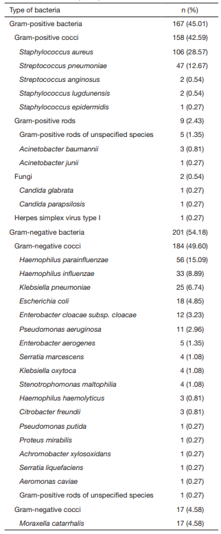
A total of 494 cases [237 (48%) boys, 257 (52%) girls; 647 eyes] of CNLDO were diagnosed based on discharge, epiphora, and mattering. Of these, 341 (69%) had unilateral dacryocystitis, 153 (31%) had bilateral infection. Among the 494 submitted specimens, 371 specimens were positive for bacteria (75.1%). Of those who tested positive, 201 (54.18%) had Gram negative bacteria (G−) bacteria and 167 (45.01%) had Gram positive bacteria (G+) bacteria. Some 28.57% of specimens showed Staphylococcus aureus infection; 15.09% were infected with Haemophilus parainfluenzae; and Streptococcus pneumoniae was present in 12.67%. Two fungal isolates (0.54%), Candida glabrata and Candida parapsilosis, were found (the proportions of the various bacteria are shown in Table 1).

Table 1 Strains classification based on bacteria culture results of lacrimal sac secretions (n=371)

Susceptibility testing results

Gram-positive organisms exhibited a high rate of sensitivity to levofloxacin (97.6%), ciprofloxacin (95.43%), and chloramphenicol (90.74%) and exhibited low sensitivity (1.2%) and strong resistance (98.2%) to erythromycin. Gram-negative bacteria exhibited a high degree of sensitivity to levofloxacin (96.02%), tobramycin (94.12%), ciprofloxacin (92.75%), and chloramphenicol (83.45%). Both Grampositive and Gram-negative bacteria were sensitive to levofloxacin, with an average effective rate of 96.81%.

Nasolacrimal duct probing

A total of 359 cases (181 boys, total 241 eyes; 178 girls, total 252 eyes; total 493 eyes) were included in this study. Of these, 261 had unilateral infection and 116 had bilateral infection. The cases were divided into two groups (children 3–6 months of age, 276 eyes; 7–12 months of age, 217 eyes). Among children from 3–6 months of age, the 1-time success rate of nasolacrimal duct probing alone was 95.65% and the 1-time success rate of probing combined with nasolacrimal duct injection of levofloxacin ophthalmic gel was 97.1% (no statistical significance, χ2 =0.104, P=0.747>0.05). Among children from 7–12 months of age, the 1-time success rate of nasolacrimal duct probing alone was 88.24% and the 1-time success rate of probing combined with nasolacrimal duct injection of levofloxacin ophthalmic gel was 96.52% (statistical significance, χ2 =5.435, P=0.02<0.05). The 1-time success rate for simple nasolacrimal duct probing alone was significantly higher in children from 3–6 months of age than for those from 7–12 months of age (statistical significance, χ2 =4.651, P=0.031<0.05). The 1-time success rates of combination therapy with nasolacrimal duct injection of levofloxacin ophthalmic gel showed no different between the two groups (no statistical significance, χ2 =0.000, P=1.000>0.05) (Tables 2-5).

Discussion

We obtained bacteria cultures of lacrimal sac secretions of children with dacryocystitis. Of the children whose secretions were cultured, 75.1% tested positive for pathogenic bacteria. The most common pathogen identified was Staphylococcus aureus, followed by Haemophilus parainfluenzae and Streptococcus pneumoniae. Susceptibility testing results demonstrated that levofloxacin was the most sensitive drug for both G− and G+ bacteria.

The distribution of pathogenic bacteria in children with dacryocystitis shows regional differences. Sun (22) reported that G+ strains were the most abundant pathogenic bacteria (74.5%) in children with dacryocystitis in Shenyang, although our results differed. The disparity may be due to different locations that vary in climate; the study by Sun took place in Northern China, whereas our facility is located in Southern China. In addition, Sun reported that coagulasenegative Staphylococcus is the most common pathogenic bacteria, followed by Staphylococcus aureus. Our results show that Staphylococcus aureus has become the primary pathogenic bacteria causing dacryocystitis in young children.

Staphylococcus aureus is the most common pathogenic bacteria for human suppurative infections; as a conditional pathogenic bacterium, it is often present in the oral cavity, nasopharynx, palpebral conjunctiva, urethral canal, and intestinal mucosa. The immune system in young children is immature, and their tears are deficient in IgA and IgM. In addition, their lacrimal gland secretion function is not perfect and their lacrimal duct is not smooth as well as their bacteriostatic ability and bacteria excretion are poor. Parents who are nursing a sick child will often wipe the child’s tears away, which can lead to infection caused by conditional pathogenic bacteria and local suppurative infection. 

Usha (23) reported the sensitivity of G+ and G− bacteria to levofloxacin as 75% and 83%, respectively. The sensitivities of Gram-positive and Gram-negative bacteria to levofloxacin, as reported by Sun (22), were 83.91% and 93.33%, respectively. Our results show that levofloxacin has become the most sensitive antibiotic for children with dacryocystitis, with an average efficacy rate of 96.81%. Thus, we believe that levofloxacin ophthalmic gel is the agent of choice for combination therapy with nasolacrimal duct probing.

According to our findings, the 1-time success rate of traditional nasolacrimal duct probing in the 3–6 months age group was 95.65% and that in the 7–12 months age group was 88.24%, which is slightly higher than the findings by Lipiec (24) (83% and 71%, respectively) and lower than those reported by Perveen (25) (100% and 94%, respectively). The 1-time success rate in the 7–12 months age group in our study is higher than that reported by Repka (13) (78%). The 1-time success rate of nasolacrimal duct probing for children from 3–6 months of age is generally higher than the rate for those older than 6 months. Therefore, the first nasolacrimal duct probing (27) should be performed in children from 3–6 months of age.

The lower success rate in children older than 6 months of age compared to children younger than 6 months is likely because children older than 6 months cry harder under local anesthesia, the duration of the procedure may cause greater tissue damage, and tissue proliferation may reduce the effectiveness of probing. However, some scholars (27) have suggested that inflammation did not promote the proliferation at the obstruction. Whether or not proliferation occurs, traditional nasolacrimal duct probing may inevitably cause mucosa edema of the lacrimal duct and even injury to mucosal epithelial cells. If inflammation lasts for a long time and postoperative tissue edema and reaction are severe, re-adhesion and obstruction may occur. Thus, the 1-time success rate in children older than 6 months is lower than those younger than 6 months.

We found that combining nasolacrimal duct injection of levofloxacin ophthalmic gel with probing significantly improves the 1-time success rate over probing alone in children aged over 6 months, from 88.24% to 96.52%.

Levofloxacin (28) is a third-generation fluoroquinolones that has fewer side effects. It kills bacteria by inhibiting their DNA gyrase action and blocking DNA reproduction. According to our results as well as previous studies, the vast majority of G+ and G− bacteria exhibit a high rate of sensitivity to levofloxacin.

Another advantage of levofloxacin ophthalmic gel is that its soft, gelatinous consistency is released slowly, thereby prolonging the duration of drug action. The main substrates are sodium hyaluronate and carbomer, the latter of which is characterized by high viscosity and good lubrication. Carbomer forms a protective film on the surface of nasolacrimal duct tissues. Sodium hyaluronate plays a role in expanding, supporting, and separating mucosal wounds and lubricating the application site. The degradation and liquification rates are rapid and will not cause residuals. One study supported the use of levofloxacin ophthalmic gel (29). Due to its strong bioadhesion, it may facilitate formation of a protective film on the surface of nasolacrimal duct tissues after probing and irrigation.

Congenital dacryostenosis (30) is a common condition in which the far end of the nasolacrimal duct underneath the inferior turbinate and epithelial cell debris blocking fails to complete its canalization in the newborn period. As the duration of the obstruction increases, serious infective complications occur. Inflammatory reaction can cause edema and hyperplasia and stimulate epithelial hyperplasia. If inflammation lasts for a long time and postoperative tissue edema and reaction are severe, re-adhesion and obstruction may occur. And in 3–6 month group, the duration of the inflammatory reaction was shorter the chance of readhesion after valve probing was low. Therefore, the support function of the gel is not needed. So the 1-time success rates of combination with levofloxacin ophthalmic gel or not showed no different in 3–6 month group. 

Table 2 Comparison of curative effect of two treatment methods in 3–6 months age group

                                       χ2 =0.104, P=0.747>0.05; the difference had no statistical significance. 

Table 3 Comparison of curative effect of two treatment methods in 7–12 months age group

                                     χ2=5.435, P=0.02<0.05; the difference had statistical significance. 

Table 4 Curative effect of simple nasolacrimal duct probing in different age groups

                                      χ2=4.651, P=0.031<0.05; the difference had statistical significance.

Table 5 Curative effect of nasolacrimal duct probing combined with nasolacrimal duct injection of levofloxacin ophthalmic gel in different age groups


                                     χ2=0.000, P=1.000>0.05; the difference had no statistical significance.

Conclusions

For children from 7–12 months of age, nasolacrimal duct probing combined with nasolacrimal duct injection of levofloxacin ophthalmic gel may increase the 1-time success rate of treatment.

1、Caputo GM, Appelbaum PC, Liu HH. Infections due to penicillin-resistant pneumococci. Clinical, epidemiologic, and microbiologic features. Arch Intern Med 1993;153:1301-10.
2、Imaizumi T, Yamashita K, Taima K, et al. Effect of peroxisome proliferator-activated receptorgamma ligands on the expression of retinoic acidinducible gene-I in endothelial cells stimulated with lipopolysaccharide. Prostaglandins Other Lipid Mediat 2005;78:46-54.
3、Eshraghi B, Akbari MR, Fard MA, et al. The prevalence of amblyogenic factors in children with persistent congenital nasolacrimal duct obstruction. Graefes Arch Clin Exp Ophthalmol 2014;252:1847-52.
4、Kim JW, Lee H, Chang M, et al. Amblyopia risk factors in infants with congenital nasolacrimal duct obstruction. J Craniofac Surg 2013;24:1123-5.
5、Ozgur OR, Sayman IB, Oral Y, et al. Prevalence of amblyopia in children undergoing nasolacrimal duct irrigation and probing. Indian J Ophthalmol 2013;61:698-700.
6、Piotrowski JT, Diehl NN, Mohney BG. Neonatal dacryostenosis as a risk factor for anisometropia. Arch Ophthalmol 2010;128:1166-9.
7、Dietrich DW, Auld DB, Mermel LA. Communityacquired methicillin-resistant Staphylococcus aureus in southern New England children. Pediatrics 2004;113:e347-52.
8、Kodsi S. Community-acquired methicillin-resistant Staphylococcus aureus in association with chronic dacryocystitis secondary to congenital nasolacrimal duct obstruction. J AAPOS 2006;10:583-4.
9、MacEwen CJ, Phillips MG, Young JD. Value of bacterial culturing in the course of congenital nasolacrimal duct (NLD) obstruction. J Pediatr Ophthalmol Strabismus 1994;31:246-50.
10、Al-Faky YH, Mousa A, Kalantan H, et al. A prospective, randomised comparison of probing versus bicanalicular silastic intubation for congenital nasolacrimal duct obstruction. Br J Ophthalmol 2015;99:246-50.
11、Kakizaki H, Takahashi Y, Kinoshita S, et al. The rate of symptomatic improvement of congenital nasolacrimal duct obstruction in Japanese infants treated with conservative management during the 1st year of age. Clin Ophthalmol 2008;2:291-4.
12、Kashkouli%20MB%2C%20Beigi%20B%2C%20Parvaresh%20MM%2C%20et%20al.%20Late%20and%20very%20late%20initial%20probing%20for%20congenital%20nasolacrimal%20duct%20obstruction%3A%20what%20is%20the%20cause%20of%20failure%3F%20Br%20J%20Ophthalmol%202003%3B87%3A1151-3.
13、Pediatric Eye Disease Investigator Group, Repka MX, Chandler DL, et al. Primary treatment of nasolacrimal duct obstruction with probing in children younger than 4 years. Ophthalmology 2008;115:577-584.e3.
14、Rajabi MT, Abrishami Y, Hosseini SS, et al. Success rate of late primary probing in congenital nasolacrimal duct obstruction. J Pediatr Ophthalmol Strabismus 2014;51:360-2.
15、Kashkouli MB, Kassaee A, Tabatabaee Z. Initial nasolacrimal duct probing in children under age 5: cure rate and factors affecting success. J AAPOS 2002;6:360-3.
16、Okumus S, Gürler B, Coskun E, et al. Efficiency of endoscopic imaging in repetitive probing following unsuccessful probing. Eur J Ophthalmol 2012;22:882-9.
17、Katowitz JA, Welsh MG. Timing of initial probing and irrigation in congenital nasolacrimal duct obstruction. Ophthalmology 1987;94:698-705.
18、Kapadia MK, Freitag SK, Woog JJ. Evaluation and management of congenital nasolacrimal duct obstruction. Otolaryngol Clin North Am 2006;39:959-77, vii.
19、Maheshwari R. Results of probing for congenital nasolacrimal duct obstruction in children older than 13 months of age. Indian J Ophthalmol 2005;53:49-51.
20、Ye YF, Wang YS, Shen ZY. National guide to Clinical Laboratory Procedures. 3rd edition. Southeast University Press, 2006.
21、Clinical and Laboratory Standards Institute. Reference method for broth dilution antifungal susceptibility testing of Yeasts; approved standard—second edition. Wayne, PA, 2002.
22、Sun H, Zhao JY, Yan QC, et al. Pathogens distribution and drug sensitivity of infantile dacryocystitis. Zhonghua Yan Ke Za Zhi 2010;46:34-7.
23、Usha K, Smitha S, Shah N, et al. Spectrum and the susceptibilities of microbial isolates in cases of congenital nasolacrimal duct obstruction. J AAPOS 2006;10:469-72.
24、Lipiec%20E%2C%20Gra%C5%82ek%20M%2C%20Niwald%20A.%20Evaluation%20of%20therapy%20outcome%20in%20congenital%20nasolacrimal%20duct%20obstruction%20in%20own%20material.%20Klin%20Oczna%202006%3B108%3A174-7.
25、Perveen S, Sufi AR, Rashid S, et al. Success rate of probing for congenital nasolacrimal duct obstruction at various ages. J Ophthalmic Vis Res 2014;9:60-9.
26、Baggio E, Ruban JM, Sandon K. Analysis of the efficacy of early probing in the treatment of symptomatic congenital lacrimal duct obstruction in infants. Apropos of 92 cases. J Fr Ophtalmol 2000;23:655-62.
27、Cahill KV, Burns JA. Management of epiphora in the presence of congenital punctal and canalicular atresia. Ophthal Plast Reconstr Surg 1991;7:167-72.
28、Segatore B, Setacci D, Perilli M, et al. Bactericidal activity of levofloxacin and ciprofloxacin on clinical isolates of different phenotypes of Pseudomonas aeruginosa. Int J Antimicrob Agents 2000;13:223-6.
29、Abraham S, Furtado S, Bharath S, et al. Sustained ophthalmic delivery of ofloxacin from an ion-activated in situ gelling system. Pak J Pharm Sci 2009;22:175-9.
30、Cassady JV. Developmental anatomy of nasolacrimal duct. AMA Arch Ophthalmol 1952;47:141-58.
上一篇
下一篇
其他期刊
  • 眼科学报

    主管:中华人民共和国教育部
    主办:中山大学
    承办:中山大学中山眼科中心
    主编:林浩添
    主管:中华人民共和国教育部
    主办:中山大学
    浏览
  • Eye Science

    主管:中华人民共和国教育部
    主办:中山大学
    承办:中山大学中山眼科中心
    主编:林浩添
    主管:中华人民共和国教育部
    主办:中山大学
    浏览
推荐阅读
出版者信息
目录