您的位置: 首页 > 2017年3月 第32卷 第1期 > 文字全文
2023年7月 第38卷 第7期11
目录

The early change of corneal vertical coma and trefoil in 2.8-mm superior incision cataract surgery

The early change of corneal vertical coma and trefoil in 2.8-mm superior incision cataract surgery

来源期刊: 眼科学报 | 2017年3月 第32卷 第1期 39-43 发布时间: 收稿时间:2024/12/30 8:16:48 阅读量:292
作者:
关键词:
Small incision cataract surgery corneal aberration vertical coma vertical trefoil
DOI:
doi: 10.3978/j.issn.1000-4432.2017.03.01
收稿时间:
2016-10-31 
修订日期:
 
接收日期:
2017-02-09 
Background: To investigate early change of corneal aberrations after 2.8-mm superior incision phacoemulsification.
Methods: This study comprised 80 eyes of 75 patients. All the patients underwent phacoemulsification with monofocal foldable intraocular lens (IOLs) implanted through a 2.8-mm superior corneal incision. The anterior corneal wavefront aberrations for the 6.0-mm pupillary diameter was measured by iTrace wavefront aberrometer (Tracey Technologies, Inc.) preoperatively and 1 month postoperatively. Changes of root mean square (RMS) values of Z(3, -3), Z(3, 3), Z(3, -1), Z(3, 1), and Z(4, 0) and total high order aberration (HOA) were evaluated.
Results: The uncorrected and corrected visual acuities improve significantly (P<0.001). No significant postoperative changes were observed in spherical aberration (P=0.652). Significant changes in vertical coma and vertical trefoil (0.005±0.214 vs. -0.049±0.242, P=0.037; -0.141±0.222 vs. -0.258±0.359, P=0.001; separately). However, the total HOAs increased after cataract surgery (0.567±0.161 vs. 0.688±0.343, P<0.001).
Conclusions: Corneal vertical coma and vertical trefoil changes significantly in 2.8 mm superior corneal incision phacoemulsification cataract surgery. In addition, those had a trend to negative direction.
Background: To investigate early change of corneal aberrations after 2.8-mm superior incision phacoemulsification.
Methods: This study comprised 80 eyes of 75 patients. All the patients underwent phacoemulsification with monofocal foldable intraocular lens (IOLs) implanted through a 2.8-mm superior corneal incision. The anterior corneal wavefront aberrations for the 6.0-mm pupillary diameter was measured by iTrace wavefront aberrometer (Tracey Technologies, Inc.) preoperatively and 1 month postoperatively. Changes of root mean square (RMS) values of Z(3, -3), Z(3, 3), Z(3, -1), Z(3, 1), and Z(4, 0) and total high order aberration (HOA) were evaluated.
Results: The uncorrected and corrected visual acuities improve significantly (P<0.001). No significant postoperative changes were observed in spherical aberration (P=0.652). Significant changes in vertical coma and vertical trefoil (0.005±0.214 vs. -0.049±0.242, P=0.037; -0.141±0.222 vs. -0.258±0.359, P=0.001; separately). However, the total HOAs increased after cataract surgery (0.567±0.161 vs. 0.688±0.343, P<0.001).
Conclusions: Corneal vertical coma and vertical trefoil changes significantly in 2.8 mm superior corneal incision phacoemulsification cataract surgery. In addition, those had a trend to negative direction.

Introduction

Cataract surgery can induce corneal changes. It has been reported that astigmatism and corneal aberration increase after cataract surgery (1,2). And surgically induced astigmatism and corneal aberrations are related to the location, type, length and whether suture incision (1,3,4)It is not seem to be smaller incision, surgically induced astigmatism (SIA) and high order aberrations decrease (5,6)
The purpose of our study was to investigate changes of corneal aberrations caused by phacoemulsification using 2.8 mm superior clear corneal incision.

Methods

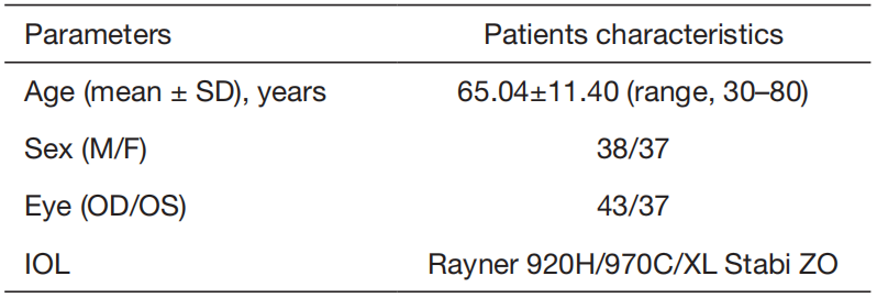
Eighty eyes of 75 patients with cataract were investigated.The sample comprised 38 males and 37 females with a mean age of 65.04±11.40 years (range, 30–80 years). All the patients underwent phacoemulsification and implantation of an acrylic aspheric intraocular lens (IOL) at Joint Shantou International Eye Center, Shantou University and the Chinese University of Hong Kong. The same surgeon (Mingzhi Zhang) performed all surgeries from July 2009 to March 2010. All patients provided informed consent. This
study followed the tenets of the Declaration of Helsinki.
The inclusion criteria included no history of refractive surgery, no systemic or ocular complications and no diseases that affect corneal refraction. The exclusion criteria included status after ocular trauma or intraocular surgery, any sign of inflammation or infection, corneal diseases, pseudoexfoliation syndrome, glaucoma, optic atrophy, diabetic retinopathy and lens dislocation.

Surgical technique

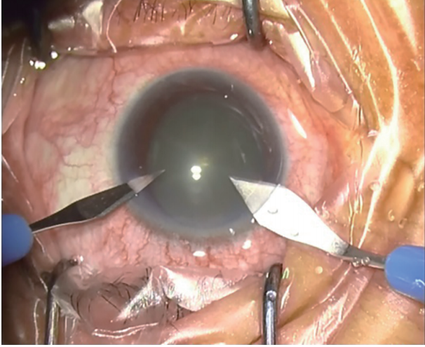
Topical anesthesia was employed with 0.05% Alcaine (Alcon). All surgeries were performed by the same surgeon uneventfully, with surgeon sitting superiorly, one-step 2.8-mm self-healing incision was made at 11 o’clock 0.5mm from limbus with a 2.8-mm corneal knife, and paracentesis at 2 o’clock (Figure 1). A continuous curvilinear capsulorhexis measuring approximately 5.5 mm in
diameter was created after anterior chamber was filled with viscoelastic. Nuclei were chopped after hydrodissection using the stop and chop phacoemulsification technique, an acrylic aspheric IOL (Rayner 920H/970C/ XL Stabi ZO) was implanted into the capsular bag after aspiration of cortical masses. The viscoelastic was completely removed from the anterior chamber. No sutures were placed at the end of the surgery. The postoperative medications included Prednisolone Acetate Eye Drops 1% and Ofloxacin Eye Drops 0.3% 6 times a day for 1 month.
Figure 1 Showing 2.8-mm self-healing incision was made at 11 o’clock and paracentesis at 2 o’clock.

Measurement

All the clinical examination data were recorded included corrected and uncorrected visual acuity, keratometry, intraocular pressure, axial length measurement, anterior chamber depth, slit lamp biomicroscopy, dilated fundus examination. Corneal aberrations in 6 mm optical zone were tested one day before and one month after cataract surgery. Because the 3rd-order and 4th-order aberrations such as trefoil, coma and spherical aberration constitute the major component of HOAs, we recorded those parameters. In this study, we used iTrace Visual Function Analyzer (Tracey Technologies, Inc., TX, USA).

Statistical analysis

Data were analyzed using SPSS 17.0 for windows software (SPSS, Inc., Chicago, IL, USA). Paired t-test was used to detect statistically significant difference in UCVA, BCVA, SE and corneal aberration between preoperative and postoperative values. The Spearman rank correlation was used to determine the relationship of corneal aberration changes between preoperatively and postoperatively. For statistical analysis of visual acuity, logarithm of minimum angle of resolution (LogMAR) values was used. A P value of <0.05 was considered statistically significant.

Results

The final analysis comprised 80 eyes of 75 patients (37 women,38 men; mean age ± SD, 65.04±11.40) (Table 1 shows basic profile of patients in this study). There were no intraoperative or postoperative complications. No wound leakage, hypotony, or intraocular pressure increase was noted during the follow-up period.
Table 2 shows the preoperative and postoperative visual acuity levels and spherical equivalent. The uncorrected and corrected visual acuities improved after surgery significantly.
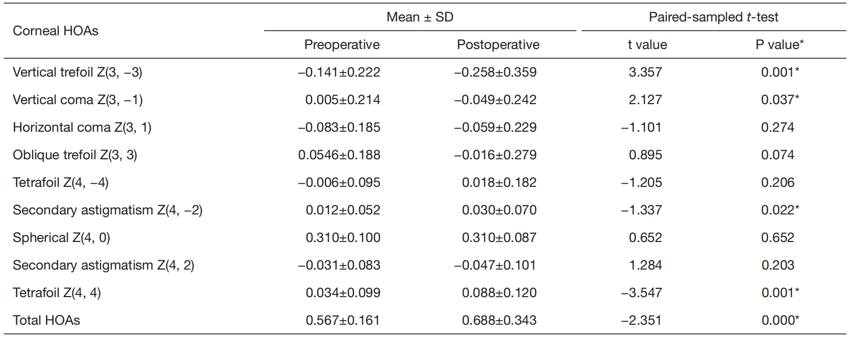
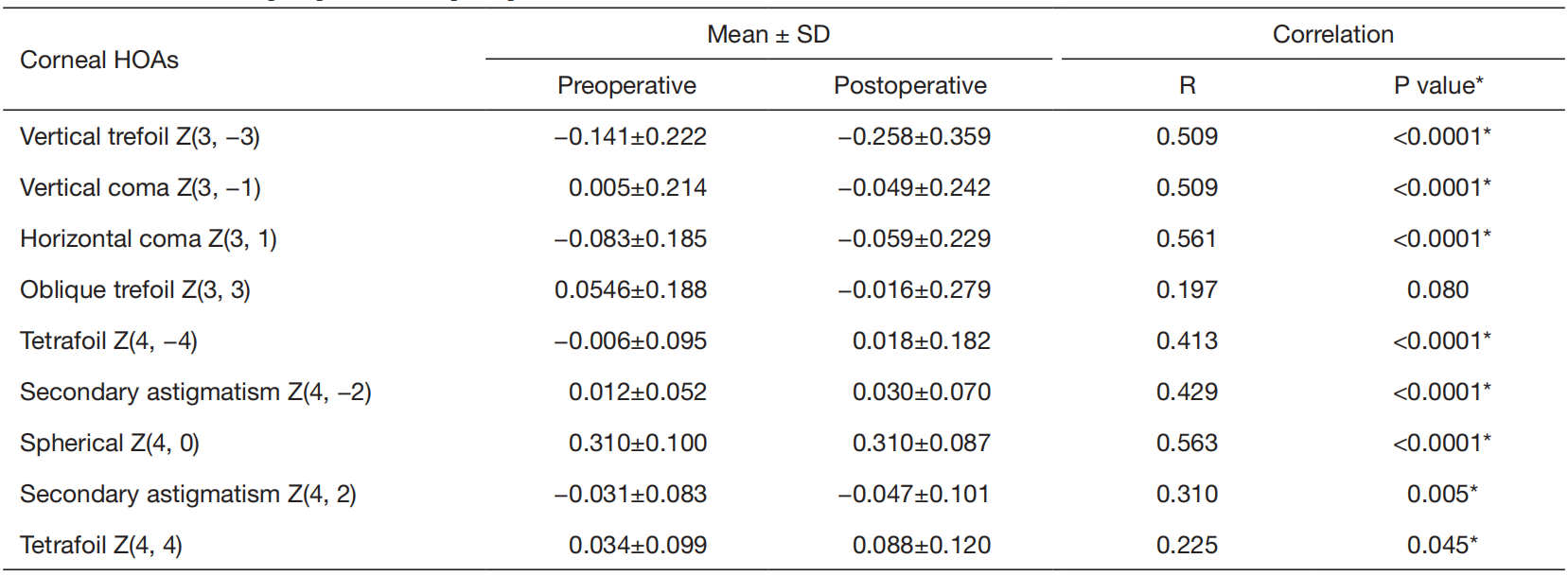
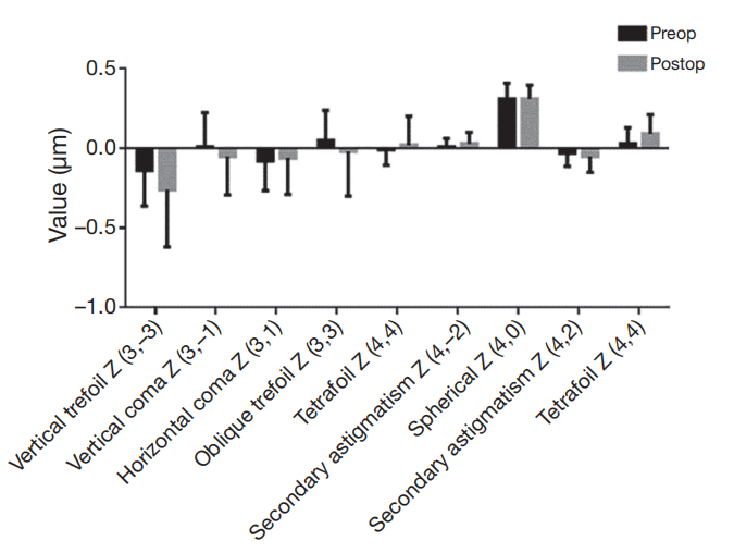
Table 3 shows changes in the Zernike terms of anterior cornea aberrations after surgery. There were significant changes in vertical coma and vertical trefoil between preoperative and postoperative (vertical coma: 0.005±0.214 vs. −0.049±0.242, P=0.037; vertical trefoil: −0.141±0.222 vs. −0.258±0.359, P=0.001). Furthermore, we found that vertical coma and vertical trefoil became more negative from preoperatively to postoperatively (Figure 2). No statistically significant difference were found in oblique trefoil (0.0546±0.188 vs. −0.016±0.279, P=0.074) and horizontal coma (−0.083±0.185 vs. −0.059±0.229, P=0.274). No significant postoperative changes were observed in spherical aberration (0.310±0.100 vs. 0.310±0.087, P=0.652), and a good correlation was found (R=0.563, P<0.001) (Table 4). However, the total HOAs increase after cataract surgery (0.567±0.161 vs. 0.688±0.343, P<0.001).
Table 1 Basic profile of patients in this study
IOL, intraocular lens; OD, right eye; OS, left eye.
Table 2 Preoperative and postoperative one month visual acuity and refraction
UCVA, uncorrected visual acuity; BCVA, best corrected visual acuity; SE, spherical equivalent; logMAR, logarithm of minimum angle of resolution. *, statistically significant (paired-sampled t-test).
Table 3 Changes in corneal HOAs preoperative and postoperative one month
HOA, high order aberration; *, statistically significant.
Table 4 Correlation of preoperative and postoperative corneal HOAs

HOA, high order aberration. Spearman rank correlation. *, statistically significant.
Figure 2 Shows anterior corneal high order aberrations before and after surgery

Discussion

We calculated only anterior corneal aberration because anterior cornea mainly contributed to the changes in the total cornea (7). There were significant changes in vertical coma and vertical trefoil between preoperative and postoperative (0.005±0.214 vs. −0.049±0.242, P=0.037; −0.141±0.222 vs. −0.258±0.359, P=0.001, separately). Our results showed no significant change in the spherical aberration before and after cataract surgery. This finding was similar to that in previous report (8). In the current study, corneal incisions located at 11 o’clock and 2 o’clock separately. The regional asymmetry in corneal shape may contribute to the ocular aberrations, asymmetry in corneal shape along the vertical principal meridian (inferior–superior) was significantly associated with the vertical trefoil and vertical coma of the cornea (9). And vertical trefoil and oblique trefoil are separated by 30 degrees and each trefoil repeats every 120 degrees vertical trefoil repeats at 90, 210, and 330 degrees, and oblique trefoil repeats at 60,180, and 300 degrees. For this reason, superior incision may induce vertical trefoil and vertical coma. The superior incision could induce a good or bad effect on optical quality, depending on the side of the vertical coma preoperatively (positive or negative) (1). It is reported that the eye’s trefoil and coma had positive coupling in retinal image quality and visual acuity (10). Previous study showed changes in trefoil, coma, astigmatism and spherical aberration with accommodative demand and increased depth of focus in several Crystal eyes (11). Also, OCT-based crystalline lens topography in accommodating eyes showed high-order irregularities, astigmatism, coma and trefoil increased with accommodation (12). Gong et al. found that ocular  aberrations such as trefoil, 2nd-order astigmatism and spherical aberration seemed to interact with objective contrast sensitivity and depth of focus (8). Furthermore, vertical coma aberration was found to be associated with better near visual acuity (13). However, coma and trefoil
with large values (approximately 1 mm) may significantly reduce visual quality (14). In our study, total HOAs increase significantly may mainly due to changes of vertical coma and trefoil.
Size and location of the incision have the greatest impact on surgery induce corneal aberrations (2,14-16). SIA and HOAs changed along with the corneal biomechanical properties changed, and corneal 3rd-order trefoil depended on incision width (17). It is reported that compared with superior and nasal incision, a temporal incision induces smaller corneal HOA changes (1,4). In our study, we found that vertical coma, and trefoil increase significantly postoperatively, and all of them became more negative (Figure 2). It also has been reported that superior incision in 2.2 mm cataract surgery cause corneal vertical coma changed to negative direction, because superior incision greater flattening effect occurred in superior cornea than in the inferior (1). Previous study showed that because CCIs cause flatten effect of the cornea, steepest-meridian CCI reduces preexisting astigmatism (18,19), but the effect of the difference was not large enough to decrease remaining astigmatism and HOAs (18).
The limitation of the current study was that we examined the postoperative cornea over a relatively short postoperative period. And we did not investigate nasal and temporal incision cataract surgery. Another limitation was that we did not calculate corneal low order aberrations and astigmatism. It is reported that 2.2-mm small-incision cataract surgery induced changes in the lower aberration [oblique astigmatism Z(2, −2)] of the total cornea (3).
In conclusion, 2.8 mm superior corneal incision cataract surgery may induce vertical coma, vertical trefoil increase, and those had a trend to negative direction. For the reason of manual CCI may induce corneal HOAs, femtosecond laser clear corneal incision (FS-CCI) for cataract surgery may benefit (20). Limbal relaxing incision may also be an effective method in reducing corneal astigmatism with neutral effect on corneal aberrations (19). Effect of corneal vertical coma and vertical trefoil changes need further research.


1、Song IS, Park JH, Park JH, et al. Corneal coma and trefoil changes associated with incision location in cataract surgery. J Cataract Refract Surg 2015;41:2145-51.
2、Park CY, Chuck RS, Channa P, et al. The effect of corneal anterior surface eccentricity on astigmatism after cataract surgery. Ophthalmic Surg Lasers Imaging 2011;42:408-15.
3、Febbraro JL, Wang L, Borasio E, et al. Astigmatic equivalence of 2.2-mm and 1.8-mm superior clear corneal cataract incision. Graefes Arch Clin Exp Ophthalmol 2015;253:261-5.
4、Maier AK, Gundlach E, Gonnermann J, et al. Superior versus temporal approach in descemet membrane endothelial keratoplasty. Am J Ophthalmol 2015;159:111-7.e1.
5、Yu YB, Zhu YN, Wang W, et al. A comparable study of clinical and optical outcomes after 1.8, 2.0 mm microcoaxial and 3.0 mm coaxial cataract surgery. Int J Ophthalmol 2016;9:399-405
6、Wei YH, Chen WL, Su PY, et al. The influence of corneal wound size on surgically induced corneal astigmatism after phacoemulsification. J Formos Med Assoc 2012;111:284-9.
7、Hidaka Y, Yamaguchi T, Saiki M, et al. Changes in corneal aberrations after cataract surgery. Jpn J Ophthalmol 2016;60:135-41.
8、Gong XH, Zheng QX, Wang N, et al. Visual and optical performance of eyes with different corneal spherical aberration implanted with aspheric intraocular lens. Int J Ophthalmol 2012;5:323-8.
9、Leung TW, Lam AK, Kee CS. Ocular Aberrations and Corneal Shape in Adults with and without Astigmatism.Optom Vis Sci 2015;92:604-14.
10、Villegas EA, Alcón E, Artal P. Impact of positive coupling of the eye's trefoil and coma in retinal image quality and visual acuity. J Opt Soc Am A Opt Image Sci Vis 2012;29:1667-72.
11、Pérez-Merino P, Birkenfeld J, Dorronsoro C, et al. Aberrometry in patients implanted with accommodative intraocular lenses. Am J Ophthalmol 2014;157:1077-89.
12、Pérez-Merino P, Velasco-Ocana M, Martinez-Enriquez E, et al. OCT-based crystalline lens topography in accommodating eyes. Biomed Opt Express 2015;6:5039-54
13、Song IS, Kim MJ, Yoon SY, et al. Higher-order aberrations associated with better near visual acuity in eyes with aspheric monofocal IOLs. J Refract Surg 2014;30:442-6.
14、Chu L, Zhao JY, Zhang JS, et al. Optimal incision sites to reduce corneal aberration variations after small incision phacoemulsification cataract surgery. Int J Ophthalmol 2016;9:540-5.
15、Kim SW, Yang H, Yoon G, et al. Higher-order aberration changes after Implantable Collamer Lens implantation for myopia. Am J Ophthalmol 2011;151:653-662.e1.
16、Hashemi H, Khabazkhoob M, Soroush S, et al. The location of incision in cataract surgery and its impact on induced astigmatism. Curr Opin Ophthalmol 2016;27:58-64.
17、Denoyer A, Ricaud X, Van Went C, et al. Influence of corneal biomechanical properties on surgically induced astigmatism in cataract surgery. J Cataract Refract Surg 2013;39:1204-10.
18、Hayashi K, Yoshida M, Yoshimura K. Effect of steepestmeridian clear corneal incision for reducing preexisting corneal astigmatism using a meridian-marking method or surgeon's intuition. J Cataract Refract Surg 2014;40:2050-6.
19、Eliwa TF, Abdellatif MK, Hamza II. Effect of Limbal Relaxing Incisions on Corneal Aberrations. J Refract Surg 2016;32:156-62.
20、Serrao S, Lombardo G, Schiano-Lomoriello D, et al. Preliminary investigation of corneal wavefront aberration following femtosecond laser clear corneal incision for cataract surgery. Eur J Ophthalmol 2014;24:842-9.
上一篇
下一篇
其他期刊
  • 眼科学报

    主管:中华人民共和国教育部
    主办:中山大学
    承办:中山大学中山眼科中心
    主编:林浩添
    主管:中华人民共和国教育部
    主办:中山大学
    浏览
  • Eye Science

    主管:中华人民共和国教育部
    主办:中山大学
    承办:中山大学中山眼科中心
    主编:林浩添
    主管:中华人民共和国教育部
    主办:中山大学
    浏览
推荐阅读
出版者信息
目录