您的位置: 首页 > 文字全文
2023年7月 第38卷 第7期11
目录

先天性葡萄膜外翻继发性青光眼:病例系列报道与文献回顾

Glaucoma secondary to congenital ectropion uveae: case series report and literature review

来源期刊: 眼科学报 | 1-8 发布时间:2024-12-02 收稿时间:2024/11/29 16:54:20 阅读量:404
作者:
关键词:
青光眼 葡萄膜外翻 虹膜外翻 继发性青光眼 小梁切除术
glaucoma congenital ectropion uveae iris ectropion secondary glaucoma trabeculectomy
DOI:
10.12419/24082701
收稿时间:
2024-08-27 
修订日期:
2024-09-18 
接收日期:
2024-10-03 

目的:分析先天性葡萄膜外翻(Congenital ectropion uveae, CEU)继发性青光眼的临床表现、影像学特点和治疗方法。方法:回顾分析就诊于中山大学中山眼科中心诊断为CEU继发性青光眼患者的电子病历资料,收集整理患者的一般情况、视力、眼压、裂隙灯检查、房角镜照相、超声生物显微镜等检查结果,以及手术治疗方式选择和术后随访眼压情况,并结合国内外文献回顾分析总结CEU继发性青光眼的临床特点和诊疗效果。结果:报告两例CEU继发性青光眼患者年龄分别为24岁和11岁,均为男性、单眼发病慢性病程,患眼都为右眼,都不伴有全身异常,最佳矫正视力为手动/40cm-0.16,就诊时平均眼压为37 mmHg,平均中央前房深度为3.45 mm。两例患者查体表现:均角膜透明,瞳孔大小约5-5.5mm,对光反应灵敏,瞳孔缘近360°葡萄膜外翻呈棕褐色,晶状体透明,眼底检查青光眼性视杯凹陷,视网膜平伏。其中24岁患者房角镜检查全周宽角开放,小梁网有色素沉着。超声生物显微镜(ultrasound biomicroscopy, UBM)检查提示右眼虹膜稍向后凹,7点位房角关闭,其余全周房角开放。而11岁患者房角镜检查以及UBM检查均为全周房角关闭。两例患者角膜内皮镜检查均正常。两例患者均诊断为右眼先天性葡萄膜外翻继发性青光眼,眼压高,最大量降眼压药物治疗下不能控制,对24岁患者行小梁切除联合术中使用抗代谢药物11岁患者行引流阀植入联合术中使用抗代谢药物,术中术后均无明显并发症,至今随访6个月,眼压控制良好。结论:CEU特征性改变为瞳孔缘葡萄膜外翻,常继发青光眼,可表现为开角型、房角发育异常(闭角型,降眼压药物治疗效果有限,通常需要手术治疗。由于CEU继发性青光眼人群以儿童和年轻患者为主,为难治性青光眼,因此抗青光眼术中联合使用抗代谢药物可获得较好的治疗效果。

Objective: To analyze the clinical manifestations, imaging characteristics, and treatment methods of secondary glaucoma associated with congenital ectropion uveae (CEU). Methods: Observational case series and literature review. A retrospective analysis was conducted on the electronic medical records of patients diagnosed with secondary glaucoma due to CEU at the Zhongshan Ophthalmic Center, Sun Yat-sen University, China. Data collected included demographics, visual acuity, intraocular pressure (IOP), slit-lamp examination, gonioscopy, ultrasound biomicroscopy (UBM), and surgical treatment methods. Postoperative IOP were also collected. A literature review was conducted to summarize the clinical characteristics and treatment outcomes of secondary glaucoma due to CEU. Results: Two male patients with secondary glaucoma due to CEU were included, aged 24 and 11 years, respectively. Both patients had a chronic course of disease affecting the right eye, with no systemic abnormalities. The best-corrected visual acuity was hand motion/40 cm to 0.16, and the average IOP at presentation was 37 mmHg, with an average central anterior chamber depth of 3.45 mm. Clinical examination revealed clear corneas, pupils measuring approximately 5-5.5 mm with brisk light reflexes, and nearly 360° of brownish ectropion of the uveae. The lenses were clear, and fundus examination showed glaucomatous optic disc cupping with a normal retina. In the 24-year-old patient, gonioscopy revealed a wide-open angle with trabecular meshwork pigmentation, while UBM indicated slight posterior bowing of the iris and angle closure at the 7 o'clock position. In the 11-year-old patient, both gonioscopy and UBM showed complete angle closure. Corneal endothelial microscopy was normal in both patients. Both were diagnosed with secondary glaucoma due to CEU in the right eye, with elevated IOP that was uncontrolled by maximal medications. The 24-year-old underwent trabeculectomy with intraoperative use of antimetabolites, while the 11-year-old received a drainage valve implantation with antimetabolites. No significant intraoperative or postoperative complications were observed, and IOP was well controlled over a six-month follow-up period. Conclusions: CEU is characterized by ectropion of the uveae and is frequently associated with secondary glaucoma, which may present as open-angle or angle-closure (goniodysgenesis) glaucoma. The effectiveness of IOP-lowering medications is limited, often necessitating surgical intervention. Given that secondary glaucoma due to CEU predominantly affects children and young adults, the use of antimetabolites during glaucoma surgery can yield favorable treatment outcomes.

葡萄膜外翻是以虹膜色素上皮过度增生覆盖于虹膜基质前表面为主要表现的罕见病,分为先天性葡萄膜外翻和继发性葡萄膜外翻。先天性葡萄膜外翻(Congenital ectropion uveae, CEU)最早由眼科医生Wicherkiewicz1891年和Spiro1896年首次报告虹膜外翻综合征,至今国内外文献报道约百余例,可合并其他眼部和全身异常,尤其是合并继发性青光眼最为多见,推测与神经外胚层和神经嵴发育异常有关。本研究回顾分析2CEU继发性青光眼病例,综合眼部检查特征,分析其可能的发病机制,并结合既往文献报道的手术方式选择,探讨CEU继发性青光眼这一罕见疾病的临床特点和诊治思考。

1 资料和方法

本研究为回顾性病例系列研究,回顾性收集了2022-2024年于中山大学中山眼科中心确诊为CEU合并继发性青光眼患者的病历资料。根据既往文献报道[1],纳入标准如下:(1)虹膜色素上皮异常增生通过瞳孔区覆盖至虹膜前表面,呈环形或扇形,界限清楚,表面扁平光滑;(2)合并继发性青光眼:经多次眼压测量,眼压>21 mmHg (1 mmHg = 0.133 kPa)。排除标准为:(1)合并外伤或虹膜新生血管等因素导致的继发性葡萄膜外翻,以及其他合并非获得性眼部异常/全身疾病或综合征的继发性儿童青光眼;(2)既往曾行内眼手术。本研究符合赫尔辛基宣言的伦理准则,通过中山大学中山眼科中心伦理委员会审查并通过豁免知情同意申请(2024KYPJ094)。本研究报告2例患者,病例资料如下。

1.1 病例1

患者男性,24岁,因右眼视力下降2年伴眼胀眼痛1个月就诊于中山大学中山眼科中心。2年前无明显诱因出现右眼视力逐渐下降,未规范诊治。近1个月来,患者右眼开始出现眼红眼胀眼痛,就诊于当地医院诊断为右眼青光眼,给予局部布林佐胺噻吗洛尔、溴莫尼定、他氟前列素滴眼液治疗,症状无明显好转。为求进一步诊疗来我院就诊。其父母非近亲,家庭成员均健康。患者否认相关疾病家族史,否认早产病史、手术史等。

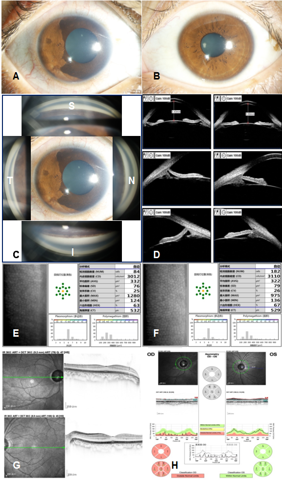
眼科检查:双眼眼球运动正常,眼睑形态和位置正常,未见明显上睑下垂。裸眼视力:右眼手动/40 cm,左眼0.5,最佳矫正视力(Best corrected visual acuity, BCVA)右眼3.50 DS/–0.50 DC×50°无提高,左眼为1.00 DS/–1.00 DC×100°1.0。眼压:右眼40.0 mmHg,左眼13 mmHg。裂隙灯显微镜检查(图1, A-B右眼结膜充血,角膜透明,角膜横径12.0 mm,角膜后沉积(keratic precipitates, KP)(-),前房中等深度,房水闪辉(-),瞳孔大小约5.5 mm,对光反应灵敏,瞳孔缘近360°葡萄膜外翻呈棕褐色,晶状体透明,晶状体前囊膜表面可见散在细小棕褐色色素颗粒沉着,眼底检查视乳头界清、色苍白,视杯与视盘直径比约1.0,视网膜平伏;左眼未见明显异常


1  病例1的术前图像

Figure 1 Preoperative Images of Case 1

(A) 右眼结膜轻度充血,角膜透明,瞳孔约5.5 mm大小,向鼻侧移位,近360°葡萄膜外翻呈棕褐色覆盖于正常虹膜表面;(B) 左眼眼前段照相,虹膜瞳孔均未见明显异常;(C) 右眼房角镜照相:左、上、右、下图分别为颞侧、上方、鼻侧、下方房角镜图像,中央为眼前段照相,可见全周房角宽角开放,未见明显粘连,上方房角和下方房角的小梁网可见棕褐色色素颗粒沉着;(D) 超声生物显微镜检查: 右眼中央前房深度3.43 mm,周边虹膜后凹(左上图),7点位虹膜根部附着点靠前、房角关闭(左下图),其余全周房角开放(左中图所示为9点位房角)。左眼中央前房深度3.50 mm(右上图),虹膜未见明显异常,9点、3点位房角结构未见明显异常(分别为右中图和右下图);(E,F) 右眼(E)角膜内皮细胞密度、形态、六边形比例与左眼(F)对比未见明显异常;(G) 双眼的黄斑区形态结构均基本正常。(H) 右眼视盘周围神经纤维层厚度各个方位均显著变薄,左眼视盘周围神经纤维层厚度正常。

(A) Anterior segment of the right eye: mild conjunctival congestion, clear cornea, pupil measuring 5.5 mm, corectopia displaced towards the nasal side, with extensive hyperpigmented ectropion uveae covering the normal iris surface; (B) Anterior segment photography of the left eye shows no obvious abnormalities in the iris and pupil; (C) Gonioscopic photographs of the right eye: The left, upper, right, and lower images represent the temporal, superior, nasal, and inferior gonioscopic views, respectively. The central image is an anterior segment photograph. It shows a wide-open angle with no obvious iridotrabecular contact. Pigment granules, brownish in color, can be seen deposited on the trabecular meshwork, primarily in the superior and inferior angles; (D) Ultrasound biomicroscopy: The central anterior chamber depth of the right eye is 3.43 mm, with posterior or concave bowing of the peripheral iris (upper left image). There is anterior iris insertion at the 7 o'clock position (lower left image), while the rest of the angles are open (middle left image shows the angle at the 9 o'clock position). The central anterior chamber depth of the left eye is 3.50 mm, and no abnormalities are observed in the iris (upper right image). The angle structures at the 9 o'clock and 3 o'clock positions show no significant abnormalities (as seen in the middle right and lower right images, respectively); (E, F) The corneal endothelial cell density, morphology, and hexagonal-shaped cell ratio of the right eye (E) show no significant abnormalities compared to the left eye (F); (G) The morphology and structure of the macular region in both eyes are normal; (H) The retinal nerve fiber layer (RNFL) thickness around the optic disc in the right eye is significantly thinned in all quadrants, while the RNFL thickness around the optic disc in the left eye is normal.

      辅助检查:房角镜检查结果
(1, C)提示,右眼全周房角宽角开放,小梁网可见色素沉着,呈浅棕色II级,左眼全周房角宽角开放,未见明显色素沉着。眼球生物测量(IOLMaster 700)检查结果提示,右眼眼轴长度为26.65 mm,左眼眼轴长度为24.88 mm超声生物显微镜(ultrasound biomicroscopy, UBM)检查(1D)显示右眼中央前房深度3.43 mm虹膜稍向后凹,全周晶状体悬韧带回声可见,7点位房角关闭,其余全周房角开放。左眼中央前房深度3.50 mm,虹膜平坦,全周房角开放。角膜内皮镜检查(1, E-F)结果提示,右眼角膜内皮细胞密度、形态和六边形细胞比例未见明显异常;相干光断层扫描 (optical coherence tomography, OCT)检查提示(1, G-H)双眼黄斑区形态结构基本正常;右眼视盘周围神经纤维厚度弥漫变薄,左眼视盘周围神经纤维厚度正常

结合患者病史及眼科检查,病例1临床诊断:右眼继发性青光眼,右眼先天性葡萄膜外翻,双眼屈光不正。

右眼在最大量局部降眼压药物治疗下布林佐胺噻吗洛尔、溴莫尼定、他氟前列素滴眼液),眼压仍不能控制(40 mmHg),故予以右眼行小梁切除术,术中使用抗代谢药物丝裂霉素C,浓度为2 mg/5 mL,同时放置调整缝线。

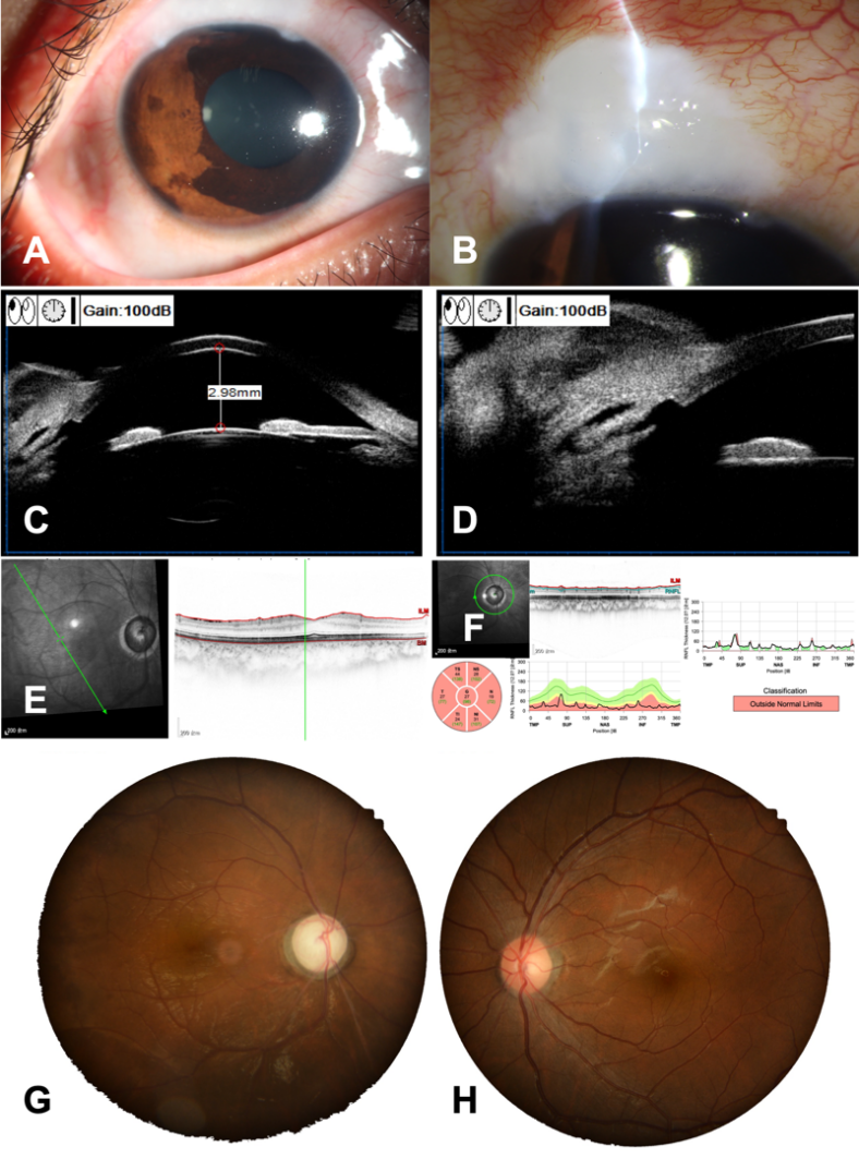
术后第1天,右眼视力从术前手动/40 cm提高至指数/20 cm,眼压6.3 mmHg,上方滤过泡弥散隆起,前房深。术后给予醋酸泼尼松龙滴眼液每天4次,妥布霉素地塞米松眼膏每晚1次。术后1个月内根据眼压、滤过泡、前房情况拆除可调整缝线,术中及术后均未出现并发症。术后1个月时,右眼裸眼视力为手动/30 cm,矫正无提高,眼压10 mmHg。术后6个月时,右眼裸眼视力为手动/30 cm,矫正无提高,眼压7 mmHg(未用药),右眼术后的眼前段照相、UBMOCT和眼底照相检查结果见图2


2  病例1的术后图像

Figure 2 Postoperative Images of Case 1
(A, B) 右眼小梁切除术后的眼前段照相示前房深、滤过泡隆起;(C, D) 右眼术后UBM示周切口通畅,中央前房深度2.98 mm(E, F) 右眼OCT提示黄斑区形态结构基本正常,视盘周围神经纤维厚度各个方位均显著变薄;(G, H) 眼底照相示:右眼青光眼性视杯凹陷,左眼未见明显异常。
(A, B) Anterior segment photography of the right eye after trabeculectomy shows a deep anterior chamber and a filtering bleb; (C, D) Postoperative UBM of the right eye shows a patent peripheral iridectomy, with a central anterior chamber depth of 2.98 mm; (E, F) OCT of the right eye indicates normal macular morphology and structure, with significant thinning of the peripapillary retinal nerve fiber layer in all quadrants; (G, H) Fundus photography shows: glaucomatous optic disc cupping in the right eye, with no significant abnormalities observed in the left eye.

病例2

患者男性,11岁,因体检发现右眼眼压高2就诊于中山大学中山眼科中心。2个月前外院体检发现右眼眼压56 mmHg,给予布林佐胺噻吗洛尔、他氟前列素滴眼液治疗,眼压仍不能控制。为求进一步诊疗来我院就诊。患者及家属否认相关疾病家族史,否认早产病史、手术史等。
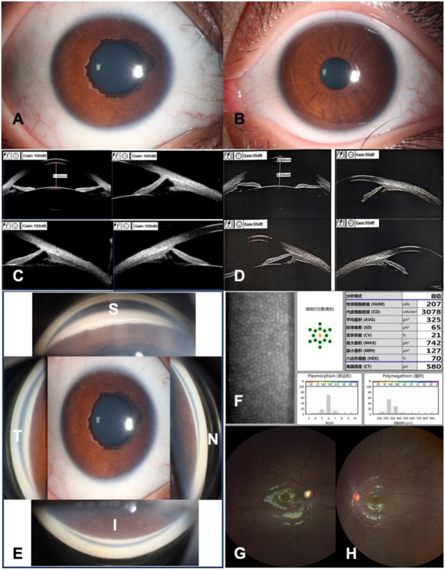
眼科检查:双眼眼球运动、眼睑形态和位置正常。裸眼视力:右眼0.16,左眼1.0验光矫正均无提高。眼压:右眼34.0 mmHg,左眼19.3 mmHg。裂隙灯显微镜检查(图3, A-B)右眼结膜无明显充血,角膜透明,角膜横径约13.0 mmKP(-),前房中等深度,房水闪辉(-),瞳孔大小约5 mm,对光反应灵敏,瞳孔缘近360°葡萄膜外翻呈棕褐色,晶状体透明,眼底检查视乳头界清、色苍白,视杯与视盘垂直径比约0.9,视网膜平伏;左眼未见明显异常。辅助检查:房角镜检查结果提示(图3, E),右眼全周房角虹膜根部附止高位,粘连关闭,房角发育异常,左眼全周房角宽角开放。超声生物显微镜(UBM)检查(3, C-D)显示右眼中央前房深度3.48 mm,全周房角关闭, 左眼中央前房深度3.54 mm,全周房角开放。角膜内皮镜检查(3, F)结果提示,右眼角膜内皮细胞密度、形态和六边形细胞比例未见明显异常。眼底照相提示(3, G-H):右眼青光眼性视杯凹陷,左眼未见明显异常。

3 病例2的术前图像

Figure 3 Preoperative Images of Case 2
(A) 右眼角膜透明,瞳孔约5 mm大小,近360°葡萄膜外翻覆盖于瞳孔缘的虹膜前表面,边界清楚;(B) 左眼眼前段照相,虹膜瞳孔均未见明显异常;(C, D) 超声生物显微镜检查: 右眼中央前房深度3.48 mm,全周虹膜根部附着点靠前、房角关闭(右上、左下、右下图分别为3点、6点、9点位房角结构);左眼中央前房深度3.54 mm,全周房角开放(右上、左下、右下图分别为3点、6点、9点位房角结构);(E) 右眼房角镜照相:左、上、右、下图分别为颞侧、上方、鼻侧、下方房角镜图像,中央为眼前段照相,可见全周房角虹膜根部附止靠前、粘连关闭、房角发育异常;(F) 右眼角膜内皮细胞密度、形态、六边形比例未见明显异常;(G, H) 眼底照相示:右眼青光眼性视杯凹陷,左眼未见明显异常
(A) The cornea of the right eye is clear, with a pupil measuring 5 mm. Nearly 360° of uveal ectropion covers the anterior surface of the iris, with clear boundaries; (B) Anterior segment photography of the left eye shows no obvious abnormalities in the iris and pupil; (C, D) Ultrasound biomicroscopy: The central anterior chamber depth of the right eye is 3.48 mm, with anterior iris insertion and angle-closure (the upper right, lower left, and lower right images show the angle structures at the 3, 6, and 9 o‘clock positions, respectively). The central anterior chamber depth of the left eye is 3.54 mm, with open angles (the upper right, lower left, and lower right images show the angle structures at the 3, 6, and 9 o‘clock positions, respectively); (E) The left, upper, right, and lower images represent the temporal, superior, nasal, and inferior gonioscopic views, respectively. Gonioscopic photography shows that the iris root is attached anteriorly, with goniodysgenesis. The central image is the anterior segment photograph; (F) The corneal endothelial cell density, morphology, and hexagonal cell ratio of the right eye are normal. (G, H) Fundus photography reveals: glaucomatous optic disc cupping in the right eye, while no significant abnormalities are observed in the left eye.
       结合患者病史及眼科检查,病例2临床诊断:右眼继发性青光眼,右眼先天性葡萄膜外翻。右眼在局部降眼压药物治疗下(布林佐胺噻吗洛尔、他氟前列素滴眼液),眼压仍不能控制(34 mmHg),予以右眼行Ahmed青光眼引流阀(FP7)植入术,术中使用抗代谢药物丝裂霉素C,浓度为2 mg/5 mL。
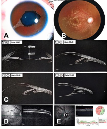
       术后第1天,右眼BCVA视力0.2,眼压13.3 mmHg, 前房深。术中及术后均未出现并发症。术后1个月时,右眼裸眼视力0.2,眼压9 mmHg。术后6个月时,右眼裸眼视力0.2,–1.50 DS/–1.75 DC×180°可矫正至0.8,眼压15.7 mmHg(未用药),右眼术后的眼前段照相、UBM和OCT检查结果见图4。

4  病例2的术后图像

Figure 4 Postoperative Images of Case 2
(A) 右眼引流阀植入术后的眼前段照相示前房深、8点位可见引流管;(B) 右眼眼底照相示青光眼性视杯凹陷; (C) 右眼术后UBM示中央前房深度3.53 mm8点位左右可见引流管回声;(D, E) 右眼OCT提示黄斑区形态结构基本正常,视盘周围神经纤维层厚度除鼻侧和鼻下方外,各个方位均显著变薄。
(A) Anterior segment photography of the right eye after surgery shows a deep anterior chamber and the tube of the glaucoma drainage device placed in the anterior chamber at the 8 o'clock position; (B) Fundus photography shows: glaucomatous optic disc cupping in the right eye; (C) Postoperative UBM of the right eye shows the Ahmed glaucoma valve tube located in the anterior chamber at the 8 o'clock position; (D, E) OCT of the right eye indicates normal macular morphology and structure, with significant thinning of the peripapillary retinal nerve fiber layer in all quadrants except for the nasal and inferonasal regions.

2 讨论

       先天性葡萄膜外翻,也称为先天性虹膜外翻或原发性虹膜色素上皮增生,是一种以虹膜色素上皮层异常增生,通过瞳孔缘覆盖至前部虹膜基质为主要表现的罕见疾病[1,2]。眼部表现为虹膜及瞳孔区呈现扇形或环形的色素上皮层外翻,受累区域宽窄不一,外翻组织表面色素增多呈棕褐色,同时由于其质地光滑平坦、凹陷及虹膜纹理减少,因此与正常虹膜的分界比较清楚。现普遍认为先天性葡萄膜外翻一词的准确描述由Wicherkiewicz于1891年以及Spiro于1896年提出[2]。既往文献指出CEU多为单眼发病,偶有双眼发病报道[1]
       目前认为CEU的发病机制可能是由于胚胎期神经外胚层发育异常导致虹膜色素上皮异常增生引起的。同时也有研究报道,CEU可合并其他眼部异常,包括虹膜缺损[3]、上睑下垂[4]等,以及Axenfeld-Rieger综合征、神经纤维瘤病1型[5]、半侧面部肥大等全身性疾病[6]。CEU中观察到的增生常仅限于虹膜色素上皮,而瞳孔括约肌和虹膜基质则未受影响,这与继发性葡萄膜外翻有所不同,这也解释了为何本文中2例CEU的瞳孔对光反射是可引出的。继发性葡萄膜外翻是由于前部虹膜表面存在牵引力导致后部虹膜色素上皮组织向外移位或外翻拉到虹膜前表面。继发性葡萄膜外翻相对更常见,多与继发因素有关,常见的病因包括眼部外伤[7]、虹膜角膜内皮综合征、Axenfeld-Rieger综合征[8]、虹膜肿瘤[9]等疾病。由于Axenfeld-Rieger异常也可表现有葡萄膜外翻,因此还需详细检查是否合并其他虹膜和房角的发育异常,如广泛的虹膜萎缩、变薄和孔洞形成,以及通过房角镜检查是否合并周边角膜后胚胎环。另外Axenfeld-Rieger异常一般为双眼发病,同时还可合并全身异常(Axenfeld-Rieger综合征),如面部发育异常(眼距过宽等)、牙齿(小牙,缺齿)、心脏、脐部以及泌尿系统的发育异常等,可进一步鉴别Axenfeld-Rieger综合征和先天性葡萄膜外翻。本研究的2例患者未合并其他眼部继发因素,根据特征性的临床表现和辅助检查结果,排除继发性葡萄膜外翻和其他合并非获得性眼部异常/全身疾病或综合征的继发性儿童青光眼[11],诊断为先天性葡萄膜外翻合并继发性青光眼。
       CEU继发性青光眼目前具体的发病机制尚不明确,可能与胚胎期神经嵴和中胚叶同时出现异常导致的小梁网、Schlemm管和前房角结构发育异常有关[10]。房水流出途径各个环节的功能障碍均会导致房水外流受阻,眼压升高,最终发生继发性青光眼。既往文献报道,此类继发性青光眼存在不同类型:开角型、房角发育异常型(表现为虹膜根部附止靠前,房角呈不同程度的粘连关闭)[11]。房角结构改变的类型可能与胚胎期神经嵴发育异常累及房角的范围和程度不同有关,开角型和房角发育异常型在各年龄段均有病例报道。例如叶天才和彭寿雄[12]在1986年最早于国内报道了3例CEU患者继发性青光眼均为开角型,平均就诊年龄为19岁(17-23岁),推测可能原因为胚胎期房角发育异常累及范围较小、程度较轻,以开角型为主,发病较迟,大多至青年时期发病,随着病情进展此时房角功能受损不足以控制眼压,患者逐渐出现高眼压导致的临床症状从而就诊,这与本研究的病例1类似。而刘学群等[13]报道11例16只眼诊断为CEU,就诊年龄中位数为11岁(3个月-30岁),除了1例2眼诊断为高眼压症外,其余10例14只眼均合并继发性青光眼,经前房角镜检查提示虹膜根部靠前附着和UBM提示前房角关闭。另一方面,Jacobson等[1]报道的8例11眼CEU继发性青光眼患者,平均就诊年龄为8.6岁(0-14.1岁),其中角膜透明能观察到房角结构的9例CEU眼中,只有1例为开角型,其余8例均为全周房角发育异常,推测可能是胚胎期房角发育异常累及范围较大,以房角发育异常型为主,房角功能损害较重,较早出现眼压增高的临床表现,就诊较早,这与我们报道的病例2类似。以上病例报道提示,房角改变类型可能是由房角发育异常的范围和程度决定的。
       近期许淑霞等[14]报道的6例6眼CEU继发性青光眼,就诊年龄中位数为10岁(3-31岁),均为房角发育异常(闭角型),关闭范围270-360°,且房角镜检查提示房角关闭范围与其葡萄膜外翻范围呈对应趋势。因此也有研究提出CEU继发性青光眼可能存在由开角型发展为闭角型的特点[1]:可能是由于发育异常的虹膜及房角表面覆盖着一层膜状物,随着疾病进展,膜状物增生逐渐牵拉引起周边虹膜前粘连,导致房角关闭[5,10,15]。然而这与绝大多数文献报道的先天性葡萄膜外翻为非进行性疾病相悖。我们认为两者之间可能并无直接关联,其原因可能与两者的发育来源不同有关:葡萄膜外翻是由于胚胎期神经外胚层发育异常导致的,由虹膜色素上皮异常增生通过瞳孔缘覆盖至虹膜表面;而房角的结构异常(周边粘连的程度和范围)是由于胚胎期神经嵴和中胚叶发育异常导致的。因此,对于葡萄膜外翻的范围与房角改变之间是否有关,目前尚无准确定论。这也可从既往文献图像展示的内容中找到相应的病例报道,比如,葡萄膜外翻范围大的患者:可表现为开角[4,26,29],也可表现为房角发育异常(闭角型)[14,30];而葡萄膜外翻范围小的患者(累及瞳孔缘的):可表现为开角[6,25],也可表现为房角发育异常(闭角型)[5,13,34]
       本研究的病例1的CEU继发性青光眼可能原因有:(1)开角型因素:本例患者的房角镜检查提示患眼全周房角基本为宽角开放,而且上方和下方小梁网可见棕褐色色素颗粒沉着,这可能与虹膜形态异常呈现后凹,虹膜后表面色素上皮与晶状体悬韧带摩擦导致色素颗粒播散至前房沉着于晶状体表面以及小梁网有关,类似于色素性青光眼的发病机制,这在既往文献中也有多例报道[10,12,16]。(2)房角发育异常因素:本患者UBM检查提示7点位虹膜根部附止靠前,但范围较小,说明房角发育异常因素不是导致本例患者出现继发性青光眼的主要原因;本例患者的房角结构在将来是否会发生变化,需对患者进行长期随访观察。总之,在开角型因素、色素颗粒阻塞小梁网因素和房角发育异常因素的共同作用下,患眼在最大量降眼压药物治疗下眼压仍为40 mmHg。本研究的病例2为儿童,就诊年龄11岁,经房角镜和UBM检查房角结构为房角发育异常(周边虹膜前粘连性房角关闭),与许淑霞等[14]和刘学群等[13]报道的就诊年龄、青光眼类型和房角结构均比较类似,中位数年龄分别为10岁和11岁。
       既往文献报道,CEU继发性青光眼对降眼压药物治疗反应差,多需要手术治疗[1,11,17],手术方式以外滤过手术联合术中使用抗代谢药物为主[2,5,18-23],术后效果较好,报道的其他手术方式还包括:深层巩膜切除术、外路小梁切开术、小梁切开联合小梁切除术、青光眼引流阀植入术以及穿透性Schlemm管成形术等,详见表1。例如Ritch等[6]治疗的7例CEU继发性青光眼均采用小梁切除术,国内叶天才[12]报道的3例中2例行小梁切除术后眼压控制,另1例行巩膜灼瘘术联合药物治疗可控制眼压。刘学群等[13]报道的10例14眼中(有2只眼因无光感未行手术治疗),12只眼均行小梁切除术,随访4个月-6年,末次随访眼压均控制良好。本病例1患者行复合式小梁切除术联合术中使用抗代谢药物丝裂霉素C,术中术后未发生并发症,至今随访5个月未使用降眼压药物下眼压控制良好。青光眼引流装置植入手术在CEU继发性青光眼中报道较少,主要用于滤过手术后滤过泡瘢痕化眼压失控时。Li等[23]报道1例行小梁切除术3年后眼压失控,再次行青光眼引流阀植入手术后眼压获得控制。而Wang等[15]报道的3例3眼CEU患儿均在引流阀植入术后4个月内发生引流阀周围纤维包裹,需要再次行手术控制眼压。这也提示对于CEU这类发病人群以儿童、青少年以及年轻患者为主的继发性青光眼患者,外滤过手术时术中联合使用抗代谢药物非常重要。本病例2患者,引流阀植入术中联合使用抗代谢药物获得眼压控制,随访至今未发生引流阀周围纤维包裹等并发症。此外,微创青光眼手术在CEU继发性青光眼患者中的应用仍在研究和验证阶段,许淑霞等[14]报道的5例无既往手术史的CEU继发性闭角型青光眼,采用穿透性Schlemm管成形术,术中光纤导管均全周穿通Schlemm管并植入10-0聚丙烯线,术后随访1年未使用降眼压药物下眼压可以控制,期待更多病例和更长随访时间以观察其手术效果。

表1  葡萄膜外翻继发性青光眼诊治的既往文献报道
Table 1 Previous publications on the management of secondary glaucoma associated with congenital ectropion uveae

作者

杂志

发表时间

例数

(人/眼)

儿童或成人

性别(男,女)

单眼/双眼

青光眼类型

治疗方式

随访时长

眼压结局/并发症

Quigley[21]

American Journal of Ophthalmology

1978

1/1

单眼

开角型

药物治疗

2年

眼压控制

Gramer[29]

Albrecht von Graefes Archiv für klinische und experimentelle Ophthalmologie

1979

2/2

儿童

1例

1例男

单眼

房角发育异常

1多次房角切开术后行青光眼环钻术;1例房角切开后行睫状体冷冻术

/

眼压控制

Ritch[6]

Ophthalmology

1984

7/7

2例成人

5例儿童

4例男

3例女

单眼

开角型

6例继发性青光眼小梁切除术;1例行激光小梁成形术

/

眼压控制

Hertzberg[19]

Australian And New Zealand Journal of Ophthalmology

1985

1/2

儿童

双眼

开角型

滤过手术

12年

眼压控制

Dowling[11]

Ophthalmology

1985

9/9

2例成人7例儿童

2例

7例

单眼

房角发育异常

7房角切开/滤过手术/后唇巩膜切除术;2眼药物治疗

4个月-13年

1眼行房角切开术后眼压控制不佳,后行眼球摘除;8例眼压控制;

[12]

眼科新进展

1986

3/4

1例成人2例儿童

1例女

2例男

2例单眼1例双眼

开角型

3眼行巩膜灼瘘术;1眼行小梁切除术

3-5年

眼压控制

Futterweit[16]

JAMA

1986

1/1

儿童

单眼

开角型

氩激光小梁成形术后行小梁切除术

/

眼压控制

Khorram[30]

Ophthalmic Surgery and Lasers

1997

1/1

儿童

单眼

闭角型

外路小梁切开术

2年5个月

眼压控制

Dietlein[23]

Ophthalmologica

1998

1/1

儿童

单眼

房角发育异常

小梁切除术

/

眼压控制

Mandal[31]

Ophthalmic Surgery and Lasers

1999

1/1

儿童

单眼

房角发育异常

小梁切开联合小梁切除术

/

眼压控制

Bansal[17]

British Journal of Ophthalmology

2002

2/2

儿童

单眼

开角型

小梁切除术

/

眼压控制

Sethi[24]

Eye

2005

1/2

成人

双眼

房角发育异常

小梁切除术

3个月

眼压控制

Harasymowycz[2]

Archives of Ophthalmology

2006

1/1

儿童

单眼

开角型

小梁切除术

3个月

眼压控制

Grieshaber[18]

Archives of Ophthalmology

2006

1/1

儿童

单眼

房角发育异常

深层巩膜切除联合小梁切开术

3个月

眼压控制

Monaco[32]

Journal of Pediatric Ophthalmology & Strabismus

2009

1/1

儿童

单眼

房角发育异常

小梁切除术

2年

眼压控制

[13]

中华眼科杂志

2009

10/14

3个月-30岁

/

6例单眼

4例双眼

闭角型

12只眼行小梁切除术

4个月-6年

10眼的眼压控制良好,1例(2合并晶状体半脱位行小梁切除术后眼压失控,双眼睫状体光凝术后控制

Seymenoğlu
[25]

International Ophthalmology

2011

1/2

儿童

双眼

房角发育异常

双眼小梁切除术

4年

眼压控制

Edward[5]

Ophthalmology

2012

2/2

儿童

1例男性1例女性

单眼

闭角型

小梁切除术

/

2例眼压均控制不佳,后行眼球摘除术

[33]

中国实用眼科杂志

2012

1/1

儿童

单眼

闭角型

小梁切除术

1年

眼压控制

[34]

眼科

2012

1/1

成人

单眼

闭角型

2次小梁切除术

6个月

眼压控制

Laaks[20]

Journal of AAPOS

2013

1/1

儿童

单眼

闭角型

小梁切开术

3个月

眼压控制

Ziakas[35]

International Ophthalmology

2014

1/1

儿童

单眼

/

药物治疗

1年

眼压控制

[36]

中国实用眼科杂志

2014

1/2

儿童

双眼

房角发育异常

右眼药物治疗,左眼行小梁切除术;

3个月

眼压控制

Kumari[26]

International Ophthalmology

2018

1/1

儿童

单眼

房角发育异常

小梁切除术

3个月

眼压控制

Wang[15]

American Journal of Ophthalmology Case Reports

2018

3/3

儿童

1例男性2例女性

单眼

闭角型

3均行引流阀植入术

/

术后3个月,3均出现引流阀周围纤维包裹:2例行睫状体冷冻术1例行小梁切除术后,眼压控制

Lim[4]

Journal of AAPOS

2020

3/3

2例儿童1例成人

1例男性2例女性

单眼

开角型

小梁切除术

5-15年

眼压控制

Venkataraman[37]

American Journal of Ophthalmology Case Reports

2020

1/1

儿童

单眼

房角发育异常伴小梁网色素沉着

药物治疗

1个月

眼压控制

[38]

中华眼视光学与视觉科学杂志

2020

1/1

成人

单眼

房角发育异常

药物治疗

/

眼压控制不佳

Kaushik[39]

American Journal of Ophthalmology

2021

10/20

新生儿

7例男性3例女性

双眼

房角发育异常

2只眼行房角切开,18只眼行小梁切开联合小梁切除术

1年

6只眼眼压控制在16mmHg以下(小于2种降眼压药物);

14只眼眼压控制不佳;

Singh[22]

European Journal of Ophthalmology

2021

1/1

儿童

单眼

房角发育异常

小梁切开联合小梁切除术

7个月

眼压控制

Chen[40]

Frontiers in Medicine

2022

1/2

成人

双眼

房角发育异常

右眼行微导管辅助下外路小梁切开术,左眼行二氧化碳激光辅助的深层巩膜切除术

2年

眼压控制

Esfandiari[41]

American Journal of Ophthalmology Case Reports

2022

1/1

儿童

单眼

闭角型

引流阀植入术

29个月

眼压控制

Jacobson[1]

American Journal of Ophthalmology

2022

8/11

儿童

2例男性6例女性

5例单眼

3例双眼

8闭角型

1开角型

7只眼行小梁切开术1只眼行小梁切开联合小梁切除术2只眼行青光眼引流阀植入术1只眼行小梁切除术

0.2-19年

除1只眼行小梁切开术后眼压控制,其余10只眼均需多次抗青光眼手术后眼压控制

Thananjeyan
[42]

Ophthalmic Genetics

2022

1/1

儿童

单眼

/

小梁切除术

3个月

眼压控制

Incandela[43]

European Journal of Ophthalmology

2023

1/1

成人

单眼

房角发育异常

氩激光小梁成形术联合药物治疗

12个月

眼压控制

Mandal[10]

European Journal of Ophthalmology

2023

21/26

6天-19岁中位数7岁)

10性,11

16例单眼5例双眼

房角发育异常

17眼行小梁切开联合小梁切除术5眼行小梁切除术3眼行经巩膜睫状体光凝术1行眼内容剜除术

随访中位数51.1个月(7-244.6个月)

除1眼行小梁切开联合切除术、2眼行经巩膜睫状体光凝术眼压控制不佳外,其余22例眼压控制

[14]

中华眼科杂志

2024

6/6

4例儿童2例成人

3例男性3例女性

单眼

闭角型

6眼均行穿透性Schlemm管成形术5眼术中全周穿通Schlemm管,1眼(3年前曾行小梁切开联合小梁切除)术中未能全周穿通Schlemm管,行Schlemm管扩张)

1年

5眼全周穿通Schlemm管,术后眼压控制1眼术中未能全周穿通Schlemm管,于术后6个月眼压失控后行青光眼引流阀植入术

注: / 为未报道。房角发育不良/闭角型为同一类型,是不同文献报道对房角发育异常(或闭角型)的描述用词有所差别。


       诊疗体会:我们报告了两例CEU继发性青光眼的诊疗过程,均为年轻男性单眼发病,就诊时视力差、眼压高,查体发现瞳孔缘近360°葡萄膜外翻覆盖于虹膜前表面,诊断为先天性葡萄膜外翻合并继发性青光眼。对于这类患者,详细术前检查评估排除继发因素尤为重要,特别是通过房角镜和UBM检查明确房角是开放还是关闭以及粘连范围等特点,这对明确诊断、治疗方式的选择以及跟踪随访具有重要的指导意义。两例患者单纯降眼压药物治疗均无法控制眼压,行抗青光眼手术治疗后眼压均控制良好。未来我们将收集整理更多患者的病例资料,长期跟踪随访观察,尤其是葡萄膜外翻的范围和程度、房角的形态结构和功能是否随时间而逐渐发生变化,为进一步探究CEU继发性青光眼的发病机制提供新的临床依据。

利益冲突

所有作者均声明不存在利益冲突。

开放获取声明

本文适用于知识共享许可协议(Creative Commons),允许第三方用户按照署名(BY)-非商业性使用(NC)-禁止演绎(ND)(CC BY-NC-ND)的方式共享,即允许第三方对本刊发表的文章进行复制、发行、展览、表演、放映、广播或通过信息网络向公众传播,但在这些过程中必须保留作者署名、仅限于非商业性目的、不得进行演绎创作。


1、Jacobson A, Moroi SE, Bohnsack BL. Characteristics and outcomes of glaucoma associated with congenital ectropion uvea[J]. Am J Ophthalmol, 2022, 241: 1-8. DOI: 10.1016/j.ajo.2021.08.023.Jacobson A, Moroi SE, Bohnsack BL. Characteristics and outcomes of glaucoma associated with congenital ectropion uvea[J]. Am J Ophthalmol, 2022, 241: 1-8. DOI: 10.1016/j.ajo.2021.08.023.
2、Harasymowycz PJ, Papamatheakis DG, Eagle RC Jr, et al. Congenital ectropion uveae and glaucoma[J]. Arch Ophthalmol, 2006, 124(2): 271-273. DOI: 10.1001/archopht.124.2.271.Harasymowycz PJ, Papamatheakis DG, Eagle RC Jr, et al. Congenital ectropion uveae and glaucoma[J]. Arch Ophthalmol, 2006, 124(2): 271-273. DOI: 10.1001/archopht.124.2.271.
3、Kumar V, Kumar K, Chanana B, et al. Congenital ectropion uveae with iris coloboma and telecanthus[J]. Cont Lens Anterior Eye, 2011, 34(3): 147-148. DOI: 10.1016/j.clae.2010.12.007. Kumar V, Kumar K, Chanana B, et al. Congenital ectropion uveae with iris coloboma and telecanthus[J]. Cont Lens Anterior Eye, 2011, 34(3): 147-148. DOI: 10.1016/j.clae.2010.12.007.
4、Lim FPM, Ho CL. Long-term treatment outcomes for congenital ectropion uveae with ptosis and glaucoma[J]. J AAPOS, 2020, 24(6): 369-371. DOI: 10.1016/j.jaapos.2020.06.015.Lim FPM, Ho CL. Long-term treatment outcomes for congenital ectropion uveae with ptosis and glaucoma[J]. J AAPOS, 2020, 24(6): 369-371. DOI: 10.1016/j.jaapos.2020.06.015.
5、Edward DP, Morales J, Bouhenni RA, et al. Congenital ectropion uvea and mechanisms of glaucoma in neurofibromatosis type 1: new insights[J]. Ophthalmology, 2012, 119(7): 1485-1494. DOI: 10.1016/j.ophtha.2012.01.027.Edward DP, Morales J, Bouhenni RA, et al. Congenital ectropion uvea and mechanisms of glaucoma in neurofibromatosis type 1: new insights[J]. Ophthalmology, 2012, 119(7): 1485-1494. DOI: 10.1016/j.ophtha.2012.01.027.
6、Ritch R, Forbes M, Hetherington J Jr, et al. Congenital ectropion uveae with glaucoma[J]. Ophthalmology, 1984, 91(4): 326-331. DOI: 10.1016/s0161-6420(84)34288-9.Ritch R, Forbes M, Hetherington J Jr, et al. Congenital ectropion uveae with glaucoma[J]. Ophthalmology, 1984, 91(4): 326-331. DOI: 10.1016/s0161-6420(84)34288-9.
7、Markovic V, Vukovic D, Radosavljevic A, et al. Acquired ectropion uveae and secondary glaucoma due to trauma: report of 3 cases[J]. Eur J Ophthalmol, 2017, 27(1): e1-e4. DOI: 10.5301/ejo.5000893.Markovic V, Vukovic D, Radosavljevic A, et al. Acquired ectropion uveae and secondary glaucoma due to trauma: report of 3 cases[J]. Eur J Ophthalmol, 2017, 27(1): e1-e4. DOI: 10.5301/ejo.5000893.
8、Li Y, Liu J, Tian Q, et al. Axenfeld-Rieger syndrome combined with ectropion uveae and pigment dispersion syndrome: a case report[J]. Medicine, 2023, 102(7): e32869. DOI: 10.1097/MD.0000000000032869.Li Y, Liu J, Tian Q, et al. Axenfeld-Rieger syndrome combined with ectropion uveae and pigment dispersion syndrome: a case report[J]. Medicine, 2023, 102(7): e32869. DOI: 10.1097/MD.0000000000032869.
9、Cherkas E, Kalafatis NE, Marous MR, et al. Iris melanoma: review of clinical features, risks, management, and outcomes[J]. Clin Dermatol, 2024, 42(1): 62-70. DOI: 10.1016/j.clindermatol.2023.10.009.Cherkas E, Kalafatis NE, Marous MR, et al. Iris melanoma: review of clinical features, risks, management, and outcomes[J]. Clin Dermatol, 2024, 42(1): 62-70. DOI: 10.1016/j.clindermatol.2023.10.009.
10、Mandal AK, Gothwal VK. Glaucoma management in congenital ectropion uveae: surgical outcomes from a large tertiary referral center in South India[J]. Eur J Ophthalmol, 2023, 33(1): 324-332. DOI: 10.1177/11206721221111595.Mandal AK, Gothwal VK. Glaucoma management in congenital ectropion uveae: surgical outcomes from a large tertiary referral center in South India[J]. Eur J Ophthalmol, 2023, 33(1): 324-332. DOI: 10.1177/11206721221111595.
11、Dowling JL Jr, Albert DM, Nelson LB, et al. Primary glaucoma associated with iridotrabecular dysgenesis and ectropion uveae[J]. Ophthalmology, 1985, 92(7): 912-921. DOI: 10.1016/s0161-6420(85)33935-0.Dowling JL Jr, Albert DM, Nelson LB, et al. Primary glaucoma associated with iridotrabecular dysgenesis and ectropion uveae[J]. Ophthalmology, 1985, 92(7): 912-921. DOI: 10.1016/s0161-6420(85)33935-0.
12、叶天才,彭寿雄. 先天性葡萄膜外翻与青光眼三例报告[J]. 眼科新进展, 1986,6(2):30-32. DOI:10.13389/j.cnki.rao.1986.02.012.
Ye TC, Peng SX. Three Cases of Congenital Ectopia Uveae and Glaucoma[J]. Recent Adv Ophthalmol,1986,(02):30-32. DOI:10.13389/j.cnki.rao.1986.02.012.
13、刘学群, 唐炘. 先天性葡萄膜外翻与青光眼[J]. 中华眼科杂志, 2009, 45(10): 888-891. DOI: 10.3760/cma.j.issn.0412-4081.2009.10.007.
Liu XQ, Tang X. Congenital ectropion uveae and secondary glaucoma[J]. Chin J Ophthalmol, 2009, 45(10): 888-891. DOI: 10.3760/cma.j.issn.0412-4081.2009.10.007.
14、许淑霞,叶雯青,张军涛,等. 先天性葡萄膜外翻继发性青光眼临床特征及穿透性Schlemm管成形术疗效初步分析[J]. 中华眼科杂志,2024,60(5):416-422. DOI:10.3760/cma.j.cn112142-20231030-00196.
Xu SX, Ye WQ, Zhang JT, et al. Clinical characteristics and surgical management outcomes of glaucoma secondary to congenital ectropion uveae: a preliminary analysis of penetrating Schlemm's canaloplasty[J]. Chin J Ophthalmol, 2024,60(5):416-422. DOI:10.3760/cma.j.cn112142-20231030-00196.
15、Wang GM, Thuente D, Bohnsack BL. Angle closure glaucoma in congenital ectropion uvea[J]. Am J Ophthalmol Case Rep, 2018, 10: 215-220. DOI: 10.1016/j.ajoc.2018.03.009.Wang GM, Thuente D, Bohnsack BL. Angle closure glaucoma in congenital ectropion uvea[J]. Am J Ophthalmol Case Rep, 2018, 10: 215-220. DOI: 10.1016/j.ajoc.2018.03.009.
16、Futterweit W, Ritch R, Teekhasaenee C, et al. Coexistence of Prader-Willi syndrome, congenital ectropion uveae with glaucoma, and factor XI deficiency[J]. JAMA, 1986, 255(23): 3280-3282. Futterweit W, Ritch R, Teekhasaenee C, et al. Coexistence of Prader-Willi syndrome, congenital ectropion uveae with glaucoma, and factor XI deficiency[J]. JAMA, 1986, 255(23): 3280-3282.
17、Bansal A, Luck J. Primary iris pigment epithelial hyperplasia and glaucoma[J]. Br J Ophthalmol, 2002, 86(3): 352-353. DOI: 10.1136/bjo.86.3.352.Bansal A, Luck J. Primary iris pigment epithelial hyperplasia and glaucoma[J]. Br J Ophthalmol, 2002, 86(3): 352-353. DOI: 10.1136/bjo.86.3.352.
18、Grieshaber MC, Orgul S, Bruder E, et al. Congenital iris ectropion and glaucoma associated with intestinal neuronal dysplasia: a manifestation of a neural crest syndrome[J]. Arch Ophthalmol, 2006, 124(10): 1495-1497. DOI: 10.1001/archopht.124.10.1495.Grieshaber MC, Orgul S, Bruder E, et al. Congenital iris ectropion and glaucoma associated with intestinal neuronal dysplasia: a manifestation of a neural crest syndrome[J]. Arch Ophthalmol, 2006, 124(10): 1495-1497. DOI: 10.1001/archopht.124.10.1495.
19、Hertzberg R. Congenital ectropion uveae and glaucoma[J]. Aust N Z J Ophthalmol, 1985, 13(1): 45-48. DOI: 10.1111/j.1442-9071.1985.tb00398.x.Hertzberg R. Congenital ectropion uveae and glaucoma[J]. Aust N Z J Ophthalmol, 1985, 13(1): 45-48. DOI: 10.1111/j.1442-9071.1985.tb00398.x.
20、Laaks D, Freeman N. Congenital iris ectropion uveae presenting with glaucoma in infancy[J]. J AAPOS, 2013, 17(2): 214-216. DOI: 10.1016/j.jaapos.2012.11.010.Laaks D, Freeman N. Congenital iris ectropion uveae presenting with glaucoma in infancy[J]. J AAPOS, 2013, 17(2): 214-216. DOI: 10.1016/j.jaapos.2012.11.010.
21、Quigley HA, Stanish FS. Unilateral congenital iris pigment epithelial hyperplasia associated with late-onset glaucoma[J]. Am J Ophthalmol, 1978, 86(2): 182-184. DOI: 10.1016/s0002-9394(14)76809-6.Quigley HA, Stanish FS. Unilateral congenital iris pigment epithelial hyperplasia associated with late-onset glaucoma[J]. Am J Ophthalmol, 1978, 86(2): 182-184. DOI: 10.1016/s0002-9394(14)76809-6.
22、Singh K, Bhattacharyya M, Gotmare N, et al. Congenital ectropion uveae (CEU) with refractory glaucoma: early trabeculectomy saves vision[J]. Eur J Ophthalmol, 2021, 31(2): NP112-NP115. DOI: 10.1177/1120672119860379. Singh K, Bhattacharyya M, Gotmare N, et al. Congenital ectropion uveae (CEU) with refractory glaucoma: early trabeculectomy saves vision[J]. Eur J Ophthalmol, 2021, 31(2): NP112-NP115. DOI: 10.1177/1120672119860379.
23、Dietlein TS, Jacobi PC, Krieglstein GK. Primary congenital ectropion uveae associated with vitreoretinal degeneration[J]. Ophthalmologica, 1998, 212(1): 63-65. DOI: 10.1159/000027263.Dietlein TS, Jacobi PC, Krieglstein GK. Primary congenital ectropion uveae associated with vitreoretinal degeneration[J]. Ophthalmologica, 1998, 212(1): 63-65. DOI: 10.1159/000027263.
24、Sethi HS, Pal N, Dada T. Bilateral juvenile glaucoma with iridotrabecular dysgenesis, congenital ectropion uveae, and thickened corneal nerves[J]. Eye, 2005, 19(12): 1347-1349. DOI: 10.1038/sj.eye.6701782.Sethi HS, Pal N, Dada T. Bilateral juvenile glaucoma with iridotrabecular dysgenesis, congenital ectropion uveae, and thickened corneal nerves[J]. Eye, 2005, 19(12): 1347-1349. DOI: 10.1038/sj.eye.6701782.
25、Seymeno%C4%9Flu%20G%2C%20Ba%C5%9Fer%20E.%20Congenital%20iris%20ectropion%20associated%20with%20juvenile%20glaucoma%5BJ%5D.%20Int%20Ophthalmol%2C%202011%2C%2031(1)%3A%2033-38.%20DOI%3A%2010.1007%2Fs10792-010-9388-6.Seymeno%C4%9Flu%20G%2C%20Ba%C5%9Fer%20E.%20Congenital%20iris%20ectropion%20associated%20with%20juvenile%20glaucoma%5BJ%5D.%20Int%20Ophthalmol%2C%202011%2C%2031(1)%3A%2033-38.%20DOI%3A%2010.1007%2Fs10792-010-9388-6.
26、Kumari R, Saha BC. Congenital ectropion uveae with glaucoma: a case report[J]. Int Ophthalmol, 2018, 38(6): 2623-2626. DOI: 10.1007/s10792-017-0727-8. Kumari R, Saha BC. Congenital ectropion uveae with glaucoma: a case report[J]. Int Ophthalmol, 2018, 38(6): 2623-2626. DOI: 10.1007/s10792-017-0727-8.
27、Hatami M, Doozandeh A, Feizi M. Glaucoma in ectropion uveae syndrome: a case report and literature review[J]. J Ophthalmic Vis Res, 2019, 14(3): 370-375. DOI: 10.18502/jovr.v14i3.4793.Hatami M, Doozandeh A, Feizi M. Glaucoma in ectropion uveae syndrome: a case report and literature review[J]. J Ophthalmic Vis Res, 2019, 14(3): 370-375. DOI: 10.18502/jovr.v14i3.4793.
28、Li H, Liu T, Chen X, et al. A rare case of primary congenital glaucoma in combination with neurofibromatosis 1: a case report[J]. BMC Ophthalmol, 2015, 15: 149. DOI: 10.1186/s12886-015-0142-8.Li H, Liu T, Chen X, et al. A rare case of primary congenital glaucoma in combination with neurofibromatosis 1: a case report[J]. BMC Ophthalmol, 2015, 15: 149. DOI: 10.1186/s12886-015-0142-8.
29、Gramer E, Krieglstein GK. Infantile glaucoma in unilateral uveal ectropion[J]. Albrecht Von Graefes Arch Klin Exp Ophthalmol, 1979, 211(3): 215-219. DOI: 10.1007/BF02387426.Gramer E, Krieglstein GK. Infantile glaucoma in unilateral uveal ectropion[J]. Albrecht Von Graefes Arch Klin Exp Ophthalmol, 1979, 211(3): 215-219. DOI: 10.1007/BF02387426.
30、Khorram KD, Mets MB. Cornea and iris changes in congenital ectropion uvea after trabeculotomy[J]. Ophthalmic Surg Lasers, 1997, 28(5): 418-421.Khorram KD, Mets MB. Cornea and iris changes in congenital ectropion uvea after trabeculotomy[J]. Ophthalmic Surg Lasers, 1997, 28(5): 418-421.
31、Mandal%20AK.%20Late-onset%20unilateral%20primary%20developmental%20glaucoma%20associated%20with%20iridotrabecular%20dysgenesis%2C%20congenital%20ectropion%20uveae%20and%20thickened%20corneal%20nerves%3A%20a%20new%20neural%20crest%20syndrome%3F%5BJ%5D.%20Ophthalmic%20Surg%20Lasers%2C%201999%2C%2030(7)%3A%20567-570.Mandal%20AK.%20Late-onset%20unilateral%20primary%20developmental%20glaucoma%20associated%20with%20iridotrabecular%20dysgenesis%2C%20congenital%20ectropion%20uveae%20and%20thickened%20corneal%20nerves%3A%20a%20new%20neural%20crest%20syndrome%3F%5BJ%5D.%20Ophthalmic%20Surg%20Lasers%2C%201999%2C%2030(7)%3A%20567-570.
32、Monaco G, Franceschin S, Cacioppo V, et al. Congenital iris ectropion associated with juvenile glaucoma[J]. J Pediatr Ophthalmol Strabismus, 2009, 46(1): 35-37. DOI: 10.3928/01913913-20090101-17.Monaco G, Franceschin S, Cacioppo V, et al. Congenital iris ectropion associated with juvenile glaucoma[J]. J Pediatr Ophthalmol Strabismus, 2009, 46(1): 35-37. DOI: 10.3928/01913913-20090101-17.
33、郑文旭, 张文松, 韩秀清, 等. 先天性葡萄膜外翻合并青光眼一例[J]. 中国实用眼科杂志, 2012, 30(4): 495-496. DOI: 10.3760/cma.j.issn.1006-4443.2012.04.040.
Zheng WX, Zhang WS, Han XQ, et al. A case of congenital uvea ectropion complicated with glaucoma[J]. Chin J Pract Ophthalmol, 2012, 30(4): 495-496. DOI: 10.3760/cma.j.issn.1006-4443.2012.04.040.
34、杨甜, 刘学群. 先天性葡萄膜外翻继发性青光眼一例[J]. 眼科, 2012, 21(2): 101.
Yang T, Liu XQ. A case of secondary glaucoma caused by congenital uvea ectropion[J]. Ophthalmol CHN, 2012, 21(2): 101.
35、Ziakas NG, Chranioti A, Malamas A, et al. Congenital ectropion uveae presenting as acute glaucoma in a 3-year-old child[J]. Int Ophthalmol, 2014, 34(1): 97-98. DOI: 10.1007/s10792-013-9735-5. Ziakas NG, Chranioti A, Malamas A, et al. Congenital ectropion uveae presenting as acute glaucoma in a 3-year-old child[J]. Int Ophthalmol, 2014, 34(1): 97-98. DOI: 10.1007/s10792-013-9735-5.
36、杨海燕, 胡萍, 王英, 等. 双眼先天性葡萄膜外翻伴继发性青光眼一例[J]. 中国实用眼科杂志, 2014, 32(11): 1398-1399. DOI: 10.3760/cma.j.issn.1006-4443.2014.011.040. Yang HY, Hu P, Wang Y, et al. A case of congenital uveal ectropion with secondary glaucoma in both eyes[J]. Chin J Pract Ophthalmol, 2014, 32(11): 1398-1399. DOI: 10.3760/cma.j.issn.1006-4443.2014.011.040.杨海燕, 胡萍, 王英, 等. 双眼先天性葡萄膜外翻伴继发性青光眼一例[J]. 中国实用眼科杂志, 2014, 32(11): 1398-1399. DOI: 10.3760/cma.j.issn.1006-4443.2014.011.040. Yang HY, Hu P, Wang Y, et al. A case of congenital uveal ectropion with secondary glaucoma in both eyes[J]. Chin J Pract Ophthalmol, 2014, 32(11): 1398-1399. DOI: 10.3760/cma.j.issn.1006-4443.2014.011.040.
37、Venkataraman P, Manapakkam M, Mohan N. A rare case of neurofibromatosis type I with unilateral congenital ectropion uveae and glaucoma[J]. Am J Ophthalmol Case Rep, 2020, 19: 100753. DOI: 10.1016/j.ajoc.2020.100753.Venkataraman P, Manapakkam M, Mohan N. A rare case of neurofibromatosis type I with unilateral congenital ectropion uveae and glaucoma[J]. Am J Ophthalmol Case Rep, 2020, 19: 100753. DOI: 10.1016/j.ajoc.2020.100753.
38、黄婧, 石小佳, 张文松. 先天性葡萄膜外翻继发青光眼一例[J]. 中华眼视光学与视觉科学杂志, 2020, 22(1): 69-71. DOI: 10.3760/cma.j.issn.1674-845X.2020.01.012.
Huang J, Shi XJ, Zhang WS. Glaucoma secondary to congenital ectropion uveae: a case report[J]. Chin J Optom Ophthalmol Vis Sci, 2020, 22(1): 69-71. DOI: 10.3760/cma.j.issn.1674-845X.2020.01.012.
39、Kaushik S, Dhingra D, Vibha B, et al. Neonatal-onset congenital ectropion uveae: A Distinct phenotype of newborn glaucoma[J]. Am J Ophthalmol, 2021, 223: 83-90. DOI: 10.1016/j.ajo.2020.10.001.Kaushik S, Dhingra D, Vibha B, et al. Neonatal-onset congenital ectropion uveae: A Distinct phenotype of newborn glaucoma[J]. Am J Ophthalmol, 2021, 223: 83-90. DOI: 10.1016/j.ajo.2020.10.001.
40、Chen M, Li Y, Cheng B, et al. CO2 laser-assisted sclerectomy vs. microcatheter-assisted trabeculotomy in the management of a bilateral congenital ectropion uveae with glaucoma: a case report and literature review[J]. Front Med, 2022, 9: 902716. DOI: 10.3389/fmed.2022.902716. Chen M, Li Y, Cheng B, et al. CO2 laser-assisted sclerectomy vs. microcatheter-assisted trabeculotomy in the management of a bilateral congenital ectropion uveae with glaucoma: a case report and literature review[J]. Front Med, 2022, 9: 902716. DOI: 10.3389/fmed.2022.902716.
41、Esfandiari H, Lasky Zeid J, Tanna AP. Progressive ectropion uveae and secondary angle-closure glaucoma in type 1 neurofibromatosis[J]. Am J Ophthalmol Case Rep, 2022, 25: 101345. DOI: 10.1016/j.ajoc.2022.101345. Esfandiari H, Lasky Zeid J, Tanna AP. Progressive ectropion uveae and secondary angle-closure glaucoma in type 1 neurofibromatosis[J]. Am J Ophthalmol Case Rep, 2022, 25: 101345. DOI: 10.1016/j.ajoc.2022.101345.
42、Thananjeyan AL, Karaconji T, Flaherty M, et al. A case of neurofibromatosis type 1 and unilateral glaucoma with ectropion uveae[J]. Ophthalmic Genet, 2022, 43(5): 709-712. DOI: 10.1080/13816810.2022.2089362. Thananjeyan AL, Karaconji T, Flaherty M, et al. A case of neurofibromatosis type 1 and unilateral glaucoma with ectropion uveae[J]. Ophthalmic Genet, 2022, 43(5): 709-712. DOI: 10.1080/13816810.2022.2089362.
43、Incandela C, Ferreri P, Sisto D, et al. Argon laser trabeculoplasty for congenital ectropion uveae: case report and review of the literature[J]. Eur J Ophthalmol, 2023, 33(6): NP35-NP40. DOI: 10.1177/11206721231156501.Incandela C, Ferreri P, Sisto D, et al. Argon laser trabeculoplasty for congenital ectropion uveae: case report and review of the literature[J]. Eur J Ophthalmol, 2023, 33(6): NP35-NP40. DOI: 10.1177/11206721231156501.
1、中山大学中央高校基本科研业务费专项资金资助(23xkjc022)。
This work was supported by the Fundamental Research Funds for the Central Universities, Sun Yat-sen University (23xkjc022).()
上一篇
下一篇
其他期刊
  • 眼科学报

    主管:中华人民共和国教育部
    主办:中山大学
    承办:中山大学中山眼科中心
    主编:林浩添
    主管:中华人民共和国教育部
    主办:中山大学
    浏览
  • Eye Science

    主管:中华人民共和国教育部
    主办:中山大学
    承办:中山大学中山眼科中心
    主编:林浩添
    主管:中华人民共和国教育部
    主办:中山大学
    浏览
推荐阅读
出版者信息
目录