您的位置: 首页 > 文字全文
2023年7月 第38卷 第7期11
目录

视网膜母细胞瘤钙化容积 CT 影像特征和临床应用价值

CT imaging features and clinical application significance of calcification volume in retinoblastoma

来源期刊: 眼科学报 | - 发布时间:2025-08-07 收稿时间:2025/8/4 14:48:10 阅读量:41
作者:
关键词:
视网膜母细胞瘤钙化计算机体层扫描
retinoblastomacalcify computed tomography
DOI:
10.12419/25052204
收稿时间:
2025-06-11 
修订日期:
2025-07-15 
接收日期:
2025-07-21 
目的:通过计算机体层扫描(computed tomography,CT)平扫和三维重建等利用图像后处理系统软件,评估视网膜母细胞瘤(Retinoblastoma,RB)病灶的钙化水平,并探究其与患者性别、眼别、年龄及影像学分期之间的相关性。方法:选取2022年5月—2024年12月经我院眼眶病与眼肿瘤科手术病理证实及临床确诊的108例RB患者127眼进行分析。应用CT重建和工作站分割技术对眼球、肿瘤、钙化进行分割和采用容积定量分析方法,运用R语言进行统计学处理,比较不同性别(男/女)、眼别(左/右)、年龄分层及影像学分期间的钙化最高CT值、肿瘤钙化容积(calcified regional volume,CRV)、肿瘤容积(tumor volume,TV)、患侧容积(eyeball volume,EV)、CRV/EV、TV/EV、CRV/TV比值的差异。结果:不同性别组其EV比较差异有统计学意义(P<0.05);不同年龄组RB其CRV、EV、CRV/EV比较差异有统计学意义(P<0.05);影像分期(眼内期、青光眼期、眼外期组)和晶状体位置变化(位置改变、形态位置密度改变、无变化组)的CRV 、TV、CRV/EV、TV/EV差异均有统计学意义(P< 0.05)。结论:RB患者的钙化沉积水平随着病程推进,在眼球内期、青光眼期至眼外期的不同阶段呈现逐级升高的态势。晶状体位置变化随病情进展、RB患者钙化程度逐渐增加。
Objective: To evaluate the calcification level of retinoblastoma (RB) lesions using image post-processing system software, such as that for computed tomography (CT) noncontrast scanning and three-dimensional reconstruction. Additionally, this study aimed to explore the relationship between retinoblastoma (RB) and patient gender (male/female) , as well as the correlation among ocular (left/right), age, and radiographic stage (e.g., international intraocular RB stage).  Methods: The study enrolled 108 patients (127 eyes) with tumors confirmed by both surgical pathology and clinical diagnosis. These patients received treatment at the Orbital Disease and Ocular Oncology Department of Zhongshan Ophthalmic Center, Sun Yat-sen University (ZOC, SYSU) from May 2022 to December 2024. CT reconstruction and workstation segmentation techniques were used to delineate the eyeball, tumour, and calcification. A quantitative volumetric analysis method was applied, and statistical processing was conducted using R language (version 4.2.1). Comparisions were made across different genders (male/female), eye segmentation (left/right), age stratifications, and imaging stages. The t-test was applied to analyze the highest CT value, calcified regional volume (CRV), tumor volume (TV), eyeball volume (EV), CRV/EV ratio, TV/EV ratio, and CRV/TV ratio in these tumor patients. Results: There was no significant differences in CT value, CRV, TV,  CRV/EV ratio, TV/EV ratio, and CRV/TV ratio among patients with different eye sides, genders and age groups (P>0.05). Only EV showed statistical significant across different age groups. Statistically significant changes were observed in CRV, TV, CRV/EV ratio, TV/EV ratio , and CRV/TV ratio with respect to both intraocular stage (including intraocular stage, glaucoma stage, extraocular stage) and lens position changes (inculding position changes, morphological position density changes, no change groups) (P< 0.05), No other statistically significant differences were found (P>0.05). Conclusions: In this group of tumor patients, the level of calcification deposition gradually increased as the disease progressed through the intraocular stage, glaucoma stage, and extraocular stage. Lens positional changes were associated with a gradual increase in the degree of calcification in patients with retinoblastoma (RB) as the disease advanced.

文章亮点

1. 关键发现关键发现

    · RB瘤内钙化沉积水平随着病程推进,在眼球内期、青光眼期至眼外期的不同阶段呈现逐级升高的态势。晶状体位置变化随病情进展、RB患者钙化程度逐渐增加。

2. 已知与发现已知与发现

    · RB患者眼球内有肿块和钙化的占比高达95%,随着RB病情进展,肿瘤及瘤内钙化生长速度较快,受到免疫反应及血液供应不足等因素影响,极易造成组织细胞的坏死,从而产生钙化复合体或钙质沉着等使肿瘤内钙化灶体积不断增大;多层螺旋CT(multi-slice computed tomography, MSCT)对钙化灶高敏感性,且具有多方位快速成像的优势,能对肿瘤内钙化程度和肿瘤的空间位置,与临近组织关系准确判断,在RB诊断和临床治疗中提供重要依据。

3. 意义与改变意义与改变

    · 本文通过容积测量实现定量分析肿瘤容积和肿瘤内钙化容积在RB病情进展过程中不同分期的影像特征。旨在通过多参数、多角度分析RB肿瘤和肿瘤内钙化容积的CT影像特征获取更多的肿瘤的生长规律,以做到早期诊断,进一步提高患者生存率,保存眼球甚至视功能。

        视网膜母细胞瘤(retinoblastoma,RB)是一种具有常染色体显性遗传特性的恶性肿瘤[1],其病理起源可追溯至视网膜发育过程中光感受器前体细胞的恶性转化[2-3]。是一种严重危害儿童视力及生命安全的恶性疾病,俗称眼癌[4]。本研究聚焦的RB是儿童眼内主要恶性肿瘤,好发于3岁以下婴幼儿[5]。随着诊断技术进步,临床检出率呈上升趋势,其典型遗传特征为本研究亚组分析提供了理论基础[6-7]。RB通常单眼首发,如累及双眼,病情严重者需行眼球摘除术治疗[7-8]
临床上,RB的特征性表现为“白瞳症”即人们常说的“猫眼”,患者眼底可见半球形灰白-黄色实性占位病灶[9]。肿块和肿块中钙化往往为RB定性的重要依据,如肿瘤属于恶性,生长速度较快,受到免疫反应及血液供应不足等因素影响,极易造成组织细胞的坏死,从而产生钙化复合体或钙质沉着等[10],使肿瘤内钙化灶体积不断增大。由于CT检查对高密度钙化灶分辨率敏感,高分辨率CT(high resolution CT,HRCT)能够发现1~2 mm的钙化灶,通过高分辨率成像结合对比度优化技术,可显著提升微小钙化灶的识别敏感度,并可借助多平面重建技术精准界定病灶与邻近组织的空间解剖关系[11]。已有临床研究证实,RB患儿眼球内有肿块和钙化的占比高达95%,形态呈多样性,可为团块状、斑片状、沙粒状、爆米花样、不规则形等,眼内肿块生长速度快、体积大的患侧眼球部分可增大,视神经可因被侵犯而增粗、毛糙、边界欠清,球后眶内段CT大多可明确诊断[12]。如果需要了解是否浸润到视神经管内,甚至浸入到颅内段视神经,需要做磁共振成像( magnetic resonance imaging, MRI)进一步检查[13],甚至需要通过脑积液检查癌细胞,判断肿瘤是否出现远处转移。本研究旨在进一步认识视网膜母细胞瘤钙化容积的CT影像特征,了解肿瘤的生长特点,以做到早期诊断,进一步提高患者生存率,保存眼球甚至视功能。

1 资料与方法

1.1 临床资料

        本研究回顾性选择2022年5月—2024年12月经中山大学中山眼科中心眼眶病与眼肿瘤科经手术病理证实及临床确诊的108例视网膜母细胞瘤患者(共127只患眼)进行临床分析。入选标准:1) 病理确诊或临床确诊为RB患者。2)术前在我院完成CT检查患者。排除标准:1)病史资料不全患者。本研究经中山眼科中心医学伦理委员会批准同意(批件号:2025KYPJ041),并免知情同意书。其中女性47例56眼,男性61例71眼;左侧67眼,右侧60眼;年龄1~27岁,其中0~<1岁组(0岁至不满1岁):61例77眼,1~<2岁组(1岁至不满2岁):28例30眼;2~<3岁组(2岁至不满3岁):11例12眼;≥3岁组(3岁及以上):8例8眼。肿瘤内钙化 122 眼,通过影像学分期将其分为4组:眼球内期组46眼,其中肿瘤内钙化42眼;青光眼期组 68 眼,其中肿瘤内钙化67眼;眼球外期组13眼,其中肿瘤内钙化13眼;。记录患者首次就诊的检查结果。

1.2 研究方法

        分析RB内钙化程度与性别、眼别、年龄、影像学分期之间的关系。 RB钙化判断标准为眼球软组织肿块内存在有一定形态,边界较清楚,密度明显高于周围组织,CT值应大于100 Hu[14]

1.3 影像检查设备及检查方法

     1.3.1 设备及参数

表1 设备及参数表 

Table 1 Equipment and parameters table

扫描设备

工作站/软件

扫描范围

扫描条件

算法/矩阵

层厚/间距

窗宽/窗位

Philips Access CT

IntelliSpaceIX/LX

/Volume软件

眶上缘至硬腭平面

120kV

/173mAs

SB/512

1mm

/0.5 mm

300Hu

/40 Hu

     1.3.2 容积测量方法
        使用荷兰飞利浦公司的飞利浦IntelliSpaceIX/LX 工作站的Volume软件检测肿瘤内钙化程度,对瘤内眼球、肿瘤、肿瘤内钙化进行分割和容积测量。肿瘤内钙化程度测量方法:在骨窗轴位图像对最高密度钙化测量CT值并记录。组织分割和容积测量方法:将1 mm软组织数据载入CT Viwer Volour中,利用Clip&3DSegmentation中3D Smanrt Segmentation Tools;眼球容积测量方法:将窗值设置为窗宽300 Hu窗位40 Hu,在轴位图上用Draw smartROI(3D)沿眼环边界圈选整个眼球范围,分别在轴位、冠状位、矢状位修正范围,使用Calculaate volume计算出容积数据并记录;肿瘤容积测量方法:将窗值设置为窗宽30 Hu窗位40 Hu,在轴位图上用Draw smartROI(3D)沿眼环边界圈选整个肿瘤范围,分别在轴位、冠状位、矢状位修正范围,使用Calculaate volume计算出容积数据并记录;肿瘤钙化容积测量方法:将窗值设置为窗宽1 Hu窗位100 Hu,在轴位图上用Draw smartROI(3D)沿眼环边界圈选眼球内肿瘤位置高密度范围,分别在轴位、冠状位、矢状位修正范围,使用Calculaate volume计算出容积数据并记录。分别测出RB患者钙化最高CT值、肿瘤内钙化区域容积(calcified region volume within the tumor,CRV)、肿瘤容积(tumor volume, TV)、眼球容积(eye volume, EV)。采用三位医生测量的数据取均值的方法,计算出CRV/EV、TV/EV、CRV/TV比值。CRV/EV是RB肿瘤内钙化区域容积总和与患侧眼球容积之间的比值,CRV/ EV 比值能够客观反映RB患者肿瘤钙化的比例,CRV/EV 比值越大,说明肿瘤内钙化体积相对较大,钙化程度越重,肿瘤生长速度越快;反之,CRV/EV比值越小,说明肿瘤内钙化体积相对较小,钙化程度越轻,肿瘤生长速度越慢。TV/EV是RB肿瘤瘤体容积总和与患侧眼球容积之间的比值。其数值反映肿瘤侵犯的范围和肿瘤对眼球的破坏程度。CRV/TV是RB肿瘤内钙化区域容积总和与肿瘤瘤体容积总和之间的比值。其数值反映RB患者肿瘤钙化占肿瘤的比例。

1.4 统计学方法

        本研究采用R语言(version 4.2.1)进行统计学分析。对于符合正态分布的连续变量,以均值±标准差(Mean±SD)的形式呈现数据特征,组间差异比较应用独立样本t检验;对于不符合正态分布的连续变量,以中位数(四分位数)[M(P₂₅,P₇₅)]的形式呈现数据特征,组间差异比较应用Wilcoxon秩和检验。上述检验均采用双侧检验,检验水准为α=0.05。

2 结 果

2.1 眼别结果

        RB患和中,左侧(OS)67眼、右侧(OD)60眼,其钙化最高CT值、CRV、TV、EV、CRV/EV、TV/EV、CRV/TV比值的比较差异均无统计学意义(P>0.05)。见表2。

2.2 性别结果

        RB患儿中,女性56眼,男性71眼,其EV比较差异有统计学意义(P<0.05),其他比较差异无统计学意义(P>0.05)。见表3。

2.3  年龄分组结果

        RB患儿中,0~<1岁组(0岁至不满1岁)77眼,1~<2岁组(1岁至不满2岁)30眼;2~<3岁组(2岁至不满3岁)12眼;≥3岁组(3岁及以上)8眼,其CRV、EV、CRV/EV比较差异有统计学意义(P<0.05),其他比较差异无统计学意义(P>0.05)。见表4。

2.4 影像分期组结果

       RB患儿中,眼内期46眼、青光眼期68眼、眼外期13眼,其钙化最高CT值、CRV、TV、EV、CRV/EV、TV/EV比较差异均有统计学意义(P<0.05),CRV/TV比较差异无统计学意义(P>0.05)。见表5。

2.5 晶状体位置变化结果

        RB患儿中,位置改变29眼、形态位置密度改变36眼、无变化57眼,其TV、CRV 、CRV/EV、TV/EV比较差异有统计学意义(P<0.05),其他比较差异无统计学意义(P>0.05)。

2.6 眼内期、青光眼期、眼外期、全身转移期不同分期的CT影像。

        见图3。

表2 不同眼别视网膜母细胞瘤患者影像学参数比较 

Table 2 Comparison of imaging parameters for retinoblastoma patients with different eye

参数

OD (n=60)

OS (n=67)

Statistic

P

钙化最高CT值

523.28 ± 285.26

 565.13 ± 350.53

t(125)= -0.73

0.470

TV单位mm³

2 338.78 ± 1 845.20

 1 927.55 ± 1 168.92

t(125)= 1.52

0.130

CRV单位mm³)

215.40 (54.05-580.15)

221.60 (78.90-702.70)

Z= -0.93

0.35

EV单位(mm³)

5 846.67 ± 1272.06

 5 637.31 ± 892.36

t(125)= 1.08

0.280

CRV/EV

0.041 (0.009-0.090)

 0.040 (0.014-0.125) 

Z= -0.88

0.38

TV/EV

0.386 ± 0.207

0.343 ± 0.201

t(125)= 1.19

0.240

CRV/TV

 0.173 ± 0.169

 0.234 ± 0.195

t(123)= -1.84

0.068

OD:右眼;OS:左眼TV肿瘤体积;CRV钙化区域体积;EV眼球总体积

OD: right eye; OS: left eye; TV: tumor volume; CRV: calcified region volume within the tumor; EV: total eye volume.

表3 不同性别视网膜母细胞瘤患者影像学参数比较 

 Table 3 Comparison of imaging parameters between genders in retinoblastoma patients

 参数

女性 (n=56)

男性 (n=71)

Statistic值

 P

钙化最高CT值

533.13 ± 297.55

555.01 ± 339.78

t(125)= -0.38

0.700

TV/mm³

1948.80 (1022.10-2527.15)

2341.00 (1250.60-3095.00)

Z= -1.53

 0.13

CRV/mm³

270.55 (96.15-641.85) 

175.00 (68.50-576.20) 

Z= 1.10

 0.27

EV/mm³

5 439.29 ± 775.23

5 970.42 ± 1 239.40

t(125)= -2.80

0.006

CRV/EV

0.047 (0.019-0.126)

0.029 (0.011-0.087)  

Z= 1.44

 0.15

TV/EV

 0.344 ± 0.193

 0.379 ± 0.212

t(125)= -0.96

0.340

CRV/TV

 0.199 (0.087-0.311)

0.099 (0.041-0.294)   

Z= 1.62

 0.11


TV:肿瘤体积;CRV:钙化区域体积;EV:眼球总体积。

TV: tumor volume; CRV: calcified region volume within the tumor; EV: total eye volume.

表4 不同年龄组间影像学参数比较 Table 4 Comparison of imaging parameters among different age groups

参数

 0~<1岁组(0岁至不满1岁)

 (n=77)

1~<2岁组(1岁至不满2岁)(n=30)

2~<3岁组(2岁至不满3岁)

(n=12)

≥3岁组(3岁及以上) (n=8)

Statistic

P

钙化最高CT值

538.14±292.03

 615.63±401.75

 530.50±334.16

373.63±162.59

F(3,123)= 1.27

0.290

TV/mm³

2179.20 (1088.00-2693.40)

1982.25 (1100.20-3022.90)

969.05 (361.55-2849.85)

3506.50 (2284.85-4104.65)

Chi2(3)= 7.95

 0.047

CRV/mm³

328.20 (102.40-642.50)

27Statistic8.25 (91.50-652.00)

92.70 (23.90-180.45)

84.80 (54.25-104.75)

Chi2(3)= 11.24

 0.010

EV/mm³

5 463.64±1 151.53

5 870.00±505.25

 6 416.67±589.04

6 837.50±1 507.07

F(3,123)= 6.90

 <0.001

CRV/EV

0.055 (0.017-0.143)

0.048 (0.014-0.111)

0.015 (0.003-0.031)

0.014 (0.008-0.018)

Chi2(3)= 13.35

 0.004

TV/EV

0.413 (0.215-0.523)

0.353 (0.216-0.509)

0.168 (0.054-0.464)

0.481 (0.382-0.626)

Chi2(3)= 5.85

 0.12

CRV/TV

0.198 (0.070-0.380)

0.179 (0.043-0.298)

0.117 (0.022-0.249)

0.030 (0.012-0.054)

Chi2(3)= 9.50

 0.023

TV:肿瘤体积;CRV:钙化区域体积;EV:眼球总体积。

TV: tumor volume; CRV: calcified region volume within the tumor; EV: total eye volume.
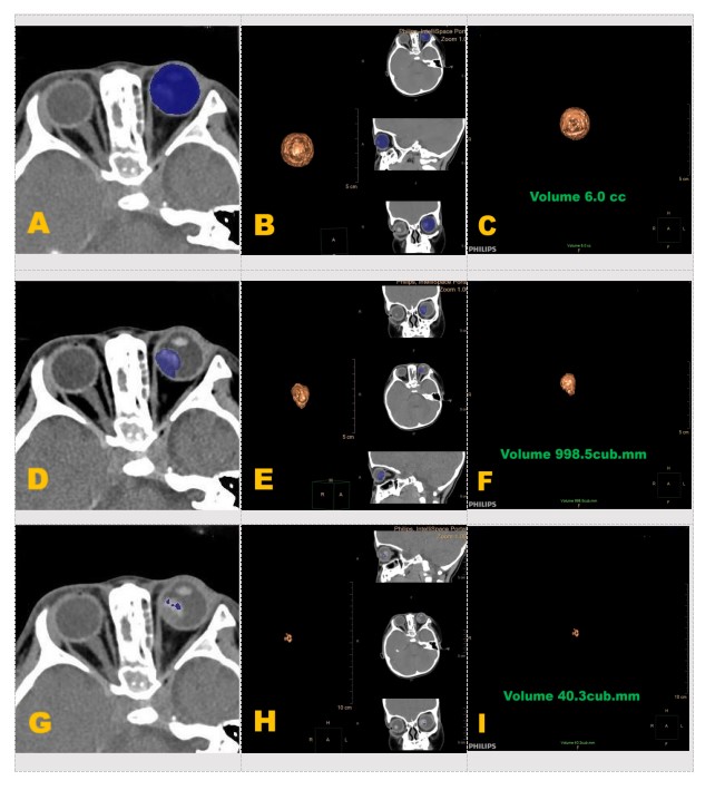
表5 不同影像分期组间影像学参数比较 

 Table 5 Comparison of imaging parameters among groups with different imaging stages

参数

眼内期组 (n=46)

青光眼期组 (n=68)

眼外期组 (n=13)

Statistic

 P

钙化最高CT值

452.09 ± 334.04

569.50 ± 303.92

749.15 ± 254.30

F(2,124)= 5.07

0.008

TV/mm³

915.65 (308.00-1407.00)

2389.00 (1967.20-3020.00)

3095.00 (2701.50-3385.20)

Chi2(2)= 47.47

<0.001

CRV/mm³

73.95 (17.50-221.60)

359.00 (126.25-745.00)

404.20 (314.40-849.50)

Chi2(2)= 28.14

<0.001

EV/mm³

5 756.52 ± 825.34

5 552.94 ± 942.76

 6 623.08 ± 1972.37

F(2,124)= 5.67

0.004

CRV/EV

0.014 (0.003-0.043)

0.062 (0.024-0.155)

0.070 (0.050-0.102)

Chi2(2)= 26.65

<0.001

TV/EV

0.157 (0.057-0.255)

0.462 (0.371-0.540)

0.544 (0.403-0.584)

Chi2(2)= 47.95

<0.001

CRV/TV

0.137 (0.025-0.248)

0.179 (0.055-0.336)

0.119 (0.084-0.250)

Chi2(2)= 1.41

 0.49

TV:肿瘤体积;CRV:钙化区域体积;EV:眼球总体积。

TV: tumor volume; CRV: calcified region volume within the tumor; EV: total eye volume.

图3 分期影像特征示图 

Figure 3 Diagram of staging CT imaging features

图3分期影像特征示图 Figure 3 Diagram of staging CT imaging features

图2容积测量步骤示图 

 Figure 2 Diagram of volume - measurement steps

图2容积测量步骤示图 Figure 2 Diagram of volume - measurement steps
(A)眼球范围选取示图:在软组织窗轴位上选取眼球感兴趣区,蓝色区域为层面内眼球组织。(B)眼球范围选取及三维修正示图:在轴位软窗中选取眼球感兴趣区,蓝色区域为层面内眼球组织,并在冠状位和矢状位中修正范围。(C)眼球容积计算示意图:黄色图像为眼球三维图,图中绿色文字为眼球容积大小。(D)肿瘤范围选取示意图:在软组织窗轴位上选取肿瘤感兴趣区,蓝色区域为层面内肿瘤组织。(E)肿瘤范围三维修正示图:在冠状位和矢状位中修正范围。(F)肿瘤容积计算示意图:黄色图像为肿瘤三维图,图中绿色文字为肿瘤容积大小。(G)肿瘤内钙化范围选取示图:在软组织窗轴位上选取肿瘤内钙化感兴趣区,蓝色区域为层面内肿瘤内钙化。(H)肿瘤内钙化范围三维修正示图:在冠状位和矢状位中修正范围。(I)肿瘤内钙化容积计算示意图:黄色图像为肿瘤内钙化三维图,图中绿色文字为肿瘤内钙化容积大小。
(A) Schematic diagram of eye range selection: The eye region of interest is selected on the axial plane of soft tissue window. The blue area represents intra-layer eye tissue. (B) Schematic diagram of eyeball range selection and three-dimensional correction: select the eyeball region of interest (ROI) on the axial soft window. The blue area represents the eyeball tissue within the slice, and the range is corrected in the coronal and sagittal planes. (C) Schematic diagram of eyeball volume calculation: the yellow image is the 3D model of the eyeball, and the green text in the figure indicates the size of the eyeball volume. (D) Schematic diagram of tumor range selection: select the tumor ROI on the axial soft tissue window. The blue area represents the tumor tissue within the slice. (E) Schematic diagram of tumor range three-dimensional correction: correct the range in the coronal and sagittal planes. (F) Schematic diagram of tumor volume calculation: the yellow image is the 3D model of the tumor, and the green text in the figure indicates the size of the tumor volume. (G) Schematic diagram of calcification range selection in tumor: select the calcification ROI within the tumor on the axial soft tissue window. The blue area represents the calcified tissue within the tumor slice. (H) Schematic diagram of three-dimensional correction of calcification range in tumor: correct the range in the coronal and sagittal planes. (I) Schematic diagram of calcification volume calculation in tumor: the yellow image is the 3D model of tumor calcification, and the green text in the figure indicates the size of the calcification volume within the tumor.

图3分期影像特征示图 

Figure 3 Diagram of staging CT imaging features

图3分期影像特征示图 Figure 3 Diagram of staging CT imaging features
(A)眼内期CT表现:眼球内肿块小,钙化少,眼环完整,无视神经增粗或眶内侵犯。 (B)青光眼期CT表现:肿块内钙化增多,玻璃体密度增高,可见散在钙化点,视神经仍无明显增粗,偶可见前房变浅征象。(C)眼外期CT表现:眼球内大片钙化灶,玻璃体密度显著增高,可伴眼球增大或变形。(D)全身转移期CT表现:眼球壁破坏,肿块不规则增大,肿瘤侵犯眼眶软组织或颅内。
(A) Intraocular CT findings: small mass in the eye, minimal calcification, intact orbital rim, no enlargement of the optic nerve or invasion into the orbit. (B) Glaucomatous phase CT findings: increased calcification within the mass, increased vitreous density, scattered calcification points visible, no significant enlargement of the optic nerve, occasional signs of shallower anterior chamber. (C) Extraocular CT findings: large areas of calcification within the eye,significantly increased vitreous density, may be accompanied by globe enlargement or deformity. (D) Systemic metastatic phase CT findings: destruction of the eye wall, irregular enlargement of the mass, tumor invasion into orbital soft tissues or intracranial space.

3 讨论

         RB是起源于视网膜内颗粒层的胚胎性恶性肿瘤,其特征性表现为视网膜母细胞蛋白分泌障碍。本研究的127例患儿中,单眼发病占主导(67.7%),与文献报道的68%~72%发病率相符[15]。作为儿童最常见的眼内恶性肿瘤,RB约占儿童全身恶性肿瘤的4%[4],同时也是5岁以下儿童后天性盲目的主要原因[3]。

3.1 RB的典型临床进程表现

        本研究显示,RB的典型临床进程表现为初期视网膜局部增厚→瘤体形成→玻璃体种植→继发性青光眼("猫眼"征)→眼外侵犯。值得注意的是,本组病例中青光眼期患者占比达53.5%(68/127),其中92.6%出现特征性钙化,这与肿瘤生长导致的缺血性坏死病理机制高度吻合[16]。多中心研究显示,RB的进展速度与年龄呈负相关(r=-0.43, P<0.01),3岁以下患儿的肿瘤体积倍增时间平均为(15.2±3.8)d[17]
        对于不同年龄的RB患者,早期诊断是非常重要的[18],如果发现眼部不适,尤其是发现视力不佳,出现“白瞳症”或“猫眼”时,首选进行眼部薄层CT检查,以更好地了解眼部内部结构的变化,了解眼内是否有肿块和肿块内的钙化灶,其中钙化灶有其独有的特点,以利于早发现、早期诊断、早治疗,有必要时进一步做MRI,进一步观察是否有颅内及远处转移,尽可能挽救患儿生命。在肿瘤防控领域,我国推行三级预防体系中的早筛、早诊、早治三早原则,通过多学科协作诊疗模式把握干预时间窗,可有效优化临床预后指标,具体表现为5年生存率显著提升(达95%以上),同时致残率和转移相关死亡率同步下降。同时,作为一种儿童罕见病,在临床诊疗中发现并诊断也非常重要。因为早期诊断和治疗,可以使患儿预后变好,甚至保存一部分视力;反之,如果出现漏诊、误诊或延误治疗,患儿的生存率影响会极大。RB还有一个重要的临床特征,不论是儿童还是成人,与瘤体的发生机制有关:由于瘤细胞生长速度较快,细胞之间的黏合能力较差,导致其一边生长一边脱落。故RB均容易在玻璃体下和视网膜下种植,但其他疾病却很少出现这种种植现象[17]

3.2 RB的影像学诊断

        多层螺旋(multi-slice computed tomography, MSCT)在RB诊断中具有不可替代的优势:1)钙化检出,本组病例钙化检出率达96.1%(122/127),显著高于超声(84.3%)和MRI(62.7%)。薄层扫描(0.625 mm)可识别最小1~2mm的钙化灶,其典型表现为 CT值>200 Hu(本组均值545.36±320.79),特征性分布为后极部(92%)>赤道部(8%),形态学分型为团块型(34%)、斑片型(28%)、微钙化型(22%)、爆米花型(12%)。2)分期评估,钙化程度与临床分期显著相关(P<0.001),其中眼内期91.3%(42/46),青光眼期98.5%(67/68),眼外期100%(13/13)。
        目前,影像学检查是诊断RB的重要手段,其中CT 与MRI及B超均有各自的优越性,三者中CT影像的作用更为明显[19]。近年来,医学影像技术的革新显著提升了视网膜母细胞瘤的诊断效能。多层螺旋CT的薄层扫描(层厚≤1 mm)联合容积重建技术,通过优化空间分辨率(可达0.3 mm³体素)使钙化检出敏感度提升40%以上。在采用标准软组织算法(窗宽350 Hu,窗位40 Hu)重建的图像上,钙化灶呈现特征性的CT值>100 Hu的致密影,其空间分布具有以下规律:约78%的病例表现为瘤周环形钙化,15%为瘤内散在钙化,7%呈中央核心型钙化。解剖学分析显示,钙化灶主要聚集于视网膜后极部(占92%),赤道前区仅占8%,这一分布特征与胚胎期光感受器细胞分化梯度高度吻合。因为高分辨率CT技术可以清晰地识别眼球肿块和微小钙化灶(1~2 mm),显著提升诊断准确性。B超可以发现病灶,对眼球内软组织肿块比较敏感,对肿块内较高密度的钙化也会产生声影,但不如CT对钙化为高密度影像显示得更明确;MRI作为一种以分辨病灶组织与正常组织间氢质子差异为显示诊断疾病的影像学检查手段,在显示钙化灶方面的灵敏度和准确度均不具优势,但MRI对视神经管内段和颅内段视神经的远处转移更有价值。
        CT在显示RB钙化方面最具优势。RB内的钙化状况在某种程度上可以反映出肿瘤的生长情况。本组108例患者127眼中,CT检测显示,122例(96.1%)患眼存在钙化。进一步分析发现,钙化率随病情进展显著升高:眼内期(91.3%)、青光眼期(98.5%)、眼外期(100%)。RB的钙化形态学分型具有高度异质性,依据2022年视网膜母细胞瘤影像诊断共识,主要可分为:团块型(>5 mm,占34%)、斑片型(2~5 mm,占28%)、微钙化型(<2 mm,占22%)以及特征性爆米花型(羟基磷灰石层状沉积,占12%),其中不规则浸润型钙化(占4%)多提示肿瘤突破Bruch膜(OR=6.32,95%CI 1.87~21.39)[20]。本研究显示,眼球外期组钙化率最高,眼球内期组钙化率最低,说明肿瘤病程进展越到后期,钙化程度越高,这与钙化产生的机理呈一致性,肿瘤小时,对血供要求并不是很高,随着肿瘤的体积增大,浸润范围的增宽增大,血供相对不足引起细胞坏死,产生钙化复合体结构[16],从而形成钙化的范围随之增大。研究表明:肿瘤CT值均值为545.362 ±320.786。 因此,RB内的钙化状况在某种程度上可以反映肿瘤的生长情况。
        通过研究RB及其钙化特征有利于与脉络膜黑色素瘤、Coats病及眼球内出血灶相鉴别点。
        1)有些成年人眼球内生长脉络膜黑色素瘤也表现为肿块和星点状高密度影,常常与RB难以鉴别,脉络膜黑色素瘤肿块多见类圆形或类椭圆形,边缘往往比较清楚,肿块内的高密度影呈散在星点状或沙粒状改变,CT值通常小于100 Hu,与RB团块状、片状、点状钙化不同;而且脉络膜黑色素瘤在MRI影像表现方面有其特征性表现,表现为T1高信号、T2低信号[21];这点可以帮助更好地与其他疾病相鉴别。
        2)Coats’病 亦名原发性视网膜血管扩张症,又名大量渗出性视网膜病,是一组以视网膜毛细血管和微血管异常扩张为特征性的一种疾病,其特点是小血管瘤的破裂或闭塞,病程反复,进程缓慢,新旧灶交替,产生增殖瘢痕组织,从而产生了钙化灶; 还有因为血管壁屏障被破坏,产生大量渗出物,可在视网膜内,也可在视网膜下。渗出物中含有大量胆固醇、胆固醇结晶、巨噬细胞及少量红细胞等,渗出量较大时可造成视网膜劈裂或视网膜脱离。视网膜组织本身可继发变性,外层可出现坏死、瘢痕组织形成,黄斑下偶见钙化灶。多呈条片状,CT值常为100 Hu左右。Coats 病为先天性,好发于儿童及青少年,发病高峰年龄为4~8岁,也可见于成年人。好发于20岁以下的男性。RB大多数在3岁以下; 性别无明显差异。CT、MRI增强扫描可以很好地鉴别。
        3)球内出血灶,多见于成年人。新鲜出血灶在CT影像上表现为高密度片状为主,边界欠清晰,CT值多为80~100 Hu。出血灶周边不会出现肿块样团块病灶。MRI上T1、T2加权像均表现为低信号改变。再结合病史,看看有没有外伤史、高血压史、糖尿病史等,因此比较容易鉴别。诊断与鉴别诊断常常借助多种影像检查手段,再结合患者年龄、病程、临床表现、眼部临床检查以及钙化的特点来进行全面分析,以保证诊断正确率[22]

3.3 研究结果

        1)人口学特征:本研究纳入127眼(男性71眼,女性56眼),性别分布与马京琪等[23]报道的244例病例趋势一致(男性56.14% vs. 女性43.86%),但组间CT钙化参数(最高CT值、CRV、TV、EV及相关比值)均无统计学差异。2) 年龄相关参数:各年龄组(0~1岁组(0<Age≤1Y)、1~2岁组(1<Age≤2Y)、2~3岁组(2<Age≤3Y)、>3岁组(Age>3Y)分析显示EV值随年龄增长呈显著递增趋势(0~1岁组(0<Age≤1Y)<1~2岁组(1<Age≤2Y)<2~3岁组(2<Age≤3Y)<>3岁组(Age>3Y)),符合眼球发育规律;CRV/EV比值随年龄增长而降低(P<0.05),提示钙化容积增速滞后于眼球发育;其余参数(TV、CRV/TV等)无显著年龄差异。3) 临床分期差异:按影像分期(眼内期/青光眼期/眼外期)比较:钙化指标中眼外期组钙化最高CT值、CRV、TV、EV显著高于其他分期,提示晚期肿瘤钙化负荷及侵袭性增加;容积比值:CRV/EV、TV/EV在眼内期最低,与刘强等[24]报道的钙化进展趋势一致; 钙化占比中CRV/TV比值在青光眼期最高,但组间无统计学差异。4) 晶状体改变关联性:晶状体状态(正常/位置改变/形态-密度改变)分析显示,钙化负荷中形态-密度改变组CRV、TV值最大,提示肿瘤前段浸润与钙化进展呈正相关;容积比值中CRV/EV、TV/EV在该组最大,反映晚期眼球扩张效应;CT值中钙化最高CT值随晶状体受累程度增加而升高。

3.4 临床启示

        基于本研究结果,笔者建议:1) 对疑似RB患儿应首选MSCT检查[25],扫描参数建议:管电压100kV-120kV,管电流200-250 mA,螺距0.4--0.6,扫描范围150mm-200mm,重建矩阵512x512,薄层重建。2) 对CT显示视神经侵犯者(本组占7.9%),需补充眼眶MRI评估视神经管内段情况。3) 建立"白瞳症"患儿的影像检查路径,可缩短确诊时间。4) 钙化与视网膜母细胞瘤的预后关系:从某种程度上来说,钙化程度较高的视网膜母细胞瘤,可能意味着肿瘤的分化程度相对较低,恶性程度较高,患者的预后相对较差。但目前也有研究者认为,视网膜母细胞瘤的钙化程度与患者的生存率并无明确的直接相关性,关键还是要根据肿瘤的具体分期、是否有转移等因素综合判断预后。5) 钙化与视网膜母细胞瘤的治疗效果评估。在视网膜母细胞瘤的治疗过程中,如化学治疗、放射治疗等,肿瘤可能会发生退变,钙化程度也可能会发生变化。如果治疗有效,肿瘤缩小,钙化灶可能会相对增多,这是因为肿瘤组织在治疗后进一步坏死、钙化所致;但如果肿瘤对治疗不敏感或出现耐药等情况,钙化灶可能不会发生明显变化,甚至肿瘤会继续生长,钙化范围也可能会扩大。
        本研究领域在现有文献中报道较少,具有重要的研究价值,这也是本研究团队开展此项工作的主要动因。近年公众健康意识显著提升。临床观察显示,大多数患者因瞳孔反光或白瞳症状在眼内期或青光眼期即就诊,故眼外期病例相对罕见,远处转移期病例则更为稀少。需说明的是,部分术后发生远处转移的病例因不符合纳入标准而未被列入本研究。随着我国医疗卫生事业的高质量发展,医疗条件和诊疗水平显著提升,为患者提供了更为便捷高效的医疗服务。后续研究将持续开展临床观察与数据收集,深化机制研究和方法探索,结合临床实践开展原创性研究。
        通过本研究再次证明钙化是RB的特征性改变。RB患者的钙化沉积水平随着病程推进,在眼球内期、青光眼期至眼外期的不同阶段呈现逐级增高的态势;晶状体位置变化随病情进展和RB患者钙化程度的增大而影响更大,符合RB生长规律和钙化形成的特点。视网膜母细胞瘤钙化容积CT影像特征研究中,钙化最高CT值、CRV、TV、EV、CRV/EV、TV/EV比值在影像分期中的特点,展示了RB患者病灶内钙化特征,可以考虑作为临床进一步诊断及制定治疗方案等提供重要的参考,值得推广应用。

声明

在本研究中,我们未使用生成式人工智能服务。论文撰写中的所有内容均由作者独立完成,并对出版物的真实性和准确性承担全部责任。

利益冲突

所有作者均声明不存在利益冲突。

开放获取声明

本文适用于知识共享许可协议(Creative Commons),允许第三方用户按照署名(BY)-非商业性使用(NC)-禁止演绎(ND)(CC BY-NC-ND)的方式共享,即允许第三方对本刊发表的文章进行复制、发行、展览、表演、放映、广播或通过信息网络向公众传播,但在这些过程中必须保留作者署名、仅限于非商业性目的、不得进行演绎创作。
1、1.高晓曼. 视网膜母细胞瘤——儿童眼内的“恶魔”[C]//广东省肿瘤康复学会. 2025年“《健康大湾区》-科普引领健康”论坛曁第2期健康科普作品征集活动作品集. 建德市第二人民医院眼科, 2025: 75-77. DOI: 10.26914/c.cnkihy.2025.011480.
Gao XM. Retinoblastoma: the "demon" in children's eyes[C]//Guangdong Anti-Cancer Association for Rehabilitation. Proceedings of 2025 "Healthy Greater Bay Area - Popular Science Leading Health" Forum and the 2nd Health Science Popularization Works Collection Activity. Department of Ophthalmology, Jiande Second People's Hospital, 2025: 75-77. DOI: 10.26914/c.cnkihy.2025.011480.
2、Abramson DH, Gobin YP, Francis JH. Good vision in retinoblastoma treated with intra-arterial chemotherapy and laser in the papillomacular bundle[J]. J Pediatr Ophthalmol Strabismus, 2023, 60(3): 228. DOI: 10.3928/01913913-20220629-01. Abramson DH, Gobin YP, Francis JH. Good vision in retinoblastoma treated with intra-arterial chemotherapy and laser in the papillomacular bundle[J]. J Pediatr Ophthalmol Strabismus, 2023, 60(3): 228. DOI: 10.3928/01913913-20220629-01.
3、Bach A, Villegas VM, Gold AS, et al. Atypical event profiling of intra-arterial chemotherapy[J]. J VitreoRetinal Dis, 2022, 6(3): 251-254. DOI: 10.1177/24741264221083411. Bach A, Villegas VM, Gold AS, et al. Atypical event profiling of intra-arterial chemotherapy[J]. J VitreoRetinal Dis, 2022, 6(3): 251-254. DOI: 10.1177/24741264221083411.
4、Venkatasamy A, Guerin E, Reichardt W, et al. Morpho-functional analysis of patient-derived xenografts reveals differential impact of gastric cancer and chemotherapy on the tumor ecosystem, affecting immune check point, metabolism, and sarcopenia[J]. Gastric Cancer, 2023, 26(2): 220-233. DOI: 10.1007/s10120-022-01359-w. Venkatasamy A, Guerin E, Reichardt W, et al. Morpho-functional analysis of patient-derived xenografts reveals differential impact of gastric cancer and chemotherapy on the tumor ecosystem, affecting immune check point, metabolism, and sarcopenia[J]. Gastric Cancer, 2023, 26(2): 220-233. DOI: 10.1007/s10120-022-01359-w.
5、崔雪皓, 李筱荣. 视网膜母细胞瘤诊断和治疗的研究进展[J]. 眼科新进展, 2022, 42(08): 634-638. DOI: 10.13389/j.cnki.rao.2022.0130.
Cui XH, LI XR. Research Progress in Diagnosis and Treatment of Retinoblastoma[J]. Recent Advances in Ophthalmology, 2022, 42(8): 634-638. DOI: 10.13389/j.cnki.rao.2022.0130.
6、Taleghani F, Ehsani M, Farzi S, et al. Nutritional challenges of gastric cancer patients from the perspectives of patients, family caregivers, and health professionals: a qualitative study[J]. Support Care Cancer, 2021, 29(7): 3943-3950. DOI: 10.1007/s00520-020-05951-7. Taleghani F, Ehsani M, Farzi S, et al. Nutritional challenges of gastric cancer patients from the perspectives of patients, family caregivers, and health professionals: a qualitative study[J]. Support Care Cancer, 2021, 29(7): 3943-3950. DOI: 10.1007/s00520-020-05951-7.
7、Ackbarali T, Janjigian YY, Van Cutsem E. The role of education in adjusting to changes in clinical practice for gastric or gastroesophageal junction adenocarcinomas[J]. J Clin Oncol, 2023, 41(4_suppl): 470. DOI: 10.1200/jco.2023.41.4_suppl.470. Ackbarali T, Janjigian YY, Van Cutsem E. The role of education in adjusting to changes in clinical practice for gastric or gastroesophageal junction adenocarcinomas[J]. J Clin Oncol, 2023, 41(4_suppl): 470. DOI: 10.1200/jco.2023.41.4_suppl.470.
8、Rong F, Shi R, Hu L, et al. Impact of risk perception and disease cognition on the willingness to participate in screening for lung cancer in a high-risk population[J]. Eur J Cancer Prev, 2024, 33(2): 141-151. DOI: 10.1097/CEJ.0000000000000843. Rong F, Shi R, Hu L, et al. Impact of risk perception and disease cognition on the willingness to participate in screening for lung cancer in a high-risk population[J]. Eur J Cancer Prev, 2024, 33(2): 141-151. DOI: 10.1097/CEJ.0000000000000843.
9、Cui RD, Ma CY, Yang GY, et al. CT and MRI features of retinoblastoma in children and their clinical application value[J]. Chin J CT MRI, 2022, 20(6): 23-25. DOI: 10.3969/j.issn.1672-5131.2022.06.008. Cui RD, Ma CY, Yang GY, et al. CT and MRI features of retinoblastoma in children and their clinical application value[J]. Chin J CT MRI, 2022, 20(6): 23-25. DOI: 10.3969/j.issn.1672-5131.2022.06.008.
10、Monroe EJ, Chick JFB, Stacey AW, et al. Radiation dose reduction during intra-arterial chemotherapy for retinoblastoma: a retrospective analysis of 96 consecutive pediatric interventions using five distinct protocols[J]. Pediatr Radiol, 2021, 51(4): 649-657. DOI: 10.1007/s00247-020-04892-x.Monroe EJ, Chick JFB, Stacey AW, et al. Radiation dose reduction during intra-arterial chemotherapy for retinoblastoma: a retrospective analysis of 96 consecutive pediatric interventions using five distinct protocols[J]. Pediatr Radiol, 2021, 51(4): 649-657. DOI: 10.1007/s00247-020-04892-x.
11、李壮, 杨凯华, 吴玲, 等. CT和MRI诊断儿童视网膜母细胞瘤的应用价值[J]. 中国校医, 2023, 37(11): 848-850.
Li Z, Yang KH, Wu L, et al. Application values of CT and MRI in diagnosis of retinoblastoma in children[J]. Chin J Sch Dr, 2023, 37(11): 848-850.
12、黄新文, 吕红彬, 高礼明, 等. 外层渗出性视网膜病变的CT诊断价值[J]. 临床放射学杂志, 2008, 27(8): 1020-1022.
Huang XW, (Lü/Lv/Lu/Lyu) HB, Gao LM, et al. CT diagnosis value of external exudative retinopathy(Coats' disease)[J]. J Clin Radiol, 2008, 27(8): 1020-1022.
13、宋庆博, 郑慧, 张玉珍, 等. 基于深度学习重建的5.0T眼眶高分辨磁共振成像在视网膜母细胞瘤中的应用价值[J].中国医学计算机成像杂志, 2025, 31(03): 338-344. DOI: 10.19627/j.cnki.cn31-1700/th.2025.03.003.
Song QB, Zheng H, Zhang YZ, et al. Application value of 5.0T orbital high-resolution MRI reconstructed based on deep learning in retinoblastoma[J]. Chinese Journal of Medical Computer Imaging, 2025, 31(03): 338-344. DOI: 10.19627/j.cnki.cn31-1700/th.2025.03.003.
14、任百超, 白芝兰, 孙乃学. 视网膜母细胞瘤28例的CT诊断[J]. 国际眼科杂志, 2003, 3(3): 42-43. DOI: 10.3969/j.issn.1672-5123.2003.03.011.
Ren BC, Bai ZL, Sun NX. Clinical value of CT in the diagnosis of 28 patients with retinoblastoma[J]. Int J Ophthalmol, 2003, 3(3): 42-43. DOI: 10.3969/j.issn.1672-5123.2003.03.011.
15、孟海燕, 杨维佳, 黄慧莲, 等. 彩色多普勒超声、MRI动态增强对眼内或眶内占位病变诊断及监测价值[J]. 中国CT和MRI杂志, 2022, 20(9): 28-29. DOI: 10.3969/j.issn.1672-5131.2022.09.011.
Meng HY, Yang WJ, Huang HL, et al. Value of color Doppler ultrasound and MRI dynamic enhancement in the diagnosis and monitoring of intraocular or intraorbital space-occupying lesions[J]. Chin J CT MRI, 2022, 20(9): 28-29. DOI: 10.3969/j.issn.1672-5131.2022.09.011.
16、王振常, 鲜军舫, 金征宇, 等. 头颈部影像学-眼科卷[M]. 北京: 人民卫生出版社, 2014: 66.
Wang ZC, Xian JF, Jin ZY, et al. Head and Neck Imaging - Ophthalmology Volume[M]. Beijing: People's Medical Publishing House, 2014: 66.
17、首都医科大学眼部肿瘤临床诊疗与研究中心, 中华医学会放射学分会头颈学组, 中华医学会放射学分会儿科学组, 等. 视网膜母细胞瘤影像检查与诊断及选择性眼动脉化疗专家共识[J]. 中华放射学杂志, 2021, 55(5): 470-477. DOI: 10.3760/cma.j.cn112149-20201214-01306.
Clinical Diagnosis and Treatment Center for Ocular Tumors, Capital Medical University, Head and Neck Radiology Group, Chinese Society of Radiology, Pediatric Radiology Group, Chinese Society of Radiology, et al. Expert consensus on imaging examination and diagnosis of retinoblastoma and selective ophthalmic artery chemotherapy[J]. Chinese Journal of Radiology, 2021, 55(5): 470-477. DOI: 10.3760/cma.j.cn112149-20201214-01306.
18、Chantada GL, Villanueva G, Abramson DH. Challenges for Early Diagnosis in Retinoblastoma in Low-and Middle-Income Countries. Pediatr Blood Cancer. 2025, 72(9): e31859. DOI: 10.1002/pbc.31859.Chantada GL, Villanueva G, Abramson DH. Challenges for Early Diagnosis in Retinoblastoma in Low-and Middle-Income Countries. Pediatr Blood Cancer. 2025, 72(9): e31859. DOI: 10.1002/pbc.31859.
19、崔若棣, 马常友, 杨光勇, 等. 儿童视网膜母细胞瘤CT、MRI影像特征及临床应用价值[J].中国CT和MRI杂志, 2022, 20(06): 23-25. DOI: 10.3969/j.issn.1672-5131.2022.06.008.
Cui RD, Ma CY, Yang GY, et al. CT and MRI imaging features and clinical application value of retinoblastoma in children[J]. Chinese Journal of CT and MRI, 2022, 20(06): 23-25. DOI: 10.3969/j.issn.1672-5131.2022.06.008.
20、王亮, 周珉, 盛茂, 等. 儿童视网膜母细胞瘤的影像诊断[J]. 江苏医药, 2011, 37(8): 934-936. DOI: 10.19460/j.cnki.0253-3685.2011.08.026.
Wang L, Zhou M, Sheng M, et al. Imaging diagnosis of retinoblastoma in children[J]. Jiangsu Med J, 2011, 37(8): 934-936. DOI: 10.19460/j.cnki.0253-3685.2011.08.026.
21、孙丰源. 黑色素瘤及顺磁性病变的影像特征和鉴别诊断[C]//中国超声医学工程学会. 中国超声医学工程学会第十三届全国眼科超声医学学术会议论文汇编. 天津医科大学眼科医院, 2024: 13. DOI: 10.26914/c.cnkihy.2024.044680.
Sun FY. Imaging Features and Differential Diagnosis of Melanoma and Paramagnetic Lesions [C]//Chinese Society of Ultrasound in Medicine. Proceedings of the 13th National Conference on Ophthalmic Ultrasound Medicine. Tianjin Medical University Eye Hospital, 2024: 13. DOI: 10.26914/c.cnkihy.2024.044680.
22、陈向东, 刘悦, 彭清华, 等. Coats病伴眼内钙化灶形成1例[J]. 临床眼科杂志, 2016, 24(5): 465-466. DOI: 10.3969/j.issn.1006-8422.2016.05.028.
Chen XD, Liu Y, Peng QH, et al. Coats disease with calcification in the eye: a case report[J]. J Clin Ophthalmol, 2016, 24(5): 465-466. DOI: 10.3969/j.issn.1006-8422.2016.05.028.
23、马京琪, 黄东生, 张谊, 等. 244例儿童视网膜母细胞瘤临床特点[J]. 眼科, 2011, 20(2): 113-115.
Ma JQ, Huang DS, Zhang Y, et al. Study on the clinical characteristics of 244 retinoblastoma cases[J]. Ophthalmol China, 2011, 20(2): 113-115.
24、刘强, 耿左军, 贾歆, 等. CT在视网膜母细胞瘤钙化研究中的价值[J]. 实用医学影像杂志, 2020(1). DOI: 10.16106/j.cnki.cn14-1281/r.2020.01.002.
Liu Q, Geng ZJ, Jia X, et al. Value of CT in the Study of Retinoblastoma Calcification[J]. Journal of Practical Medical Imaging, 2020, 21(1). DOI: 10.16106/j.cn14-1281/r.2020.01.002.
25、李婷, 鲜军舫. 眼眶和颅脑磁共振成像促进视网膜母细胞瘤精准诊疗——视网膜母细胞瘤影像检查与诊断专家共识解读[J]. 磁共振成像, 2021, 12(11): 74-79. DOI: 10.12015/issn.1674-8034.2021.11.016.
Li T, Xian JF. Orbital and Cranial Magnetic Resonance Imaging Promotes Precision Diagnosis and Treatment of Retinoblastoma—Interpretation of the Expert Consensus on Imaging Examination and Diagnosis of Retinoblastoma[J]. Magnetic Resonance Imaging, 2021, 12(11): 74-79. DOI: 10.12015/issn.1674-8034.2021.11.016.
上一篇
下一篇
其他期刊
  • 眼科学报

    主管:中华人民共和国教育部
    主办:中山大学
    承办:中山大学中山眼科中心
    主编:林浩添
    主管:中华人民共和国教育部
    主办:中山大学
    浏览
  • Eye Science

    主管:中华人民共和国教育部
    主办:中山大学
    承办:中山大学中山眼科中心
    主编:林浩添
    主管:中华人民共和国教育部
    主办:中山大学
    浏览
目录

点击右上角菜单,浏览器打开下载

我知道了