您的位置: 首页 > 2024年9月 第39卷 第9期 > 文字全文
2023年7月 第38卷 第7期11
目录

腰椎感染致双眼内源性眼内炎一例及文献回顾

Endophthalmitis caused by lumbar infection: a case report and literature review

来源期刊: 眼科学报 | 2024年9月 第39卷 第9期 481-488 发布时间:2024-09-28 收稿时间:2024/10/18 11:38:40 阅读量:530
作者:
关键词:
腰椎感染念珠菌眼内炎葡萄膜炎误诊
lumbar spine infection Candida endophthalmitis Uveitis misdiagnosis
DOI:
10.12419/24061904
收稿时间:
2024-06-19 
修订日期:
2024-07-30 
接收日期:
2024-08-19 
内源性真菌性眼内炎(endogenous fungal endophtalmitis, EFE)是最具破坏性的眼部感染之一,在临床上较少见。如诊断和治疗不及时,可严重损害患者视力,甚至需摘除眼球。由于EFE发病隐匿,病程较长,病原学涂片和培养阳性率较低,早期临床症状与葡萄膜炎相似,极易被误诊和漏诊,延误治疗时机。EFE最常见的感染灶来源为肝脏、肺、尿路、脑膜炎、胃肠道、心内膜以及骨髓。文章报道了一例腰椎感染致双眼内源性念珠菌性眼内炎的男性患者,66岁,因“右眼视力下降1周”首诊于眼科,专科检查见右眼玻璃体炎性混浊,初诊为右眼葡萄膜炎,予抗炎等治疗症状无好转,右眼视力持续下降,右眼前房穿刺抽液送检提示:热带念珠菌感染,之后左眼视力也逐渐下降,加之患者近期于骨科住院,术中腰椎间盘退变的纤维软骨组织DNA-病原微生物宏基因组检测结果示热带念珠菌,考虑双眼EFE,予全身及局部使用抗真菌药物联合双眼玻璃体切割手术,治疗后患者视力恢复良好,随访1年无复发。该病例及相关文献回顾,有助于加深临床医生对此类疾病的认识,为今后临床诊疗提供一定思路,也起到一定警示作用。
Fungal endophthalmitis is one of the most destructive eye infections and is relatively rare in clinical practice.If not diagnosed and treated promptly, it can severely damage vision and even lead to enucleation.Due to its insidious onset, long course, low positive rates in smears and cultures, and early clinical symptoms similar to uveitis, it is prone to misdiagnosis and missed diagnosis, leading to delayed treatment. A review of the literature indicates that the most common sources of EFE infection are the liver, lung, urinary tract, meningitis, gastrointestinal tract, endocarditis and osteomyelitis.In this paper, we report a case of lumbar spine infection causing bilateral candidal endophthalmitis in a 66-year-old male patient.He initially presented to the ophthalmology department of our hospital with a one-week history of decreased vision in the right eye, specialized examination revealed inflammatory opacity in the vitreous of the right eye, initially diagnosed as uveitis and treated with anti-inflammatory therapy without improvement.As the vision in the right eye continued to decline, aqueous humor aspiration from the anterior chamber of the right eye indicated infection with tropical Candida.Subsequently, the vision in the left eye also gradually decreased.Considering the recent hospitalization in the orthopedic department for lumbar disc degeneration, metagenomics analysis of fibrous cartilage tissue DNA during surgery detected tropical Candida, suggesting bilateral endogenous fungal endophthalmitis,The patient was treated with systemic and local antifungal medications in combination with bilateral vitrectomy surgery.After treatment, the vision recovered well, and there was no recurrence during a one-year follow-up.The objective of this thesis is to deepen the understanding of clinicians on this type of disease by reporting this case and reviewing relevant literature, providing some insights for future clinical diagnosis and treatment, and serving as a warning.

 文章亮点

1. 关键发现

• 通过查阅文献尚未发现继发于腰椎感染的内源性真菌性眼内炎。

2. 已知与发现

• 内源性真菌性眼内炎早期症状与葡萄膜炎相似,极易漏诊和误诊。
• 内源性真菌性眼内炎最常见的感染灶来源为肝脏、肺、泌尿道、脑膜炎、胃肠道、心内膜以及骨髓。

3. 意义与改变

• 本文报道了一例腰椎感染继发内源性真菌性眼内炎的患者,经过治疗后视力恢复良好。因初诊时临床医生诊断意识的不足,同时目前缺乏相关的病例报道,导致误诊为葡萄膜炎,希望通过该报道,有助于加深临床医生对此类疾病的认识,为今后临床诊疗提供思路。

       EFE为病原菌由全身或其他脏器感染通过血源性
传播进入眼内,导致眼部化脓性全葡萄膜炎,是一种威胁视力的全身性真菌血症并发症,是全世界视力丧失的主要原因之一,发病率较低。早期症状与葡萄膜炎相似,可出现眼红、眼痛、视力模糊、视力下降或飞蚊症等,极易漏诊和误诊,如果诊断和治疗不及时,有眼球摘除的风险。因此,本文对2023年4月13日就诊于成都市第一人民医院的一例腰椎感染后继发双眼内源性真菌性眼内炎的病例进行报道,并回顾整理相关文献资料,希望能为今后临床诊疗提供一定思路。

1 临床资料

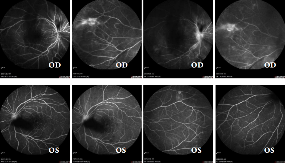
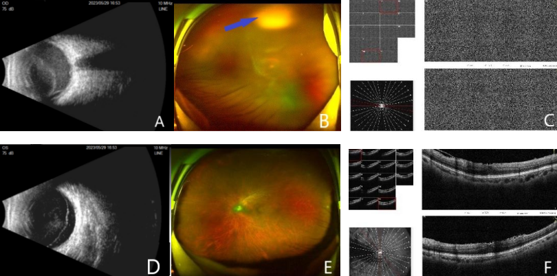
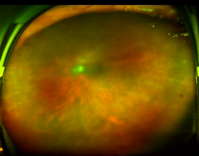
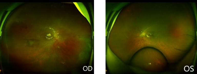
       患者为男性,66岁,2023年4月13日因“右眼视力下降1周”于成都市第一人民医院门诊就诊,眼科专科检查:最佳矫正视力,右眼为0.25,左眼为0.6;眼压,右眼为15.0 mmHg(1 mmHg =0.133 kPa),左眼为16.0 mmHg(1 mmHg =0.133 kPa);右眼结膜正常,角膜透明,角膜后沉着物(keratic preciptates, KP)(—),房水闪辉(Tyndall现象)(—),晶状体轻度混浊,玻璃体炎性混浊,眼底未见明显异常;左眼前后节未见明显异常。考虑:右眼葡萄膜炎予荧光素眼底血管造影(fluorescein fundus angiography, FFA)检查,结果提示双眼葡萄膜炎(图1)。之后患者因腰痛于骨科就诊,其间曾使用醋酸泼尼松龙滴眼液点双眼,症状无缓解。患者于2023年5月30日再次至眼科就诊,以“右眼视力下降1月余,加重5天”收入院。眼科专科检查:最佳矫正视力,右眼HM/40 cm,左眼0.4;眼压,右眼为10.0 mmHg,左眼为19.0 mmHg;右眼结膜混合充血,角膜透明,KP(++,尘状KP,呈弥漫性分布),前房深度正常,Tyn(++),前房细胞(++),瞳孔药物性散大,直径4 mm×3 mm,对光反射消失,虹膜后粘连,晶状体混浊程度为C1N1P0,晶体表面色素沉着,玻璃体混浊;眼底模糊可见视网膜上方团状白色实性混浊;左眼晶状体混浊程度为C1N1P0眼底:黄斑区可见锡箔样反光,余未见明显异常(图2)。既往史:高血压9年、冠状动脉粥样硬化性心脏病7年、糖尿病3年,均规律服用药物治疗。手术史:2023年3月因腰椎椎管狭窄、腰椎不稳定,于外院C臂下行经皮穿刺L4/5、L5/S1椎间盘切除术与臭氧消融术;2023年5月8日因腰椎椎管狭窄、腰椎不稳定,且腰椎感染不能明确再次于成都市第一人民医院骨科行腰椎手术治疗,术中见:L4-5间隙感染积脓,术后口服左氧氟沙星0.5 g/d。术中腰椎间盘退变的纤维软骨组织送检培养结果回报:少量白念珠菌生长,DNA-病原微生物宏基因组检测:疑似人体微生态细菌群:热带念珠菌。实验室检查:糖化血红蛋白 6.2%,余未见临床有意义的异常。初步诊断:双眼葡萄膜炎。治疗:双眼滴妥布霉素地塞米松滴眼液每2小时1次、右眼滴复方托吡卡胺滴眼液每日3次、右眼用硫酸阿托品凝胶每晚1次。患者既往有全身疑似真菌感染,眼部体征符合感染性葡萄膜炎临床表现,因此行右眼前房穿刺抽液送检,2023年6月3日结果回报:真菌(热带念珠菌)。修改诊断:右眼真菌性眼内炎,予右眼玻璃体腔注射伏立康唑,同时静脉滴注伏立康唑,症状未见好转,于2023年6月6日行右眼玻璃体腔切割术、视网膜前膜剥除联合硅油填充术,术中见玻璃体大量积脓,明显混浊,视盘前大片视网膜前增殖膜,累及视盘、黄斑,大量视网膜血管周边白鞘形成,全视网膜隆起。术后第1天,患者自觉左眼视力下降,左眼专科检查:玻璃体混浊,玻璃体炎性细胞(+),视盘前可见白色团絮状物,周边玻璃体可见散在小片状白色漂浮物,左眼底较前模糊(图3),为控制左眼病情进一步进展,行左眼玻璃体腔注射伏立康唑,继续予抗炎、活动瞳孔、全身及局部抗真菌等治疗。2023年6月12日患者最佳矫正视力:右眼为0.10,左眼为0.05,左眼有进一步加重趋势,2023年6月13日行左眼玻璃体切割术联合玻璃体腔注药术(伏立康唑)+硅油填充术,术中见玻璃体大量炎性混浊,视网膜可见大量散在灰白色菌落样沉积物,周边视网膜白鞘。左眼玻璃体切割术后第6天,最佳矫正视力:右眼为0.4,左眼为0.4,双眼玻璃体腔硅油填充,眼底视网膜平伏(图4、图5),玻璃体液腔穿刺液培养:未培养出真菌,未生长细菌,未生长真菌。患者于2023年6月20日出院,出院后继续口服伏立康唑 200 mg每日2次,监测肝肾功能。2024年3月12日、2024年4月23日分别行双眼白内障超声乳化抽吸、人工晶体植入联合硅油取出术。2024年5月8日门诊复诊,眼科专科检查:最佳矫正视力,右眼为0.5,左眼为0.5,双眼前节阴性(图6),眼底视网膜平伏(图7)。

图1 双眼FFA
Figure 1 FFA of both eyes
右眼视网膜前大量不规则可移动遮挡荧光,视盘毛细血管扩张渗漏,视网膜末梢血管广泛渗漏,后期视盘呈强荧光,边界不清。左眼视盘前可见片状遮蔽荧光,鼻上视盘毛细血管可见渗漏,边界不清,周边视网膜末梢可见团状荧光,后期黄斑区血管末梢轻度荧光素渗漏。
In the right eye, the anterior retina was filled with irregular moveable blocks of fluorescence, the optic disc was Telangiectasia, and the peripheral blood vessels of the retina were widely leaky. In the left eye, there was a patchy occlusion of fluorescence in front of the optic disc. In the upper nasal capillary, there was leakage with unclear border. Round fluorescence was seen in peripheral retinal endings, and there was a slight leakage of fluorescein in vascular endings of macular area in late stage.

图2 2023年5月30日双眼检查
Figure 2 Examination of both eyes on May 30,2023
(A)右眼B超:右眼玻璃体内可探及连续条带状回声,带状回声区与球壁间见大量弱点状回声,不与球壁回声连接。(B)右眼激光扫描检眼镜(Scanninglaser ophthalmoscope, SLO):模糊可见视网膜上方团状白色实性混浊。(C)右眼黄斑光学相干断层扫描(Optical coherence tomography, OCT):窥不进。(D)左眼B超:左眼璃体腔内探及大量弱点状回声及光滑带状回声,球壁形态基本平整。(E)左眼SLO:眼底豹纹状改变,黄斑区锡箔样反光。(F)左眼OCT:视网膜内层表面黏附一条高反射光带,黄斑厚度增加,视网膜结构欠连续,局部低反射。
(A)B-mode ultrasonography of the right eye: Continuous band echo can be detected in the vitreous body of right eye. There are a lot of weak point echo between the band echo area and the bulbar wall, which is not connected with the bulbar wall echo. (B)Scanning laser ophthalmoscope (SLO) of the right eye: A blurry white mass of solid opacity is visible over the retina. (C)Optical coherence tomography(OCT)of the right eye: Cannot see into. (D)B-mode ultrasonography of the left eye: A lot of weak echo and smooth band echo were detected in the vitreous cavity of the left eye. (E)SLO of the left eye:Leopard-print change in the fundus, tinfoil-like reflection in the macular region. (F)OCT of the left eye: There is a high reflection band on the inner surface of retina, the thickness of macula increases, the retinal structure is less continuous and the local low reflection.

图3 2023年6月9日左眼SLO
Figure 3 SLO of the left eye on June 9,2023
图示较前模糊。
The picture is more blurred than before.

图4 2023年6月16日出院前双眼SLO
Figure 4 SLO of both eyes before discharge from hospital on June 16, 2023
图示视网膜平伏,硅油反光。
The retina is flattened, and silicone oil reflects light.

图5 2023年6月16日出院前双眼黄斑OCT
Figure 5 Macular OCT of both eyes before discharge from hospital on June 16,2023
右眼视网膜内表面局部高反射,视网膜结构欠连续;左眼视网膜内表面黏附一条带状高反射,黄斑中心凹厚度增加,视网膜结构欠连续。
The inner surface of the retina in the right eye was highly reflective and the structure of the retina was not continuous, while the inner surface of the retina in the left eye adhered to a strip of high reflection, the thickness of fovea increased and the structure of the retina was not continuous.


图6 2024年5月8日双眼前节照相
Figure 6 Anterior segment images of both eyes on May 8,2024

图7 2024年5月8日双眼SLO
Figure 7 SLO of both eyes on May 8,2024

2 讨论

       眼内炎是一种较为罕见但严重致盲的眼部化脓性全葡萄膜炎,是全世界视力丧失的主要原因之一。致病性微生物包括细菌和真菌,其中真菌性眼内炎发病率低于细菌性眼内炎。一项研究显示,所有眼内炎中,约有85.1%由革兰阳性菌引起,10.3%由革兰阴性菌引起,4.6%是由真菌引起的[1]。内源性眼内炎占所有眼内炎的2%~15%,其中多数由真菌引起。EFE是一种威胁视力的全身性真菌血症并发症,发病亚急性,平均潜伏期为30 d,只有3%的患者在一周内发病,早期可出现眼红、眼痛、视力模糊、视力下降或飞蚊症等,可能同时出现发烧等系统性症状,右眼更易发病,这可能是因为循环系统的特殊性,血液从心脏到达右颈动脉更直接,约70%可累及双眼[2]。典型表现为黄白色视网膜浸润病灶,病灶周围视网膜血管鞘形成,玻璃体串珠状或团状混浊,可出现虹膜睫状体炎或前房积脓[2-3]。EFE主要为全身或其他脏器感染,病原菌通过血源性传播进入眼内,其危险因素较多,包括近期住院、糖尿病、恶性肿瘤、肾衰竭、中心静脉导管、肝脏疾病、静脉注射药物、获得性免疫缺陷综合征、器官移植、尿路感染和近期手术史等[1,4-5]。国外曾报道EFE较多,而我国则是外源性真菌性眼内炎较多,可能由于我国人群劳动时大多缺乏眼部防护[6]虽然EFE较少见,但是近年来随着免疫抑制剂治疗、静脉内注射药物的广泛使用以及糖尿病患者不断增加等因素,发病率明显上升[7]。一项研究报道,在11例眼内炎患者中,有9例合并糖尿病[8]
       EFE的最常见病因是念珠菌属,其次是曲霉菌属[9]。白念珠菌是念珠菌性眼内炎的首要原因,其次是热带念珠菌[10]。念珠菌为机会致病菌,是寄居在人体内的一种共生菌,通常存在于胃肠道、泌尿生殖道和呼吸道中,EFE是由正常的共生菌念珠菌在一定条件下引起的,一般在扰乱免疫系统、抵抗力低下时发病[11]
       EFE的治疗仍然是一个较大挑战,其预后通常较差,主要分为药物治疗和手术治疗。一旦确诊,应及时局部和全身使用抗真菌药物。在药物的选择上,全身用药必须能够穿过血视网膜屏障并达到较高的眼部浓度,伏立康唑是第二代三唑类药物,穿透力强,生物利用度高,不良反应少,是首选药物。与氟康唑相比,伏立康唑的肝毒性更高。一般推荐联合局部玻璃体腔注射抗真菌药物,常使用两性霉素B或伏立康唑,玻璃体内注射伏立康唑的视网膜毒性风险低,抗菌谱更广,是目前的治疗方法[12-13]。对于全身及局部用药无应答的患者,必要时联合玻璃体切割术,目前认为真菌性眼内炎行玻璃体切割术的指征:诊断性玻璃体切割、感染治疗和感染引起的并发症的处理。目前尚无关于玻璃体切割在真菌性眼内炎治疗作用中的共识指南,手术时机的选择没有统一标准,也没有证据表明玻璃体切割比单纯药物治疗效果更好。但玻璃体切割在获取玻璃体标本以诊断和治疗并发症(如持续性玻璃体混浊和牵引性视网膜脱离)的作用已得到证实[14]。然而,玻璃体切割对感染控制的益处还有待商榷,但至少在理论上能够清除病灶,并增加抗真菌药物进入视网膜的机会。Sallam等[15]对患有EFE的44眼进行了回顾性研究,在发病后1周内进行玻璃体切割术,并未降低严重视力丧失的风险,但将视网膜脱离的风险减少至原来的1/5。硅油能使微生物生长受到抑制,在体外试验中显示出良好的抗菌活性,可作为EFE患者玻璃体切割术后的眼内填充物[16]
       在EFE的治疗中,糖皮质激素(激素)的应用存在争议。激素能抑制真菌感染引起的炎症反应,也会导致真菌繁殖加速,加重病情。既往研究表明,激素可以抑制真菌生长中的某个环节,对真菌的生长有短暂的抑制作用,但对其整个生长周期不起主要作用[17]。激素会抑制巨噬细胞膜对抗原的吞噬和处理、抑制细胞免疫,导致免疫系统活性降低。此外,激素可以与真菌细胞内的蛋白质受体结合,参与真菌细胞的代谢,引起真菌细胞内发生一系列改变,促进真菌的生长繁殖,增强致病力,还会使单核细胞的抗真菌活性受到影响,进而加重炎症反应[18]。然而,在Coats等[19]的研究中,没有证据表明玻璃体内注射地塞米松会削弱抗真菌活性或增强真菌增殖。甚至有研究认为,在抗真菌治疗有效时,局部使用激素可减轻过度炎症引起的眼部组织破坏,减少增生性病变的发生,对视力的恢复有一定帮助[20]。一项回顾性研究也表明,玻璃体内注射地塞米松可促进真菌性眼内炎症更快恢复[21]。虽然玻璃体内注射激素的继发性宿主毒性有待进一步研究,但与局部和静脉注射相比,玻璃体内注射可能是较为安全的。总之,目前激素在真菌性眼内炎中的作用还没有明确结论,临床中使用激素治疗真菌性眼内炎应在评估风险后,谨慎使用。
       回顾本例病史,患者2023年3月曾于外院行腰椎手术,推测感染可能在此之前就已存在,也可能是此次手术所导致。该患者基础疾病多,全身情况差,既往糖尿病3年余,长期的高糖状态为真菌的生长繁殖提供了良好环境,同时会削弱中性粒细胞的募集、杀伤能力,降低巨噬细胞的黏附、抗菌活性和吞噬作用,导致免疫功能下降[22],为真菌感染提供了条件;加之腰椎手术后长期使用抗生素,导致菌群失衡,有利于真菌的生长繁殖,增加了真菌感染的机会。鉴于患者无眼外伤史及其他感染来源,考虑眼部念珠菌感染很大可能来源于腰椎念珠菌血源性播散,进入视网膜脉络膜血管,从而引起眼内炎。
       本例患者经全身及局部使用伏立康唑联合玻璃体切割术治疗后,感染得到控制,视力明显改善,从中总结出如下经验:1)该患者初诊时高度怀疑葡萄膜炎,之后于骨科行腰椎手术的术中送检结果提示真菌感染,但骨科医生当时并未考虑到眼部真菌感染的可能,没有及时予抗真菌治疗,眼科医生在接诊时也没有仔细询问病史,导致误诊,以致患者病情持续恶化,虽最终视力恢复良好,但也说明临床诊断意识的不足。在临床诊疗过程中,应注重全身病史的询问,这是帮助临床医生诊断的重要一环。2)查阅文献,内源性眼内炎最常见的感染灶来源为肝脏、肺、泌尿道、脑膜炎、胃肠道、心内膜以及骨髓,了解EFE与单个组织和器官感染的相关性,能帮助临床医生更快地做出诊断并实施治疗[23-24],本病例为腰椎感染后出现眼内炎,今后临床医生应警惕腰椎感染后真菌性眼内炎的发生,一旦出现相应眼部症状应立即积极治疗。3)对于初诊为葡萄膜炎,予以激素等治疗后未见好转,病情反而加重的患者,应高度怀疑眼内炎,尤其是全身情况差,具有上述高危因素患者,尽早抽取眼内液进行微生物学检查,以往多采用普通前房及玻璃体穿刺液涂片和培养的方法,但阳性检出率较低,约为38%。在一项回顾性研究中,房水或玻璃体培养物的灵敏度仅为17%[25],有条件者可采用聚合酶链反应(polymerase chain reaction, PCR)技术进行眼内液中的DNA检测,该方法灵敏、快速、阳性率高,或采用宏基因组测序技术,其灵敏度更高,并且无需培养[26],一旦患者被确诊为EFE,应立即对其进行全身及局部抗真菌治疗,根据病原学检测结果选择敏感的抗真菌药物,若疗效不佳,应联合玻璃体切割术彻底清除病灶,以期最大程度挽救患者视力,患者病情稳定后应继续服用抗真菌药物巩固治疗,以防复发。

利益冲突

所有作者均声明不存在利益冲突。

开放获取声明

本文适用于知识共享许可协议 ( Creative Commons),允许第三方用户按照署名(BY)-非商业性使用(NC)-禁止演绎(ND)(CC BY-NC-ND)的方式共享,即允许第三方对本刊发表的文章进行复制、发行、展览、表演、放映、广播或通过信息网络向公众传播,但在这些过程中必须保留作者署名、仅限于非商业性目的、不得进行演绎创作。
1、Haseeb%20AA%20%2C%20Elhusseiny%20AM%20%2C%20Siddiqui%20MZ%20%2C%20et%20al%20.%20Fungal%20%0AEndophthalmitis%3A%20A%20Comprehensive%20Review%5B%20J%5D.%C2%A0J%20Fungi%20(Basel)%2C%202021%2C%20%0A7(11)%3A%20996.%20DOI%3A10.3390%2Fjof7110996.Haseeb%20AA%20%2C%20Elhusseiny%20AM%20%2C%20Siddiqui%20MZ%20%2C%20et%20al%20.%20Fungal%20%0AEndophthalmitis%3A%20A%20Comprehensive%20Review%5B%20J%5D.%C2%A0J%20Fungi%20(Basel)%2C%202021%2C%20%0A7(11)%3A%20996.%20DOI%3A10.3390%2Fjof7110996.
2、Sheu%20SJ.Endophthalmitis%5B%20J%5D.%C2%A0Korean%20J%20Ophthalmol%2C%202017%2C%2031(4)%3A%20283-%0A289.%20DOI%3A%2010.3341%2Fkjo.2017.0036.Sheu%20SJ.Endophthalmitis%5B%20J%5D.%C2%A0Korean%20J%20Ophthalmol%2C%202017%2C%2031(4)%3A%20283-%0A289.%20DOI%3A%2010.3341%2Fkjo.2017.0036.
3、Relhan N, Forster RK, Jr Flynn HW. Endophthalmitis: then and now[ J]. Am J Ophthalmol, 2018, 187: xx-xxvii. DOI: 10.1016/ j.ajo.2017.11.021.Relhan N, Forster RK, Jr Flynn HW. Endophthalmitis: then and now[ J]. Am J Ophthalmol, 2018, 187: xx-xxvii. DOI: 10.1016/ j.ajo.2017.11.021.
4、Gurnani%20B%2C%20Kaur%20K.%20Endogenous%20Endophthalmitis.%20In%3A%C2%A0%20StatPearls.%20%0ATreasure%20Island%20(FL)%3A%20StatPearls%20Publishing%3B%20June%2011%2C%202023.Gurnani%20B%2C%20Kaur%20K.%20Endogenous%20Endophthalmitis.%20In%3A%C2%A0%20StatPearls.%20%0ATreasure%20Island%20(FL)%3A%20StatPearls%20Publishing%3B%20June%2011%2C%202023.
5、Danielescu C, Stanca HT, Iorga RE, et al. The diagnosis and treatment of fungal endophthalmitis: an update[ J]. Diagnostics (Basel), 2022,12(3):679. DOI:10.3390/diagnostics12030679.Danielescu C, Stanca HT, Iorga RE, et al. The diagnosis and treatment of fungal endophthalmitis: an update[ J]. Diagnostics (Basel), 2022,12(3):679. DOI:10.3390/diagnostics12030679.
6、Fan JC, Niederer RL, von Lany H, et al. Infectious endophthalmitis: clinical features, management and v isual outcomes[ J]. Clin Exp Ophthalmol, 2008, 36(7): 631-636. DOI: 10.1111/j.1442- 9071.2008.01813.x.Fan JC, Niederer RL, von Lany H, et al. Infectious endophthalmitis: clinical features, management and v isual outcomes[ J]. Clin Exp Ophthalmol, 2008, 36(7): 631-636. DOI: 10.1111/j.1442- 9071.2008.01813.x.
7、Wang H, Chang Y, Zhang Y, et al. Bilateral endogenous fungal endophthalmitis: a case report[ J]. Medicine, 2023, 102(16): e33585. DOI: 10.1097/MD.0000000000033585.Wang H, Chang Y, Zhang Y, et al. Bilateral endogenous fungal endophthalmitis: a case report[ J]. Medicine, 2023, 102(16): e33585. DOI: 10.1097/MD.0000000000033585.
8、罗广娥, 崔仁哲, 田莲姬, 等. 内源性眼内炎11例临床分析 [ J]. 吉林医学, 2018, 39(4): 711-712. DOI: 10.3969/j.issn.1004- 0412.2018.04.051.
Luo GE, Cui RZ, Tian LJ, et al. Clinical analysis of 11 cases with endogenous endophthalmitis[ J]. Jilin Med J, 2018, 39(4): 711-712. DOI: 10.3969/j.issn.1004-0412.2018.04.051.
9、Danielescu C, Anton N, Stanca HT, et al. Endogenous endophthalmitis: a review of case series published between 2011 and 2020[ J]. J Ophthalmol, 2020, 2020: 8869590. DOI: 10.1155/2020/8869590.Danielescu C, Anton N, Stanca HT, et al. Endogenous endophthalmitis: a review of case series published between 2011 and 2020[ J]. J Ophthalmol, 2020, 2020: 8869590. DOI: 10.1155/2020/8869590.
10、Phongkhun K, Pothikamjorn T, Srisurapanont K, et al. Prevalence of ocular candidiasis and candida endophthalmitis in patients with candidemia: a systematic review and meta-analysis[ J]. Clin Infect Dis, 2023, 76(10): 1738-1749. DOI: 10.1093/cid/ciad064.Phongkhun K, Pothikamjorn T, Srisurapanont K, et al. Prevalence of ocular candidiasis and candida endophthalmitis in patients with candidemia: a systematic review and meta-analysis[ J]. Clin Infect Dis, 2023, 76(10): 1738-1749. DOI: 10.1093/cid/ciad064.
11、Chee YE, Eliott D. The role of vitrectomy in the management of fungal endophthalmitis[ J]. Semin Ophthalmol, 2017, 32(1): 29-35. DOI: 10.1080/08820538.2016.1228396.Chee YE, Eliott D. The role of vitrectomy in the management of fungal endophthalmitis[ J]. Semin Ophthalmol, 2017, 32(1): 29-35. DOI: 10.1080/08820538.2016.1228396.
12、陈星, 杨勋. 真菌性眼内炎的药物和手术治疗进展[ J ] . 国际眼科杂志, 2019, 19(12): 2064-2067. DOI: 10.3980/ j.issn.1672-5123.2019.12.15.
Chen X, Yang X. Advances in drugs and surgical treatment of fungal endophthalmitis[ J]. Int Eye Sci, 2019, 19(12): 2064-2067. DOI: 10.3980/j.issn.1672-5123.2019.12.15.
13、Abu Talib DN, Yong MH, Nasaruddin RA, et al. Chronic endogenous fungal endophthalmitis: diagnostic and treatment challenges: a case report[ J]. Medicine, 2021, 100(14): e25459. DOI: 10.1097/ MD.0000000000025459.Abu Talib DN, Yong MH, Nasaruddin RA, et al. Chronic endogenous fungal endophthalmitis: diagnostic and treatment challenges: a case report[ J]. Medicine, 2021, 100(14): e25459. DOI: 10.1097/ MD.0000000000025459.
14、Ly V, Sallam A. Fungal Endophthalmitis.[M]. Treasure Island (FL): StatPearls Publishing, 2023.Ly V, Sallam A. Fungal Endophthalmitis.[M]. Treasure Island (FL): StatPearls Publishing, 2023.
15、Sallam A, Taylor SRJ, Khan A, et al. Factors determining visual outcome in endogenous Candida endophthalmitis[ J]. Retina, 2012, 32(6): 1129-1134. DOI: 10.1097/IAE.0b013e31822d3a34.Sallam A, Taylor SRJ, Khan A, et al. Factors determining visual outcome in endogenous Candida endophthalmitis[ J]. Retina, 2012, 32(6): 1129-1134. DOI: 10.1097/IAE.0b013e31822d3a34.
16、Weber C, Stasik I, Herrmann P, et al. Early Vitrectomy with Silicone Oil Tamponade in the Management of Postoperative Endophthalmitis[ J]. J Clin Med, 2023,12(15):5097. DOI:10.3390/jcm12155097.Weber C, Stasik I, Herrmann P, et al. Early Vitrectomy with Silicone Oil Tamponade in the Management of Postoperative Endophthalmitis[ J]. J Clin Med, 2023,12(15):5097. DOI:10.3390/jcm12155097.
17、段正芳,张雅杰,宋馥香,等.糖皮质类固醇激素对常见致病真菌 生长及形态的影响[ J]. 吉林医学, 1996, 17(3): 145-146.
Duan ZF, Zhang YJ, Song FX, et al. Effects of corticosteroid on the growth and morphology of common pathogenic fungal[ J]. Jilin Med J, 1996,17(3):145-146.
18、Sallam A , Jayakumar S, Lightman S. Intraocular deliver y of anti-infective drugs-bacterial, v iral, fungal and parasitic[ J]. Recent Pat Antiinfect Drug Discov, 2008, 3(1): 53-63. DOI: 10.2174/157489108783413164.Sallam A , Jayakumar S, Lightman S. Intraocular deliver y of anti-infective drugs-bacterial, v iral, fungal and parasitic[ J]. Recent Pat Antiinfect Drug Discov, 2008, 3(1): 53-63. DOI: 10.2174/157489108783413164.
19、Coats ML, Peyman GA. Intravitreal corticosteroids in the treatment of exogenous fungal endophthalmitis[ J]. Retina, 1992, 12(1): 46-51. DOI: 10.1097/00006982-199212010-00010.Coats ML, Peyman GA. Intravitreal corticosteroids in the treatment of exogenous fungal endophthalmitis[ J]. Retina, 1992, 12(1): 46-51. DOI: 10.1097/00006982-199212010-00010.
20、林晓峰, 袁敏而. 重视真菌性眼内炎诊疗规范性[ J]. 中华 实验眼科杂志, 2019, 37(5): 321-325. DOI: 10.3760/cma. j.issn.2095-0160.2019.05.001.
Lin XF, Yuan ME. Focus on the standardization of diagnosis and treatment of fungal endophthalmitis[ J]. Chin J Exp Ophthalmol, 2019, 37(5): 321-325. DOI: 10.3760/cma.j.issn.2095-0160.2019.05.001.
21、Ching Wen Ho D, Agarwal A, Lee CS, et al. A review of the role of intravitreal corticosteroids as an adjuvant to antibiotics in infectious endophthalmitis[ J]. Ocul Immunol Inflamm, 2018, 26(3): 461-468. DOI: 10.1080/09273948.2016.1245758.Ching Wen Ho D, Agarwal A, Lee CS, et al. A review of the role of intravitreal corticosteroids as an adjuvant to antibiotics in infectious endophthalmitis[ J]. Ocul Immunol Inflamm, 2018, 26(3): 461-468. DOI: 10.1080/09273948.2016.1245758.
22、Pari B, Gallucci M, Ghigo A, et al. Insight on Infections in Diabetic Setting[ J]. Biomedicines, 2023,11(3):971. DOI:10.3390/ biomedicines11030971.Pari B, Gallucci M, Ghigo A, et al. Insight on Infections in Diabetic Setting[ J]. Biomedicines, 2023,11(3):971. DOI:10.3390/ biomedicines11030971.
23、Gajdzis%20M%2C%20Figu%C5%82a%20K%2C%20Kami%C5%84ska%20J%2C%20et%20al.%20Endogenous%20endophthalmitis%02the%20clinical%20significance%20of%20the%20primary%20source%20of%20infection%5B%20J%5D.%20J%20Clin%20%0AMed%2C%202022%2C%2011(5)%3A%201183.%20DOI%3A%2010.3390%2Fjcm11051183.Gajdzis%20M%2C%20Figu%C5%82a%20K%2C%20Kami%C5%84ska%20J%2C%20et%20al.%20Endogenous%20endophthalmitis%02the%20clinical%20significance%20of%20the%20primary%20source%20of%20infection%5B%20J%5D.%20J%20Clin%20%0AMed%2C%202022%2C%2011(5)%3A%201183.%20DOI%3A%2010.3390%2Fjcm11051183.
24、Steeples LR, Jones NP. Staphylococcal endogenous endophthalmitis in association with pyogenic vertebral osteomyelitis[ J]. Eye, 2016, 30(1): 152-155. DOI: 10.1038/eye.2015.200.Steeples LR, Jones NP. Staphylococcal endogenous endophthalmitis in association with pyogenic vertebral osteomyelitis[ J]. Eye, 2016, 30(1): 152-155. DOI: 10.1038/eye.2015.200.
25、Sandhu HS, Hajrasouliha A, Kaplan HJ, et al. Diagnostic utility of quantitative poly merase chain reaction versus culture in endophthalmitis and uveitis[ J]. Ocul Immunol Inflamm, 2019, 27(4): 578-582. DOI: 10.1080/09273948.2018.1431291.Sandhu HS, Hajrasouliha A, Kaplan HJ, et al. Diagnostic utility of quantitative poly merase chain reaction versus culture in endophthalmitis and uveitis[ J]. Ocul Immunol Inflamm, 2019, 27(4): 578-582. DOI: 10.1080/09273948.2018.1431291.
26、Chen KJ, Sun MH, Chen YP, et al. Endogenous fungal endophthalmitis: causative organisms, treatments, and visual outcomes[ J]. JoF, 2022, 8(6): 641. DOI: 10.3390/jof8060641.Chen KJ, Sun MH, Chen YP, et al. Endogenous fungal endophthalmitis: causative organisms, treatments, and visual outcomes[ J]. JoF, 2022, 8(6): 641. DOI: 10.3390/jof8060641.
上一篇
下一篇
其他期刊
  • 眼科学报

    主管:中华人民共和国教育部
    主办:中山大学
    承办:中山大学中山眼科中心
    主编:林浩添
    主管:中华人民共和国教育部
    主办:中山大学
    浏览
  • Eye Science

    主管:中华人民共和国教育部
    主办:中山大学
    承办:中山大学中山眼科中心
    主编:林浩添
    主管:中华人民共和国教育部
    主办:中山大学
    浏览
推荐阅读
出版者信息
目录