您的位置: 首页 > 2025年9月 第40卷 第9期 > 文字全文
2023年7月 第38卷 第7期11
目录

14例眼眶泪腺导管囊肿的临床特征及手术治疗

Clinical characteristics and surgical management of 14 cases with orbital lacrimal duct cysts

来源期刊: 眼科学报 | 2025年9月 第40卷 第9期 700-707 发布时间:2025-09-28 收稿时间:2025/9/25 14:33:35 阅读量:94
作者:
关键词:
泪腺导管囊肿临床特征手术
lacrimal duct cyst clinical features surgery
DOI:
10.12419/24123104
收稿时间:
2025-01-02 
修订日期:
2025-03-13 
接收日期:
2025-07-24 
目的:探讨眼眶泪腺导管囊肿的临床特征及手术治疗方式,提高对该疾病诊疗的认识。方法:回顾性分析天津医科大学第二医院1991年1月—2023年12月经手术后病理证实的14例眼眶泪腺导管囊肿患者的临床资料。结果:14例患者中,男性9例,女性5例;年龄8个月~59岁,平均年龄为26.5岁;均为单眼,其中左眼9例,右眼5例;多以发现眼眶肿物或眼睑肿胀隆起就诊。B超/彩色多普勒超声或CT均提示囊性占位性病变。所有患者均接受了囊肿切除术,全身麻醉手术4例,局部麻醉手术10例;其中前路皮肤切口6例,结膜切口8例,辅助外眦切开3例。术中6例囊肿破裂,囊腔内注入医用透明质酸钠凝胶,辅助完整摘除囊壁。术后病理均提示泪腺导管囊肿,其中睑叶泪腺导管囊肿4例,眶叶泪腺导管囊肿4例,副泪腺导管囊肿5例,异位泪腺导管囊肿1例。术后复查未见并发症。结论:泪腺导管囊肿是临床较少见的疾病,可以发生在眼部的任何部位,结合典型的临床及影像学表现,主泪腺导管囊肿可基本明确诊断,但仍需与上皮/结膜包含性囊肿及皮样囊肿等鉴别,最终需病理确诊。手术完整切除囊肿是治疗该病的主要方法,切除不完全,有复发可能;术中辅助应用医用透明质酸钠凝胶囊内注射有助于囊肿完整摘除。
Abstract Objective:To explore the clinical characteristics and surgical treatment for lacrimal duct cyst, aiming to enhance the diagnosis and therapeutic outcomes of this disease. Method: We conducted a retrospective analysis of the clinical data from 14 patients with lacrimal duct cysts all of which were confirmed by postoperative pathology. These patients were treated at our hospital from January 1991 to December 2023. Results: Among the 14 patients,  there were 9 male and 5 females, with age ranging from 8 months to 59 years (mean: 26.5 years). All cases involved unilateral involvement, with 9 affecting the left eye and 5 affecting the right eye. The majority of patients sought medical care due to the presence of an orbital mass or eyelid swelling and protrusion. Imaging studies, including B-ultrasound, color Doppler ultrasound, or CT scans, consistently revealed cystic space-occupying lesions. All patients underwent cystectomy, with 4 cases performed under general anesthesia and 10 cases under local anesthesia.Surgical approahces included anterior skin incision in 6 cases, conjunctival incision in 8 cases, and  auxiliary lateral canthotomy in 3 cases involving. During surgery, cyst rupture occurred in 6 cases, necessitating the injection of medical sodium hyaluronate gel into the cyst cavity to facilitate the complete removal of the cyst wall. Postoperative pathological analysis confirmed the presence of lacrimal gland duct cysts, which were further classified as follows: 4 cases of eyelid lobe lacrimal gland duct cysts, 4 cases of orbital lobe lacrimal gland duct cysts, 5 cases of accessory lacrimal gland duct cysts, and 1 case of ectopic lacrimal gland duct cyst. No postoperative complications were observed in any the patients. Conclusions: Lacrimal duct cysts are relatively uncommon in clinical practice and can arise in any part of the eye. Given their typical clinical and imaging features, a primary lacrimal duct cyst can generally be diagnosed with reasonable degree of certainty. However, it is still necessary to be differentiated it from epithelial/conjunctival inclusion cysts and dermoid cysts, and other similar conditions., with the final diagnosis relying on pathological confirmation. Complete surgical resection of the cyst is the primary treatment approach for this disease, as incomplete resection may result in recurrence. Intra-capsular injection of medical sodium hyaluronate gel during surgery can aid in the complete remval of the cyst.

文章亮点

1. 关键发现

· 通过分析眼眶泪腺导管囊肿的临床特征及手术方式的选择,使我们能够更充分的认识这种少见的疾病及其处理原则。

2. 已知与发现 

· 眼眶泪腺导管囊肿临床少见,多表现为眼睑肿胀隆起及穹隆部结膜下肿物,手术切除时术中囊肿容易破裂,致囊壁部分残留,术后复发。在囊肿破裂后使用透明质酸钠囊内注射恢复其囊肿的完整性有助于囊肿的完整摘除。

3. 意义与改变

· 充分认识了眼眶泪腺导管囊肿的临床特征,术中辅助使用透明质酸钠提高了完整摘除囊肿的成功率,减少了复发。

        泪腺导管囊肿是一种较为少见的眼眶良性病变,可发生于主泪腺、副泪腺或异位泪腺组织。该病可出现在任何年龄,临床表现多样,常表现为无痛性眼睑肿胀、结膜下肿物或眼球突出移位,影响外观,并易与其他眼眶占位性病变混淆,导致误诊或延迟治疗。目前,泪腺导管囊肿的发病机制尚未完全明确,可能与泪腺导管阻塞、慢性炎症或外伤等因素相关。影像学检查虽有助于初步诊断,但最终确诊仍需依赖组织病理学检查。手术完整切除囊肿是主要的治疗方式,但手术入路选择、囊壁完整性保持及复发预防仍是临床难点。因此,术前精准评估、术中精细操作及术后病理验证对提高诊疗效果至关重要。本研究回顾性分析1991年1月—2023年12月经手术及病理证实的14例泪腺导管囊肿患者的临床资料,探讨其临床特征及手术方式的选择,旨在提高对该病的认识,减少误诊及复发,并为临床诊疗提供参考。

1 资料与方法

        收集1991年1月—2023年12月在我院眼科住院,经手术治疗并病理证实的14例眼眶泪腺导管囊肿患者的临床资料,包括主诉、既往史、临床表现、影像学表现、术中所见、术中术后并发症及随访结果等(表1)。本研究已通过天津医科大学第二医院伦理委员会审查(批件号:KY2023K164),所有患者在手术前被告知手术风险及手术后可能出现的后果,并获得患者书面知情同意。
        纳入标准:1)眼眶肿物行手术切除并经病理证实为泪腺导管囊肿的患者;2)随访6个月以上。排除标准:合并有其他眼眶疾病的患者。

表1 14例眼眶泪腺导管囊肿的临床资料

Table 1 Clinical data of 14 cases of lacrimal duct cysts in the orbit

病例

性别

年龄

眼别

主诉

发病时间

囊肿位置

手术关键步骤

病理

1

29岁

左眼外眦部肿物

1年

眶外上方

局部麻醉,结膜切口;囊肿周围粘连紧密

睑叶泪腺导管囊肿

2

22岁

发现左眼肿物

3周

眶外上方

局部麻醉,皮肤切口;囊肿基底部与泪腺相连,完整摘除

睑叶泪腺导管囊肿

3

28岁

发现左眼肿物

1年

眶内上方

局部麻醉,结膜切口伴外眦切开,囊肿与周围组织粘连,术中破裂,囊内注入透明质酸钠后完整摘除

异位泪腺导管囊肿

4

27岁

发现右眶肿物

4年

眶上方

局部麻醉,皮肤切口;囊肿基底部与泪腺相连,囊肿破裂,应用透明质酸钠后完整摘除

眶叶泪腺导管囊肿

5

51岁

发现左眶肿物

10年

眶外上方

局部麻醉,皮肤切口;囊肿基底部与泪腺相连

眶叶泪腺导管囊肿

6

8岁

右眼上睑内侧隆起

6

眶内上方

全身麻醉,结膜切口;囊肿基底部与睑板上缘粘连紧密

副泪腺导管囊肿

7

41岁

偶然发现左眼眶肿物

10 d

眶内侧

局部麻醉,皮肤切口;囊肿与睑板上缘粘连紧密;泪小管断裂,行泪小管吻合+人工泪管置入术;囊肿破裂,注入透明质酸钠后完整摘除

副泪腺导管囊肿

8

12岁

鼻窦CT发现左眼眶肿物

3周

眶外上方

全身麻醉,皮肤切口;囊肿与周围组织轻度粘连,基底部与泪腺相连,完整摘除

眶叶泪腺导管囊肿

9

21岁

发现右眼眶肿物

1年

眶外上方

局部麻醉,皮肤切口;肿物与周围组织粘连紧密,基底部与泪腺相连,完整摘除

眶叶泪腺导管囊肿

10

1岁

发现下睑皮肤局限性隆起

1

眶下方

全身麻醉,结膜切口;囊肿与睑板下缘粘连紧密,完整摘除

副泪腺导管囊肿

11

28岁

偶然发现左眼内侧肿物

10 d

眶内上方

局部麻醉,结膜切口伴外眦切开;肿物与周围组织粘连紧密,分离中囊肿破裂,注入透明质酸钠后完整摘除

副泪腺导管囊肿

12

59岁

偶然发现左眼肿物

6

眶外上方

局部麻醉,结膜切口;囊肿基底部与睑部泪腺相连,分离中囊肿破裂,注入透明质酸钠后完整摘除

睑叶泪腺导管囊肿

13

44岁

发现右眼睑肿胀隆起

2

眶外上方

局部麻醉,结膜切口;囊肿基底部与睑部泪腺相连,完整摘除

睑叶泪腺导管囊肿

14

5月

发现左眼睑肿胀隆起

3

眶内上方

5月龄时行囊内液抽吸术;8月龄时复查发现囊肿复发增大行全身麻醉手术切除,结膜切口伴外眦切开;囊肿与周围组织粘连,分离中囊肿破裂,注入透明质酸钠后完整摘除

副泪腺导管囊肿

2 结果

2.1 一般情况

       共14例患者,男性9例,女性5例;年龄8个月~59岁,平均为26.5岁;均为单眼,其中左眼9例,右眼5例。主诉发现眼部肿物10例,发现眼睑肿胀隆起4例,其中2例遇到冷风刺激后肿物增大,按摩后泪液流出肿物减小。病程最短10 d,最长10年,从发病到就诊平均时间为1.36年。1例有外伤史,1例有手术切除史,1例有眼眶囊肿穿刺抽吸病史。

2.2 临床表现

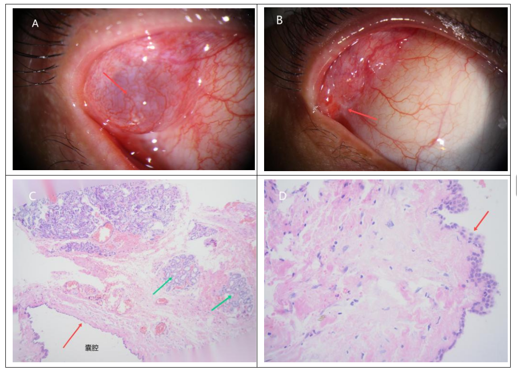
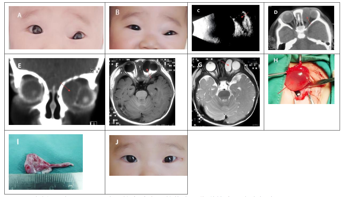
       结膜下囊性肿物5例(图1A),眼睑肿胀隆起7例(图2A、B),眼球突出2例,上睑下垂1例,眼球运动障碍1例。14例患者均不伴有视力下降。

2.3 影像学表现

       病变位于眼眶外上方7例,眼眶内上方4例,上方1例,内侧1例,下方1例。B超13例提示病变呈囊性,圆形或椭圆形,边清,囊内为无回声或低回声区(图2C)。彩色多普勒超声(彩超)4例提示肿瘤内未见血流信号,囊肿边缘可见少许红蓝血流信号。CT 12例提示(图2 D、E)病变呈软组织密度影,圆形或椭圆形,边界清晰,其中有2例患者检测的CT值分别为24 Hu和30 Hu,其中1例出现泪腺区额骨局限性轻度凹陷。磁共振成像(magnetic resonance imaging,MRI)1例提示病变呈囊性,T1加权成像(T1-weighted imaging,T1WI)呈低信号(图2F),T2加权成像(T2-weighted imaging,T2WI)呈高信号(图2G)。

2.4 手术情况

       14例患者均行手术治疗。其中全身麻醉4例,局部麻醉10例;根据病变位置,行前路皮肤切口6例,前路结膜切口8例,辅助外眦切开3例。术中6例囊肿破裂,囊内液为透明水样液体,然后向囊腔内注入透明质酸钠,辅助完整摘除囊壁(图2 H、I)。发生于主泪腺的泪腺导管囊肿,囊肿基底部通常与正常泪腺组织相连。1例位于眼眶内上方的肿瘤,行前路皮肤切口进入眶内,摘除肿瘤过程中致泪总管断裂,术中行泪总管断裂吻合术同时置入人工泪管。

2.5 术后病理  

       14例的术后病理提示均为泪腺导管囊肿,其中发生于睑叶泪腺导管囊肿4例,眶叶泪腺导管囊肿4例,副泪腺导管囊肿5例,异位泪腺导管囊肿1例。病理检查显示囊腔周围可见由纤维组织包裹的立方柱状上皮衬里,部分可见正常泪腺组织小叶及邻近松散的纤维基质内少量炎症细胞(图1C、D)。

2.6 术后复查

       术后早期1例患者出现轻度上睑下垂,随访3个月后上睑睑缘位置恢复正常。结膜切口处可见少量瘢痕(图1B)。术中泪总管断裂患者,随访6个月后拔除人工泪管,未诉泪溢。所有病例随访至少半年,未见复发,无复视及视力下降,未诉眼干表现。

3 讨论

       眼眶泪腺导管囊肿是临床上较少见的良性疾病,由Schmidt等在1803年首次报道[1],约占泪腺良性上皮病变的4%~9%[2-3]。多见单眼发病,偶见双眼发病[4-7,23];无明显性别差异。本组病例均为单眼发病。本病可发生于各年龄段,本组患者的平均年龄为26.5岁,低于Alsarhani等[7]报道的41.2岁,可能与本组中18岁以下患者占4例(占28.6%,其中年龄最小者仅5个月),且样本量偏少有关。
       泪腺导管囊肿好发于主泪腺、副泪腺(Wolfring腺和Krause腺),少部分在泪阜的Popoff腺[7-10]。Wolfring腺多位于睑板上缘附近,而Krause腺集中于上下穹隆结膜(上睑数量约为下睑的2倍)。因主泪腺位于眼眶外上方且副泪腺上睑分布较多,囊肿好发于眼眶外上方(本组占比85.7%,12/14)。根据泪腺导管囊肿发生的位置可将其分为4类:睑叶型、眶叶型、副泪腺型及异位型[11]。本组病例中发生于主泪腺的泪腺导管囊肿占57.2%(8/14),较Alsarhani 等[7]报道的32.8%(19/55)存在差异,可能与种族及样本量等有关。
       泪腺导管囊肿的形成机制尚未明确,主要涉及两方面因素:先天性泪腺排泄管异常及分泌成分改变(如囊内液中高浓度IgA引发的渗透压失衡)[12-13];继发性因素包括创伤、感染或慢性炎症等,这些因素通过削弱泪腺导管的神经肌肉收缩性,导致泪腺导管被动扩张形成囊肿[4,14]
       睑叶泪腺导管囊肿,典型表现为外上方眼睑无痛性肿胀隆起,表面光滑,可透光,结膜面可见白-蓝色囊性肿物,部分伴有压痛,偶见继发感染导致脓肿、瘘管或眼眶蜂窝织炎[15-17]。特征性表现为哭泣、寒冷刺激后囊肿短暂增大,或按压后泪液溢出致囊肿缩小(本组2例具有此特征),此动态变化有助于鉴别诊断。眶叶泪腺导管囊肿,因病变位置较深及占位效应,可造成眼球突出、移位,严重者可出现复视。副泪腺导管囊肿,主要发生于上下穹隆近睑板缘处,可表现为眼睑肿胀隆起,穹隆部结膜可见近透明囊肿;眼眶上方的病变,可致上睑下垂,儿童患者需警惕弱视及屈光异常风险,建议早期干预。
       发生在主泪腺的泪腺导管囊肿,临床诊断相对容易;而发生在副泪腺及异位泪腺导管囊肿易误诊,需与上皮/结膜包含性囊肿、汗腺囊瘤及皮样囊肿等鉴别[18]。对于非典型或疑似异位病例,需要进行眼部B超、CT/MRI等影像学辅助定位及定性评估[19]
       泪腺导管囊肿体积小且无症状者,可观察随访;体积大,影响外观,有美容需求或有临床症状(如上睑下垂、眼球突出移位),则需要手术切除,而单纯的囊内液抽吸易复发。本组病例13于初次就诊时(5月龄)接受囊内液抽吸术(抽吸量1.5 mL),但术后3个月复查示囊肿复发增大,最终改行手术完整切除。依据囊肿所在位置,手术入路可选择前路皮肤切口和结膜切口,必要时联合外眦切开。
       发生在眶叶的泪腺导管囊肿,推荐外上方眉弓下皮肤切口,有利于暴露深部病变且避免损伤提上睑肌。本组中4例眶叶泪腺导管囊肿均采用了眉弓下皮肤切口,未见上睑下垂等手术并发症。而睑叶的泪腺导管囊肿多可直接经外上穹隆结膜面显露,结膜切口入路可实现直视下切除,但存在损伤穹隆部泪腺导管开口、继发干眼及穹隆部变浅的风险;而重睑皮肤切口入路,虽需要分离囊肿表面的肌层组织(存在提上睑肌损伤可能),但其优势在于对Müller肌或提上睑肌撕裂可同期修复。本组中4例睑叶泪腺导管囊肿均采用了结膜入路切除囊肿,术后复查未见干眼和穹隆部变浅等并发症。睑叶泪腺导管囊肿手术中重睑皮肤切  口与结膜切口入路的优劣尚需进一步验证,受限于现有样本量,其手术相关并发症的差异仍有待多中心对照研究明确。
       另外,睑叶和眶叶泪腺导管囊肿基底部通常与正常泪腺组织毗邻,术中需精细分离并保留正常泪腺组织,以规避术后泪液分泌功能障碍风险。发生在副泪腺的泪腺导管囊肿,本组中3例采用结膜切口入路未见并发症,1例皮肤切口入路发生泪总管断裂。但文献显示皮肤切口入路(30例)相较结膜切口入路(18例)切除囊肿,术中囊肿破裂更少,需要结膜移植及术后残留上睑下垂的比例更低[20]
       泪腺导管囊肿术中囊壁易破裂导致残留,从而增加复发及瘘管形成风险,研究者一直寻求不同方法摘除囊肿。Nakauchi等[21]使用视网膜冷冻探头冷冻固定囊壁并辅助整个囊肿切除。Weatherhead等[22]建议发生在副泪腺的泪腺导管囊肿切除宽为1~2 mm的长形睑板条,以降低破裂率。而术中一旦囊肿破裂,完整摘除囊壁,成为一个难点。无色透明的医用透明质酸钠凝胶是眼科白内障手术中常用的维持前房的物质,无毒、无刺激,具有高黏弹性、良好的假塑性和高黏合性的特性。通过破裂囊壁口将其注入囊腔后能够有效维持囊肿的形状,为术中提供清晰的解剖边界,帮助术中精准分离囊壁,避免过度切除周围正常组织及囊壁残留。刘立民等[23]指出在不同的囊性肿物破裂后使用医用透明质酸钠亦可辅助完整摘除囊肿。我们亦借助此种方法处理破裂的囊肿,也证实这种方法简易、可行,不受特殊器械的限制。此外,对发生在眼眶前部的泪腺导管囊肿,有学者采用囊肿造袋术[24-25],也取得了一定治疗效果,而不必担心囊肿破裂的问题。
       泪腺导管囊肿术后复发率低,文献报道为0~3.5% [7, 14,26]。精准微创操作可有效减少临近泪腺、眼外肌及提上睑肌损伤相关的并发症。
       总之,泪腺导管囊肿作为较少见的眼眶良性病变,其发病机制尚未完全阐明,需要进一步探索。结合特征性的临床表现及影像学检查,发生于主泪腺的泪腺导管囊肿诊断准确率较高。手术完整摘除囊肿是首选的治疗策略,术中囊肿破裂后辅助应用医用透明质酸钠囊腔内注射,可显著提高囊壁的辨识度与整体囊肿的切除率。

图1 病例13右眼睑叶泪腺导管囊肿患者的临床及病理检查 

Figure 1 Clinical and pathological examination of a lacrimal duct cyst in the palpebral lobe of the right eye of Case 13

图1 病例13右眼睑叶泪腺导管囊肿患者的临床及病理检查 Figure 1 Clinical and pathological examination of a lacrimal duct cyst in the palpebral lobe of the right eye of Case 13
(A)术前右眼外上方穹隆部可见近蓝紫色囊肿(红色箭),伴局部结膜充血;(B)术后2年复查未见囊肿复发,可见少量结膜瘢痕(红色箭头);(C)病理组织检查(苏木精-伊红染色,×100)囊腔周围可见由纤维组织包裹的立方柱状上皮衬里(红色箭头),并可见正常泪腺组织小叶(绿色箭头)及邻近松散的纤维基质内少量炎症细胞;(D)病理组织检查(苏木精-伊红染色,×400)可见双层立方柱状上皮(红色箭头)。
(A) A bluish-purple cyst (red arrow) with localized conjunctival congestion is observed in the superotemporal fornix of the right eye preoperatively.(B) Two-year postoperative follow-up revealed no cyst recurrence, with mild conjunctival scarring observed (red arrow). (C)Histopathological examination (hematoxylin-eosin [HE] staining, ×100) demonstrates a cuboidal-columnar epithelial lining (red arrow) surrounded by ffbrous tissue encapsulation. Normal lacrimal gland lobules (green arrow) and scattered inffammatory cells within adjacent loose fibrous stroma are noted. (D) Histopathological examination (HE staining, ×400) reveals a bilayer of cuboidal-columnar epithelium (red arrow).

图2:病例14左眼眶副泪腺导管囊肿患者的临床影像学检查和术中标本

Figure 2 Clinical imaging and intraoperative specimens of an accessory lacrimal duct cyst in the left orbit of Case 14

图2:病例14左眼眶副泪腺导管囊肿患者的临床影像学检查和术中标本 Figure 2 Clinical imaging and intraoperative specimens of an accessory lacrimal duct cyst in the left orbit of Case 14
(A) 患儿5月龄时第1次就诊的外观像,左眼上睑内侧皮肤肿胀隆起,睑缘呈“S”型;B.患儿8月龄(囊肿穿刺抽液后3个月)时第2次就诊的术前外观像,可见左眼上睑内侧皮肤仍肿胀隆起;(C) 术前肿物B型超声间接探查可见肿物呈囊性(红色箭头),部分边界不显示;(D) 术前眼眶轴位CT(软组织窗)显示左眼眶内上方近椭圆形肿物(红色箭头),均质,边界清晰;(E)术前眼眶冠状位CT(软组织窗)显示左眼眶内上方近椭圆形肿物(红色箭头),均质,边界清晰,眼球受压变形,眶顶骨质较对侧压迫变薄;(F) 术前眼眶MR显示肿物TIWI上呈低信号(红色箭头);(G) 术前眼眶MRI显示肿物T2WI上呈高信号(红色箭头);(H) 术中摘除的囊性肿物(红色箭头),伴囊内注入部分透明质酸钠;(I) 术中摘除的囊性肿物大体标本,囊壁破裂,内液已完全溢出;( J)术后10 d外观像,可见左眼上睑睑缘形态恢复正常,外眦部皮肤切口痕迹。
(A) Initial clinical photograph at 5 months of age showing swelling of the medial upper eyelid skin and an "S-shaped" deformity of the leff eyelid margin. (B) Preoperative photograph at the second visit (8 months of age, 3 months post-cyst aspiration) demonstrating persistent swelling of the medial upper eyelid skin on the leff side.(C) Postoperative photograph at 10 days affer surgery revealing restoration of normal eyelid contour and a skin incision scar at the lateral canthus. (D) Preoperative B-scan ultrasonography (indirect mode) depicting a cystic lesion (red arrow) with partially indistinct boundaries. (E) Preoperative axial orbital CT (soff tissue window) showing a homogeneous, well-deffned, ovalshaped mass in the superomedial leff orbit (red arrow). (F) Preoperative coronal orbital CT (soff tissue window) conffrming the superomedial oval mass (red arrow), homogeneous with sharp margins, accompanied by globe displacement and thinning of the orbital roof compared to the contralateral side. (G) Preoperative orbital MRI (T1-weighted image, T1WI) displaying hypointense signal of the mass (red arrow). (H)Preoperative orbital MRI (T2-weighted image, T2WI) illustrating hyperintense signal of the mass (red arrow). (I) Intraoperative view of the excised cystic mass (red arrow) partially fflled with sodium hyaluronate. ( J) Gross specimen of the excised cyst, showing a ruptured cyst wall with complete evacuation of intracystic ffuid.

声明

在论文撰写中无使用生成式人工智能。论文撰写中的所有内容均由作者独立完成,并对出版物的真实性和准确性承担全部责任。

利益冲突

所有作者均声明不存在利益冲突。

开放获取声明

本文适用于知识共享许可协议(Creative Commons),允许第三方用户按照署名(BY)-非商业性使用(NC)-禁止演绎(ND)(CC BY-NC-ND)的方式共享,即允许第三方对本刊发表的文章进行复制、发行、展览、表演、放映、广播或通过信息网络向公众传播,但在这些过程中必须保留作者署名、仅限于非商业性目的、不得进行演绎创作。
1、Rootman J. Inflammatory diseases[M]//Diseases of the Orbit: a multidisciplinary aproach. 2nd Ed. Philadelphia, PA: Lippincott Williams & Wilkins, 2003: 431-432. Rootman J. Inflammatory diseases[M]//Diseases of the Orbit: a multidisciplinary aproach. 2nd Ed. Philadelphia, PA: Lippincott Williams & Wilkins, 2003: 431-432.
2、 Shields JA, Shields CL, Scartozzi R. Survey of 1264 patients with orbital tumors and simulating lesions: The 2002 Montgomery Lecture, part 1[J]. Ophthalmology, 2004, 111(5): 997-1008. DOI:10.1016/j.ophtha.2003.01.002. Shields JA, Shields CL, Scartozzi R. Survey of 1264 patients with orbital tumors and simulating lesions: The 2002 Montgomery Lecture, part 1[J]. Ophthalmology, 2004, 111(5): 997-1008. DOI:10.1016/j.ophtha.2003.01.002.
3、 Shields CL, Shields JA, Eagle RC, et al. Clinicopathologic review of 142 cases of lacrimal gland lesions[J]. Ophthalmology, 1989, 96(4): 431-435. DOI:10.1016/s0161-6420(89)32873-9. Shields CL, Shields JA, Eagle RC, et al. Clinicopathologic review of 142 cases of lacrimal gland lesions[J]. Ophthalmology, 1989, 96(4): 431-435. DOI:10.1016/s0161-6420(89)32873-9.
4、 Tsiouris AJ, Deshmukh M, Sanelli PC, et al. Bilateral dacryops: correlation of clinical, radiologic, and histopathologic features[J]. AJR Am J Roentgenol, 2005, 184(1): 321-323. DOI:10.2214/ajr.184.1.01840321. Tsiouris AJ, Deshmukh M, Sanelli PC, et al. Bilateral dacryops: correlation of clinical, radiologic, and histopathologic features[J]. AJR Am J Roentgenol, 2005, 184(1): 321-323. DOI:10.2214/ajr.184.1.01840321.
5、Feijó ED, Alencastro Landim G, de Melo Dias M, et al. Giant bilateral cysts of the accessory lacrimal glands of wolfring in a child[J]. Ophthalmic Plast Reconstr Surg, 2020, 36(1): e4-e6. DOI:10.1097/IOP.0000000000001494. Feijó ED, Alencastro Landim G, de Melo Dias M, et al. Giant bilateral cysts of the accessory lacrimal glands of wolfring in a child[J]. Ophthalmic Plast Reconstr Surg, 2020, 36(1): e4-e6. DOI:10.1097/IOP.0000000000001494.
6、Tsai FF, Mukhopadhyay C, Zeng J, et al. Bilateral marked dacryops following trauma[J]. Orbit, 2012, 31(6): 435-437. DOI:10.3109/01676830.2012.711889. Tsai FF, Mukhopadhyay C, Zeng J, et al. Bilateral marked dacryops following trauma[J]. Orbit, 2012, 31(6): 435-437. DOI:10.3109/01676830.2012.711889.
7、 Alsarhani WK, Al-Sharif EM, Al-Faky YH, et al. Dacryops and clinical diagnostic challenges[J]. Can J Ophthalmol, 2022, 57(6): 388-393. DOI:10.1016/j.jcjo.2021.06.014. Alsarhani WK, Al-Sharif EM, Al-Faky YH, et al. Dacryops and clinical diagnostic challenges[J]. Can J Ophthalmol, 2022, 57(6): 388-393. DOI:10.1016/j.jcjo.2021.06.014.
8、Jakobiec FA, Roh M, Stagner AM, et al. Caruncular dacryops[J]. Cornea, 2015, 34(1): 107-109. DOI:10.1097/ico.0000000000000287. Jakobiec FA, Roh M, Stagner AM, et al. Caruncular dacryops[J]. Cornea, 2015, 34(1): 107-109. DOI:10.1097/ico.0000000000000287.
9、Stern K, Jakobiec FA, Harrison WG. Caruncular dacryops with extruded secretory globoid bodies[J]. Ophthalmology, 1983, 90(12): 1447-1451. DOI:10.1016/s0161-6420(83)34376-1. Stern K, Jakobiec FA, Harrison WG. Caruncular dacryops with extruded secretory globoid bodies[J]. Ophthalmology, 1983, 90(12): 1447-1451. DOI:10.1016/s0161-6420(83)34376-1.
10、Vaidya PR, D’Aquila ML, Ramey NA, et al. Caruncular dacryops after cataract surgery with histopathological characterization[J]. Am J Dermatopathol, 2021, 43(2): e27-e29. DOI:10.1097/DAD.0000000000001804. Vaidya PR, D’Aquila ML, Ramey NA, et al. Caruncular dacryops after cataract surgery with histopathological characterization[J]. Am J Dermatopathol, 2021, 43(2): e27-e29. DOI:10.1097/DAD.0000000000001804.
11、Bullock JD, Fleishman JA, Rosset JS. Lacrimal ductal cysts[J]. Ophthalmology, 1986, 93(10): 1355-1360. DOI:10.1016/s0161-6420(86)33566-8. Bullock JD, Fleishman JA, Rosset JS. Lacrimal ductal cysts[J]. Ophthalmology, 1986, 93(10): 1355-1360. DOI:10.1016/s0161-6420(86)33566-8.
12、Lam K, Brownstein S, Jordan DR, et al. Dacryops: a series of 5 cases and a proposed pathogenesis[J]. JAMA Ophthalmol, 2013, 131(7): 929-932. DOI:10.1001/jamaophthalmol.2013.1885. Lam K, Brownstein S, Jordan DR, et al. Dacryops: a series of 5 cases and a proposed pathogenesis[J]. JAMA Ophthalmol, 2013, 131(7): 929-932. DOI:10.1001/jamaophthalmol.2013.1885.
13、Jakobiec FA, Zakka FR, Perry LP. The cytologic composition of dacryops: an immunohistochemical investigation of 15 lesions compared to the normal lacrimal gland[J]. Am J Ophthalmol, 2013, 155(2): 380-396.e1. DOI:10.1016/j.ajo.2012.07.028. Jakobiec FA, Zakka FR, Perry LP. The cytologic composition of dacryops: an immunohistochemical investigation of 15 lesions compared to the normal lacrimal gland[J]. Am J Ophthalmol, 2013, 155(2): 380-396.e1. DOI:10.1016/j.ajo.2012.07.028.
14、Galindo-Ferreiro A, Alkatan HM, Muinos-Diaz Y, et al. Accessory lacrimal gland duct cyst: 23 years of experience in the Saudi population[J]. Ann Saudi Med, 2015, 35(5): 394-399. DOI:10.5144/0256-4947.2015.394. Galindo-Ferreiro A, Alkatan HM, Muinos-Diaz Y, et al. Accessory lacrimal gland duct cyst: 23 years of experience in the Saudi population[J]. Ann Saudi Med, 2015, 35(5): 394-399. DOI:10.5144/0256-4947.2015.394.
15、Park HJ, Park SJ, Chi M. Lacrimal gland ductal cyst infection presenting as acute orbital cellulitis with abscess[J]. J Craniofac Surg, 2021, 32(5): e429-e432. DOI:10.1097/SCS.0000000000007239. Park HJ, Park SJ, Chi M. Lacrimal gland ductal cyst infection presenting as acute orbital cellulitis with abscess[J]. J Craniofac Surg, 2021, 32(5): e429-e432. DOI:10.1097/SCS.0000000000007239.
16、Sadek H, Mirani N, Lee HJ, et al. Lacrimal gland ductal cyst complicated by abscess formation[J]. Ophthalmic Plast Reconstr Surg, 2020, 36(2): e32-e34. DOI:10.1097/IOP.0000000000001539.Sadek H, Mirani N, Lee HJ, et al. Lacrimal gland ductal cyst complicated by abscess formation[J]. Ophthalmic Plast Reconstr Surg, 2020, 36(2): e32-e34. DOI:10.1097/IOP.0000000000001539.
17、Eifrig CW, Chaudhry NA, Tse DT, et al. Lacrimal gland ductal cyst abscess[J]. Ophthalmic Plast Reconstr Surg, 2001, 17(2): 131-133. DOI:10.1097/00002341-200103000-00011. Eifrig CW, Chaudhry NA, Tse DT, et al. Lacrimal gland ductal cyst abscess[J]. Ophthalmic Plast Reconstr Surg, 2001, 17(2): 131-133. DOI:10.1097/00002341-200103000-00011.
18、Tanaboonyawat S, Idowu OO, Copperman TS, et al. Dacryops–A review[J]. Orbit, 2020, 39(2): 128-134. DOI:10.1080/01676830.2019.1608564. Tanaboonyawat S, Idowu OO, Copperman TS, et al. Dacryops–A review[J]. Orbit, 2020, 39(2): 128-134. DOI:10.1080/01676830.2019.1608564.
19、于文玲, 刘中林, 朴颖实, 等. 泪腺导管囊肿的CT、MRI表现 [J]. 临床放射学杂志, 2014, 33(2): 182-184. DOI:10.13437/j.cnki. jcr.2014.02.007.
YU Wenling,LIU Zhonglin,PIAO Yingshi,et al.CT and MRI Findings of Lacrimal Duct Cyst[J]. Journal of Clinical Radiology, 2014, 33(2): 182-184. DOI:10.13437/j.cnki.jcr.2014.02.007.
20、Diab MM, Allen RC, Mohammed KK, et al. Clinical characteristics and surgical outcomes of transcutaneous versus transconjunctival excision of Wolfring gland ductal cysts[J]. BMC Ophthalmol, 2024, 24(1): 164. DOI:10.1186/s12886-024-03420-x. Diab MM, Allen RC, Mohammed KK, et al. Clinical characteristics and surgical outcomes of transcutaneous versus transconjunctival excision of Wolfring gland ductal cysts[J]. BMC Ophthalmol, 2024, 24(1): 164. DOI:10.1186/s12886-024-03420-x.
21、 Nakauchi K, Katori N, Imagawa Y, et al. A case report on lacrimal ductal cyst causing unilateral blepharoptosis[J]. Br J Ophthalmol, 2009, 93(9): 1143-1145. DOI:10.1136/bjo.2009.160812. Nakauchi K, Katori N, Imagawa Y, et al. A case report on lacrimal ductal cyst causing unilateral blepharoptosis[J]. Br J Ophthalmol, 2009, 93(9): 1143-1145. DOI:10.1136/bjo.2009.160812.
22、Weatherhead RG. Wolfring dacryops[J]. Ophthalmology, 1992, 99(10): 1575-1581. DOI:10.1016/S0161-6420(92)31764-6. Weatherhead RG. Wolfring dacryops[J]. Ophthalmology, 1992, 99(10): 1575-1581. DOI:10.1016/S0161-6420(92)31764-6.
23、刘立民, 赵伟, 任明玉, 等. 应用透明质酸钠凝胶术中辅助破裂 囊肿摘除临床观察[J]. 中国实用眼科杂志, 2016, 34(3): 276-277. DOI:10.3760/cma.j.issn.1006-4443.2016.03.018.
Liu Limin,Zhao Wei,Ren Mingyu, et al.Clinical observation of medical sodium hyaluronate gel aid in removal of ruptured periocular cyst[J]. China J Pract Ophthalmol,2016, 34(3): 276-277. DOI:10.3760/cma.j.issn.1006-4443.2016.03.018.
24、Salam A, Barrett AW, Malhotra R, et al. Marsupialization for lacrimal ductular cysts (dacryops): a case series[J]. Ophthalmic Plast Reconstr Surg, 2012, 28(1): 57-62. DOI:10.1097/IOP.0b013e31822ddda7. Salam A, Barrett AW, Malhotra R, et al. Marsupialization for lacrimal ductular cysts (dacryops): a case series[J]. Ophthalmic Plast Reconstr Surg, 2012, 28(1): 57-62. DOI:10.1097/IOP.0b013e31822ddda7.
25、Harris GJ. Marsupialization of a lacrimal gland cyst[J]. Ophthalmic Surg, 1983, 14(1): 75-78. Harris GJ. Marsupialization of a lacrimal gland cyst[J]. Ophthalmic Surg, 1983, 14(1): 75-78.
26、Ozgonul C, Uysal Y, Ayyildiz O, et al. Clinical features and management of dacryops[J]. Orbit, 2018, 37(4): 262-265. DOI:10.1080/01676830.2017.1423081. Ozgonul C, Uysal Y, Ayyildiz O, et al. Clinical features and management of dacryops[J]. Orbit, 2018, 37(4): 262-265. DOI:10.1080/01676830.2017.1423081.
上一篇
下一篇
其他期刊
  • 眼科学报

    主管:中华人民共和国教育部
    主办:中山大学
    承办:中山大学中山眼科中心
    主编:林浩添
    主管:中华人民共和国教育部
    主办:中山大学
    浏览
  • Eye Science

    主管:中华人民共和国教育部
    主办:中山大学
    承办:中山大学中山眼科中心
    主编:林浩添
    主管:中华人民共和国教育部
    主办:中山大学
    浏览
目录

点击右上角菜单,浏览器打开下载

我知道了