您的位置: 首页 > 2023年2月 第38卷 第2期 > 文字全文
2023年7月 第38卷 第7期11
目录

囊袋上经巩膜缝线固定IOL植入术治疗球形晶状体的有效性和安全性研究

The efficacy and safety of the application of phacoemulsification combined with supra-capsular and scleral-fixated intraocular lens implantation in microspherophakia surgery

来源期刊: 眼科学报 | 2023年2月 第38卷 第2期 92-100 发布时间:2023-02-01 收稿时间:2023/2/23 10:11:29 阅读量:6589
作者:
关键词:
球形晶状体囊袋超声乳化吸出术改良囊袋张力环
microspherophakia capsular bag phacoemulsification modified capsular tension ring
DOI:
10.12419/j.issn. 1000-4432.2023.02.03
收稿时间:
 
修订日期:
 
接收日期:
 
目的:探讨超声乳化晶状体吸除联合囊袋上经巩膜缝线固定人工晶状体(intraocular lens,IOL)植入术治疗球形晶状体(microspherophakia,MSP)的有效性和安全性。方法:采用回顾性分析,选取2019年1月至 2020年6月期间在复旦大学附属眼耳鼻喉科医院进行治疗的MSP患者37例(37眼),随机分为两组,纳入行超声乳化晶状体吸除联合囊袋上巩膜缝线固定IOL植入术(supra-capsular and scleral-fixated intraocular lens implantation,SCSF-IOL)的MSP患者20例(20眼)和行超声乳化晶状体吸除联合改良型囊袋张力环植入术(transscleral-fixated modified capsular tension ring and in-the-bag intraocular lens implantation,MCTR-IOL)的MSP患者17例(17眼),观察两组术后最佳矫正视力及并发症等情况。结果:两组术后最佳矫正视力比术前均有改善(P<0.001),而组间比较差异无统计学意义(P=0.326)。两组的IOL倾斜度相当(P=0.216)。预防性Nd:YAG激光后囊膜切开术在SCSFIOL术后1周至1个月进行。在SCSF-IOL组,2眼(10.00%)需要重复激光治疗,1眼(5.00%)出现囊口偏心。后囊膜混浊是MCTR组最常见并发症(6眼,35.29%)。随访期间两组均未出现IOL脱位、继发性青光眼和视网膜脱离。结论:SCSF-IOL是治疗球形晶状体的简单易行的手术方式,疗效与MCTR-IOL相当。Nd:YAG激光后囊膜切开术是预防SCSF-IOL术后囊袋并发症的必要手段。
Objective: To investigate the efficacy and safety of phacoemulsification combined with supra-capsular and scleral-fixated intraocular lens (IOL) implantation in the treatment of microspherophakia (MSP). Methods: by retrospective analysis, 37 MSP patients (37 eyes) who were treated in our hospital from January 2019 to June 2020 were randomly divided into two groups, including 20 MSP patients (20 eyes) who treated by SCSF-IOL and 17 MSP patients (17 eyes) who treated by transscleral-fixated modified capsular tension ring and in-the-bag intraocular lens implantation (MCTR-IOL). The best corrected vision and complications were observed. Results: the best corrected vision was significantly improved in both groups (P < 0.001), but there was no remarkable difference between the two groups (P = 0.326). The IOL tilt was also comparable (P = 0.216). Prophylactic Nd: YAG laser posterior capsulotomy was performed from 1 week to 1 month after the SCSF-IOL procedure. In the SCSF-IOL group, two eyes (10.00%) needed repeated laser treatment, and one eye (5.00%) had a decentered capsule opening. Posterior capsular opacification was the most common complication (6, 35.29%) in the MCTR group. No IOL dislocation, secondary glaucoma, or retinal detachment was observed during follow-up. Conclusions: SCSF-IOL is a simple and viable surgical option for managing MSP and is comparable with the MCTR-IOL. Nd: YAG laser posterior capsulotomy is a necessary mean to prevent residual capsule complications after the SCSF-IOL procedure.
球形晶状体(microspherophakia,MSP)是一种罕见的先天性发育异常性疾病,由于晶状体悬韧带发育不全、张力不足,导致晶状体生长受限[1]。MSP患者的晶状体较小,形状更接近于球形,因此常并发晶状体半脱位(44%)和晶状体源性高度近视(84.6%),并有发生继发性青光眼倾向(44.4%~51%)[2]。MSP独特的临床表现和潜在的并发症对该病的治疗提出了挑战,MSP的治疗以手术为主,曾尝试运用过的术式包括前房角支撑人工晶状体(intraocular lens,IOL)[3]、虹膜夹持型IOL[4]、中周部虹膜缝合IOL[5]和缝线[6]或无缝线[7-8]巩膜固定IOL。然而,以上手术大都采用晶状体切除术或囊袋切除术,而前部玻璃体切割术通常是必要的步骤,这不仅会增加视网膜脱离的风险[9],而且会增加IOL植入后的倾斜、偏心甚至脱位的风险[10]。随着囊袋辅助装置的临床应用,为减少术中、术后并发症,现代晶状体不全脱位手术治疗的核心是最大限度地保留和重塑晶状体囊袋悬韧带隔,囊袋拉钩联合Cionni双钩改良囊袋张力环(modified capsular tension ring,MCTR)植入是比较符合解剖结构的手术选择,但国内近年来暂无MCTR供应,2019年开始我们尝试应用超声乳化晶状体吸除联合囊袋上巩膜缝线固定式IOL植入术(supra-capsular and scleral-fixated intraocular lens implantation,SCSF-IOL)治疗球形晶状体取得了较好的效果,并与经巩膜固定改良囊袋内张力环植入术与IOL囊袋内植入术(transscleral-fixated MCTR and in-the-bag intraocular lens implantation,MCTR-IOL)的患者进行对比和评估,探讨SCSF-IOL术式的有效性和安全性。

1 对象与方法

1.1 对象

采用回顾分析方法,选择2019年1月至 2020年6月复旦大学附属眼耳鼻喉科医院眼科收治的MSP患者为研究对象,患者均已签署知情同意书。本研究获复旦大学眼耳鼻喉科医院伦理委员会审批通过(no. 2020126-1)所有病例均由同一位经验丰富的眼
科医生在完全扩瞳的情况下进行裂隙灯检查,采用国际标准Snellen视力表行最佳矫正视力检查(best-corrected visual acuity,BCVA)。其他检查包括光学相干生物测量仪(IOLMaster)、非接触眼压、以及B超检查。波前像差仪(OPD-ScanⅢ)用于采集眼前段后照法图片及眼像差:若后照法图片显示同轴角膜映光点落在撕囊口外或残留囊袋上,则定义为囊袋偏心;倾斜(tilt)、慧差(coma)和三叶草差(trefoil)等用于间接反映IOL的倾斜程度[11]
收集患者术前资料,包括年龄、性别、眼别、BCVA、眼压等。手术眼即为患者术前登记的单侧需进行手术的眼,如果患者登记为双侧均需进行手术,则随机选择其中一只眼。纳入标准:在扩瞳下能够观察到整个晶状体赤道部,以及AS-OCT对瞳孔直径有限眼的检查补充诊断。手术适应证如下:1)BCVA (LogMAR)低于0.5;2) 无法矫正的晶状体源性散光;3) 晶状体脱位致瞳孔阻滞;4)弱视进展风险高。具有以下特征的患者未入选:1)晶状体脱位至前房或玻璃体;2) 有眼外伤或眼内手术史;3)合并视网膜脱离、视网膜色素变性、终末期青光眼或角膜内皮失代偿。

1.2 方法

本研究回顾性分析了2019年至2021年于复旦大学眼耳鼻喉科医院医院行晶状体手术的球形晶状体患者,所有患者手术均由同一医师在显微镜下主刀完成。由于MCTR在中国的注册证于2020年6月过期,对于2020年6月之前的球形晶状体患者我们行MCTR-IOL手术治疗,对于2020年6月之后的球形晶状体患者我们采用SCSF-IOL手术方案。随访观察两组术后最佳矫正视力及并发症等情况。
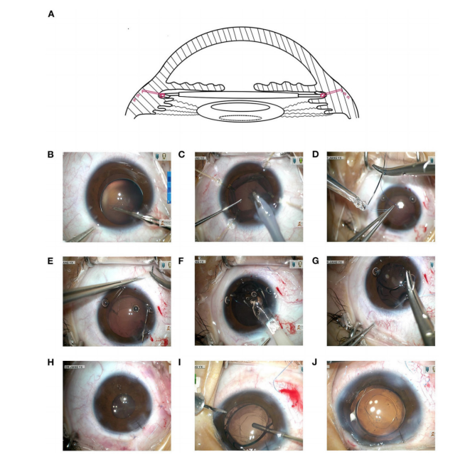
SCSF-IOL:做2.6 mm的透明角膜隧道切口,注入粘弹剂,仔细行4.5~5.0 mm的连续环形撕囊,囊袋拉钩固定。使用Centurion超声乳化器通过(I/A)模式在低负压、低流速、低灌注下吸除晶状体核及皮质,再次注入粘弹剂,用9-0聚丙烯线套结IOL(Superflex Aspheric 920H 或C-flex Aspheric 970C, Rayner Surgical Group Ltd.,West Sussex , UK)的下襻,缝针在角膜缘后方1.5~2.0mm处出针,并通过2.6mm透明角膜隧道切口将IOL植入前房,9-0聚丙烯线套结IOL的上襻,上襻于下襻180度对应处出针,无结Z形缝合技术将聚丙烯线固定在巩膜层间,关闭角膜切口和结膜瓣,在手术结束时,检查IOL的居中性、前囊和后囊撕囊口。由于SCSF-IOL手术将IOL固定在睫状沟,采用如下方式进行睫状沟度数调整:IOL屈光力在+35.00~+27.5 D者IOL度数减小1.50 D,+27.00~+17.50 D者度数减小1.00 D,+17.00~+9.50 D者减小0.50 D,+9.00~-5.00 D者不予调整。预防性Nd:YAG激光后囊膜切开术在SCSF-IOL术后1周至1个月进行。对于预计不配合激光治疗的儿童在术中使用角膜缘入路行后囊切开术和前玻璃体切割术(图1)。
MCTR-IOL:做2.6 mm的透明角膜隧道切口,注入粘弹剂,仔细行4.5~5.0 mm的连续环形撕囊,囊袋拉钩固定。使用Centurion超声乳化器通过(I/A)模式在低负压、低流速、低灌注下吸除晶状体核及皮质,再次注入粘弹剂,将9-0聚丙烯缝合线预置双钩MCTR,然后通过主切口将MCTR植入囊袋,缝针于角膜缘后1.5~2.0 mm处穿出眼外,采用改良的无结Z形缝合技术将MCTR牵引固定在巩膜上,在囊袋内植入IOL(Superflex Aspheric 920H或C-flex Aspheric 970C, Rayner Surgical Group Ltd. ,West Sussex,UK)。对于该组患者,仅对后囊膜浑浊或前囊收缩明显者,行Nd:YAG激光后囊或前囊切开术。
20230224082056_2981.png
图 1 SCSF-IOL治疗MSP的手术设计及手术过程
Figure 1. The demonstration of the principles of the SCSF-IOL procedure and detailed process
(A)IOL悬吊于囊袋前方,后囊在术后由Nd:YAG激光切开或术中切开。(B~H)SCSF-IOL手术步骤展示,连续环形撕囊后在囊袋拉钩的固定下吸除晶状体核及皮质,通过2.6mm透明角膜切口将预置缝线的人工晶体植入眼内,并将缝线进行巩膜层间固定,(I~J)对于预计不配合激光治疗的儿童在术中使用角膜缘入路行后囊切开术和前玻璃体切除术。IOL,人工晶状体;SCSF-IOL,囊袋上经巩膜缝线固定IOL植入术;MSP,球形晶状体。
(A) The IOL sutured with 9–0 polypropylene was sutured above the preserved capsule, which was opened by Nd:YAG laser postoperatively or by intraoperative capsulotomy. (B-H) The details of SCSF-IOL procedure. The lens was aspired after continuous circular capsulorhexis in the aid of four capsular hooks. Preloaded and loop-sutured IOL was implanted through 2.6 mm clear corneal tunnel incision and was sutured intrasclerally using Z-suturing. (I-J) Primary regional capsulotomy was performed in young patients with expected poor cooperation. IOL, intraocular lens; SCSF-IOL, supra-capsular and scleral-fixated intraocular lens implantation; MSP, microspherophakia. 

1.3 统计学处理

采用SPSS25.0统计学软件进行数据分析,所有定量数据均以均数±标准差(x±s)或中位数(下四分位数,上四分位数),采用t检验或Mann–Whitney U检验对两个独立组之间进行比较,采用配对样本t检验或配对Wilcoxon检验比较术前和术后BCVA等测量结果和并发症等情况;定性数据以百分比表示,采用Fisher确切概率法进行组间比较。 P<0.05为差异有统计学意义。

2 结果

2.1 基线特征

SCSF组男女比例为12/8, MCTR组为10/7,二者比较差异无统计学意义(Fisher确切概率法,P=1.000);SCSF组年龄为12.00(IQ:5.00,21.00)岁,MTCR组为6.50(IQ:5.50,27.50)岁,二者比较差异无统计学意义 (Mann-Whitney U检验,P=0.562);术前BCVA(LogMAR)在SCSF-IOL组和MCTR-IOL组分别为0.70(0.40,0.80)和0.70(0.40,1.00),二者比较差异无统计学意义(Mann-Whitney U检验,P=0.405);术前眼轴在SCSF-IOL组和MCTR-IOL组分别为(26.17±2.97)mm和(24.54±2.04)mm,二者比较差异无统计学意义(t检验,P=0.065);术前前房深度在SCSF-IOL组和MCTR-IOL组分别为(3.03±0.73)mm和(2.89±0.75)mm,二者比较差异无统计学意义(t检验,P=0.600);角膜散光在SCSF-IOL组和MCTR-IOL组分别为(?1.65±1.06)D和(?1.97±1.14)D,二者比较差异无统计学意义(t检验,P=0.385)。在SCSF-IOL组中,88.2%的患者近视度数大于?6.0 D,这一比例在MCTR-IOL组中为84.6%,二者比较差异无统计学意义(Fisher确切概率法,P=1.000)。SCSF组患者术后随访时间2.25~6.50个月,位数为4.50 个月;行MCTR组患者术后随访时间2.00~7.00个月,中位数为4.00个月,二者随访时间比较差异无统计学意义(Mann-Whitney U检验,P=0.988)。

2.2 术后视力及眼压

视力:末次随访评估患者BCVA。大多数眼的BCVA均有改善,SCSF-IOL组(P<0.001)和MCTR-IOL组(P<0.001)与治疗前比较差异均有统计学意义。SCSF-IOL组和MCTR-IOL组的术后BCVA(LogMAR)分别为0.13(0.02,0.28)和0.15(0.10,0.46),二者比较差异无统计学意义(P=0.326)(表1)。
眼压:两种手术均未降低眼压(SCSF组P=0.196;MCTR组P=0.824),两组术后眼压相似(P=0.755。术后未观察到继发性青光眼发生(表1)。
IOL倾斜程度:对两组患者的眼像差进行评估,间接比较IOL倾斜的严重程度。倾斜(P=0.216)、彗差和(P=0.151)和三叶草差(P=0.264)在组间比较差异均无统计学意义(表1)。
屈光状态:SCSF-IOL患者平均球镜度数为(?0.80±1.56)D,MCTR-IOL组为(?1.51±2.07)D,二者比较差异无统计学意义(P=0.248);中位柱镜度数在SCSF-IOL组为?1.50(?2.00,?0.75)D,在MCTR-IOL组为?1.50(?2.75,?1.25)D,二者比较差异无统计学意义(P=0.379)。

2.3 术后囊膜变化

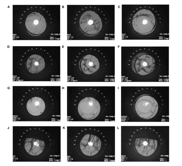
在 SCSF-IOL 组中,术后 1 周的囊膜清晰平整 ( 图 2A),术后 1 个月开始缩小 ( 图 2B),行预防性激光囊膜切开术 ( 图 2C)。在 1 年的随访中,可以见部分患者的后囊激光切开口位置是稳定的 ( 图2D~F)。2 眼 (10.00%) 接受 2 次激光治疗,患者均在 8 岁以下,再次激光治疗时间均在术后第 2 个月内,并最终达到满意的后囊切开效果。2 例年轻患者术中行局部后囊切开术和前部玻璃体切割术,囊口保持中心、收缩小,无需进一步激光治疗 ( 图2G~I)。1 眼 (5.00%) 在激光治疗后 5 个月意外出现了偏心的囊膜开口 ( 图 2J~L)。在 MCTR-IOL 组中,6 眼 (35.29%) 术后出现明显的后囊浑浊 (PCO),发生时间为术后 3.00 (2.50,9.00) 个月,均行 Nd:YAG激光囊切开术。
表 1. SCSF-IOL 和 MCTR-IOL 两种手术方式的疗效比较
Table 1. Comparison of surgical outcomes of SCSF-IOL and MCTR-IOL in eyes with MSP
20230224082421_2447.png
注:BCVA,最佳矫正视力;IOP,眼内压;IOL,人工晶状体,SCSF-IOL,囊袋上经巩膜缝线固定联合IOL植入术;LogMAR,最小分辨角对数,MCTR-IOL,改良囊袋张力环巩膜缝线固定联合IOL植入术;RMS,均方根;
1 mmHg=0.133 kPa。 a. Normally distributed data are shown in the mean ± standard deviation, while skewed data are shown in the median (interquartile). BCVA, best-corrected visual acuity; IOP, intraocular pressure; SCSF-IOL, supra-capsular and scleral-fixated intraocular lens implantation; LogMAR, logarithm of the minimal angle of resolution; MCTR-IOL, transscleral-fixated modified capsular tension ring and in-the-bag intraocular lens implantation. RMS, root mean square. 

图 2 SCSF-IOL组术后囊膜变化
Figure 2 The capsular change after SCSF-procedure
(A~C)术后早期囊袋改变(术后1周至1个月)。(D~F)术后中远期囊袋改变(术后1周~1年)。(G~I)行一期囊袋切开的患者,囊袋保持稳定和居中。(J~L)1例患者在Nd:YAG激光后出现撕囊口偏心。
(A-C) The early stage of capsular change with 1 month. (D-F) The mid and long-term capsular changes after operation (1-week postoperatively to 1-year postoperatively). (G-I) The residual capsule remained stable and centered during the follow-up after primary capsulotomy. ( J-L) Unexpected decentered capsulorhexis opening after Nd:YAG capsulotomy in one patient. 

2.4 术后并发症

两组患者在激光治疗后 3 位患者出现短暂的眼前黑影飘动感外,均未出现其他并发症。SCSF-IOL 组 的 2 眼 (10.00%) 和 MCTR 组 的 4 眼(23.53%) 在术前被诊断为青光眼,术后均需要辅以抗青光眼滴眼液治疗,但滴眼的次数减少。两组均未观察到术后继发性青光眼。两组均无出现缝线暴露、人工晶状体脱位,黄斑囊样水肿或视网膜脱离。

3 讨论

晶状体源性屈光不正和继发于晶状体脱位的并发症严重威胁着MSP患者的长期视力预后,因此临床上早期进行手术干预是必要的,但晶状体摘除联合IOL植入术往往受到晶状体悬韧带张力不足以及囊袋过小的综合影响,手术复杂、并发症多,导致目前尚缺乏针对MSP患者的治疗金标准。
20世纪70年代,晶状体切割或囊内或囊外摘除异位晶状体术中玻璃体丢失和术后视网膜脱离的发生率较高[12-13]。保留囊袋和残留的悬韧带尽管困难,但仍是值得尝试的,因为眼后节保持完整,可降低了有关玻璃体视网膜的并发症,如视网膜脱离、玻璃体脱出、脉络膜上腔和玻璃体积血等。既往文献显示,囊袋切除和前玻璃体切割术后巩膜固定IOL的视网膜脱离的发生率相对较高(4.1%~17.2%)[9,14-17]。但视网膜脱离在保留囊袋的手术如囊膜张力环(CTR)、囊膜张力段(CTS)和MCTR中发生率较低(0~2.40%)[18-20]。以上现象或许可以部分解释本研究中两组患者均未出现视网膜脱离的现象。同时,保留的囊袋和完整的前部玻璃体可能提供额外的支撑,以稳定人工晶状体的位置,从而减少人工晶状体坠入玻璃体腔的可能性[21-22]
除晶状体摘除外,另一个需要解决的问题是如何在囊膜支撑不足的情况下长期固定IOL。已有在MSP病例中通过CTR进行稳定的囊内IOL成功植入的报道[23]。然而在MSP中,标准CTR可能并不稳定,MCTR植入是较好的方法。本研究在先天性晶状体不全脱位手术中应用MCTR取得了较好的疗效,但MCTR在MSP中的应用的报道仍然较少[24]。另外,将标准尺寸的MCTR植入本已过小、过松的囊袋对术者技术要求较高,且难以重复[25]
为此,笔者提出了一种新的微创手术,即SCSF-IOL,即先行连续环形撕囊,吸出晶状体核及皮质,保留囊袋,通过囊袋上植入IOL并巩膜缝线固定。SCSF-IOL手术可显著改善视力,改善IOL稳定性,并可减少玻璃体视网膜一系列并发症。手术使用双股9-0聚丙烯缝线无结Z形巩膜层间穿行缝合技术,可以减少缝线暴露做巩膜瓣的并发症,同时IOL固定的稳定性也有很好的保证。虽然与MCTR-IOL组比较差异无统计学意义,但对于熟悉前段手术的外科医生而言,SCSF-IOL也不失为一种简单可行的手术方式。
在SCSF-IOL手术中,由于保留的囊袋前后囊很快粘连、与IOL光学区接触不紧密,囊膜纤维化浑浊和收缩几乎是不可避免。因此,在SCSF-IOL手术后1个月内,Nd:YAG激光后囊膜切开术是常规处理,对于年轻患者,可在1周随访时进行。虽然少部分儿童进行了多次激光后囊膜切开术,但术后效果满意。Nd:YAG激光后囊膜切开术被证明对年轻患者有效且安全(43例),对于一些合作性不佳的年轻患者,初次手术需要进行中轴部后囊膜切开术和局限性的玻璃体切割术。需要注意的是,无MSP或MSP伴严重脱位的晶状体脱位患者可能不宜行SCSF-IOL手术,因残余悬韧带力量的不对称性增加了囊袋位置的不可预测性,一旦赤道部囊膜收缩遮挡视轴,视力可能明显受损,此时激光后囊膜切开术疗效欠佳。
综上所述,SCSF-IOL手术结合超声乳化技术对晶状体进行吸除,并以相对简单的方式保留残留的晶状体囊袋,预后良好,并发症少,与MCTR-IOL疗效相当,是治疗MSP一种可行的选择,具有较高的有效性和良好的安全性。

开放获取声明

    本文适用于知识共享许可协议(Creative Commons),允许第三方用户按照署名(BY)-非商业性使用(NC)-禁止演绎(ND)(CCBY-NC-ND)的方式共享,即允许第三方对本刊发表的文章进行复制、发行、展览、表演、放映、广播或通过信息网络向公众传播,但在这些过程中必须保留作者署名、仅限于非商业性目的、不得进行演绎创作。详情请访问:https://creativecommons.org/licenses/by-nc-nd/4.0/
1、Kumar B, Chandler H L, Plageman T, et al. Lens Stretching Modulates Lens Epithelial Cell Proliferation via YAP Regulation[ J]. Invest Ophthalmol Vis Sci, 2019, 60(12): 3920-3929.Kumar B, Chandler H L, Plageman T, et al. Lens Stretching Modulates Lens Epithelial Cell Proliferation via YAP Regulation[ J]. Invest Ophthalmol Vis Sci, 2019, 60(12): 3920-3929.
2、Yu X , Chen W , Xu W. Diagnosis and treatment of microspherophakia[ J]. J Cataract Refract Surg, 2020, 46(12): 1674- 1679.Yu X , Chen W , Xu W. Diagnosis and treatment of microspherophakia[ J]. J Cataract Refract Surg, 2020, 46(12): 1674- 1679.
3、Khokhar S, Pillay G, Sen S, et al. Clinical spectrum and surgical outcomes in spherophakia: a prospective interventional study[ J]. Eye, 2018, 32(3): 527-536.Khokhar S, Pillay G, Sen S, et al. Clinical spectrum and surgical outcomes in spherophakia: a prospective interventional study[ J]. Eye, 2018, 32(3): 527-536.
4、Burakgazi A Z, Ozbek Z, Rapuano C J, et al. Long-term complications of iris-claw phakic intraocular lens implantation in Weill-Marchesani syndrome[ J]. Cornea, 2006, 25(3): 361-363.Burakgazi A Z, Ozbek Z, Rapuano C J, et al. Long-term complications of iris-claw phakic intraocular lens implantation in Weill-Marchesani syndrome[ J]. Cornea, 2006, 25(3): 361-363.
5、Fouda S M, Al A M, Ibrahim B M, et al. Retropupillary iris-claw intraocular lens for the surgical correction of aphakia in cases with microspherophakia[ J]. Indian J Ophthalmol, 2016, 64(12): 884-887.Fouda S M, Al A M, Ibrahim B M, et al. Retropupillary iris-claw intraocular lens for the surgical correction of aphakia in cases with microspherophakia[ J]. Indian J Ophthalmol, 2016, 64(12): 884-887.
6、Subbiah S, Thomas P A, Jesudasan C A. Scleral-fixated intraocular lens implantation in microspherophakia[ J]. Indian J Ophthalmol, 2014, 62(5): 596-600.Subbiah S, Thomas P A, Jesudasan C A. Scleral-fixated intraocular lens implantation in microspherophakia[ J]. Indian J Ophthalmol, 2014, 62(5): 596-600.
7、Yamane S, Inoue M, Arakawa A, et al. Sutureless 27-gauge needleguided intrascleral intraocular lens implantation with lamellar scleral dissection[ J]. Ophthalmology, 2014, 121(1): 61-66.Yamane S, Inoue M, Arakawa A, et al. Sutureless 27-gauge needleguided intrascleral intraocular lens implantation with lamellar scleral dissection[ J]. Ophthalmology, 2014, 121(1): 61-66.
8、Nowomiejska K, Haszcz D, Onyszkiewicz M, et al. Double-needle yamane technique using flanged haptics in ocular trauma: a retrospective survey of visual outcomes and safety[ J]. J Clin Med, 2021, 10(12): 2562.Nowomiejska K, Haszcz D, Onyszkiewicz M, et al. Double-needle yamane technique using flanged haptics in ocular trauma: a retrospective survey of visual outcomes and safety[ J]. J Clin Med, 2021, 10(12): 2562.
9、Fan F, Luo Y, Liu X, et al. Risk factors for postoperative complications in lensectomy–vitrectomy with or without intraocular lens placement in ectopia lentis associated with Marfan syndrome[ J]. Br J Ophthalmol, 2014; 98(10): 1338-1342.Fan F, Luo Y, Liu X, et al. Risk factors for postoperative complications in lensectomy–vitrectomy with or without intraocular lens placement in ectopia lentis associated with Marfan syndrome[ J]. Br J Ophthalmol, 2014; 98(10): 1338-1342.
10、Ye H, Liu Z, Cao Q, et al. Evaluation of Intraocular Lens Tilt and Decentration in Congenital Ectopia Lentis by the Pentacam Scheimpflug System[ J]. J Ophthalmol, 2022, 2022: 7246730.Ye H, Liu Z, Cao Q, et al. Evaluation of Intraocular Lens Tilt and Decentration in Congenital Ectopia Lentis by the Pentacam Scheimpflug System[ J]. J Ophthalmol, 2022, 2022: 7246730.
11、He W, Qiu X, Zhang S, et al. Comparison of long-term decentration and tilt in two types of multifocal intraocular lenses with OPD-Scan III aberrometer[ J]. Eye (Lond), 2018, 32(7): 1237-1243.He W, Qiu X, Zhang S, et al. Comparison of long-term decentration and tilt in two types of multifocal intraocular lenses with OPD-Scan III aberrometer[ J]. Eye (Lond), 2018, 32(7): 1237-1243.
12、Jarrett W I. Dislocation of the lens. A study of 166 hospitalized cases[ J]. Arch Ophthalmol, 1967, 78(3): 289-296.Jarrett W I. Dislocation of the lens. A study of 166 hospitalized cases[ J]. Arch Ophthalmol, 1967, 78(3): 289-296.
13、Cross H E, Jensen A D. Ocular manifestations in the Marfan syndrome and homocystinuria[ J]. Am J Ophthalmol, 1973, 75(3): 405-420.Cross H E, Jensen A D. Ocular manifestations in the Marfan syndrome and homocystinuria[ J]. Am J Ophthalmol, 1973, 75(3): 405-420.
14、Wang A, Fan Q, Jiang Y, et al. Primary scleral-fixated posterior chamber intraocular lenses in patients with congenital lens subluxation.Wang A, Fan Q, Jiang Y, et al. Primary scleral-fixated posterior chamber intraocular lenses in patients with congenital lens subluxation.
15、Oh J, Smiddy W E. Pars plana lensectomy combined with pars plana vitrectomy for dislocated cataract[ J]. J Cataract Refract Surg, 2010, 36(7): 1189-1194.Oh J, Smiddy W E. Pars plana lensectomy combined with pars plana vitrectomy for dislocated cataract[ J]. J Cataract Refract Surg, 2010, 36(7): 1189-1194.
16、Heo H, Lambert S R. Incidence of retinal detachment after lens surgery in children and young adults with nontraumatic ectopia lentis[ J]. J Cataract Refract Surg, 2021, 47(11): 1454-1459.Heo H, Lambert S R. Incidence of retinal detachment after lens surgery in children and young adults with nontraumatic ectopia lentis[ J]. J Cataract Refract Surg, 2021, 47(11): 1454-1459.
17、Sen P, Attiku Y, Bhende P, et al. Outcome of sutured scleral fixated intraocular lens in Marfan syndrome in pediatric eyes[ J]. Int Ophthalmol, 2020, 40(6): 1531-1538.Sen P, Attiku Y, Bhende P, et al. Outcome of sutured scleral fixated intraocular lens in Marfan syndrome in pediatric eyes[ J]. Int Ophthalmol, 2020, 40(6): 1531-1538.
18、Kim E J, Berg J P, Weikert M P, et al. Scleral-fixated capsular tension rings and segments for ectopia lentis in children[ J]. Am J Ophthalmol, 2014, 158(5): 899-904.Kim E J, Berg J P, Weikert M P, et al. Scleral-fixated capsular tension rings and segments for ectopia lentis in children[ J]. Am J Ophthalmol, 2014, 158(5): 899-904.
19、Vasavada A R, Praveen M R, Vasavada V A, et al. Cionni Ring and In-the-Bag Intraocular Lens Implantation for Subluxated Lenses: A Prospective Case Series[ J]. Am J Ophthalmol, 2012, 153(6): 1144- 1153.Vasavada A R, Praveen M R, Vasavada V A, et al. Cionni Ring and In-the-Bag Intraocular Lens Implantation for Subluxated Lenses: A Prospective Case Series[ J]. Am J Ophthalmol, 2012, 153(6): 1144- 1153.
20、Vasavada V, Vasavada V A, Hoffman R O, et al. Intraoperative performance and postoperative outcomes of endocapsular ring implantation in pediatric eyes[ J]. J Cataract Refract Surg, 2008, 34(9): 1499-1508.Vasavada V, Vasavada V A, Hoffman R O, et al. Intraoperative performance and postoperative outcomes of endocapsular ring implantation in pediatric eyes[ J]. J Cataract Refract Surg, 2008, 34(9): 1499-1508.
21、Lee G I, Lim D H, Chi S A, et al. Risk Factors for Intraocular Lens Dislocation After Phacoemulsification: A Nationwide PopulationBased Cohort Study[ J]. Am J Ophthalmol, 2020, 214: 86-96.Lee G I, Lim D H, Chi S A, et al. Risk Factors for Intraocular Lens Dislocation After Phacoemulsification: A Nationwide PopulationBased Cohort Study[ J]. Am J Ophthalmol, 2020, 214: 86-96.
22、Fan Q, Han X, Luo J, et al. Risk factors of intraocular lens dislocation following routine cataract surgery: a case-control study[ J]. Clin Exp Optom, 2021, 104(4): 510-517.Fan Q, Han X, Luo J, et al. Risk factors of intraocular lens dislocation following routine cataract surgery: a case-control study[ J]. Clin Exp Optom, 2021, 104(4): 510-517.
23、Yang J, Fan Q, Chen J, et al. The efficacy of lens removal plus IOL implantation for the treatment of spherophakia with secondary glaucoma[ J]. Br J Ophthalmol, 2016, 100(8): 1087-1092.Yang J, Fan Q, Chen J, et al. The efficacy of lens removal plus IOL implantation for the treatment of spherophakia with secondary glaucoma[ J]. Br J Ophthalmol, 2016, 100(8): 1087-1092.
24、Chen Z, Zhang M, Deng M, et al. Surgical outcomes of modified capsular tension ring and intraocular lens implantation in Marfan syndrome with ectopia lentis[ J]. Eur J Ophthalmol, 2021: 484513540.Chen Z, Zhang M, Deng M, et al. Surgical outcomes of modified capsular tension ring and intraocular lens implantation in Marfan syndrome with ectopia lentis[ J]. Eur J Ophthalmol, 2021: 484513540.
25、Khokhar S, Gupta S, Nayak B, et al. Capsular hook-assisted implantation of modified capsular tension ring[ J]. BMJ Case Rep, 2016, 2016: bcr2015214274.Khokhar S, Gupta S, Nayak B, et al. Capsular hook-assisted implantation of modified capsular tension ring[ J]. BMJ Case Rep, 2016, 2016: bcr2015214274.
1、上海市科学技术委员会“科技创新行动计划”医学创新研究项目 (20Y119110);上海市科学技术委员会“科技创新行动计划”医学创新研究项目 (22Y11910400)。
This work was supported by the Shanghai Science and Technology Commission Scientific Innovation Project (20Y119110); Shanghai Science and Technology Commission Scientific Innovation Project (22Y11910400).()
上一篇
下一篇
其他期刊
  • 眼科学报

    主管:中华人民共和国教育部
    主办:中山大学
    承办:中山大学中山眼科中心
    主编:林浩添
    主管:中华人民共和国教育部
    主办:中山大学
    浏览
  • Eye Science

    主管:中华人民共和国教育部
    主办:中山大学
    承办:中山大学中山眼科中心
    主编:林浩添
    主管:中华人民共和国教育部
    主办:中山大学
    浏览
推荐阅读
出版者信息
目录