您的位置: 首页 > 2023年2月 第38卷 第2期 > 文字全文
2023年7月 第38卷 第7期11
目录

改良的“Z”形无线结经巩膜缝线固定后房型人工晶状体手术治疗先天性晶状体脱位的临床疗效评价

Clinical evaluation of modified "Z"-shaped knotless transscleral suture fixation posterior chamber intraocular lens surgery for Congenital ectopia lentis

来源期刊: 眼科学报 | 2023年2月 第38卷 第2期 83-91 发布时间: 收稿时间:2023/2/27 15:47:31 阅读量:7396
作者:
关键词:
巩膜缝线固定先天性晶状体脱位缝线暴露
Transscleral suture fixation Congenital ectopia lentis Suture exposed
DOI:
10.12419/j.issn. 1000-4432.2023.02.02
收稿时间:
 
修订日期:
 
接收日期:
 
目的:通过对改良“Z”形无线结经巩膜缝线固定人工晶状体手术和传统有线结巩膜缝线固定人工晶状体手术治疗先天性晶状体脱位的比较来评价改良术式的临床疗效。方法:回顾性病例研究。纳入2018年1月—2021年3月期间于中山大学中山眼科中心行手术治疗的先天性晶状体脱位患者73例73眼,按手术方式不同将患者分为无线结组36例36眼和有线结组37例37眼。比较两组患者术前和术后1年的球镜度(DS)、柱镜度(DC)、等效球镜(SE)、最佳矫正视力(BCVA)、眼压(IOP)、眼轴长度(AL)、角膜内皮细胞计数和术后并发症的发生率。结果:两组患者术前各项观察指标组间比较差异无统计学意义(均P>0.05)。两组患者术后1年 BCVA 均较术前提高(均P<0.05),SE均较术前降低(均P<0.05)。两组患者术后1年 BCVA 、DS、DC、SE、IOP、AL、角膜内皮细胞丢失率组间比较差异均无统计学意义(均P>0.05)。术后1年,有线结组有5例(13.5%)出现缝线暴露,无线结组未出现缝线暴露,组间比较差异有统计学意义(P<0.05)。结论:改良无线结 IOL 巩膜缝线固定手术可改善CEL患者的最佳矫正视力和屈光不正,有效减少缝线暴露及相关并发症。
Objective: To evaluate the clinical efficacy of modified “Z”-shaped knotless transscleral suture fixation intraocular lens (IOL) and traditional knotted transscleral suture fixation IOL in congenital ectopia lentis. Methods: A retrospective case study. A total of 73 eyes of 73 patients with congenital ectopia lentis who underwent surgical treatment in our hospital from January 2018 to March 2021 were included. According to different surgical methods, the patients were divided into the knotless group (36 eyes) and knotted group (37 eyes). Preoperative and postoperative of 1-year diopter sphere (DS), diopter cylinder (DC), spherical equivalent (SE), best corrected visual acuity (BCVA), intraocular pressure (IOP), and axial length (AL), corneal endothelial cell counts and the occurrence of postoperative complications rate were analyzed among two groups. Results: There was no significant difference in preoperative outcome measures between the two groups (P>0.05). BCVA at 1-year postoperative was significantly better (P<0.05), and SE at 1-year postoperative was significantly lower (P<0.05). There was no significant difference in BCVA, DS, DC, SE, IOP, AL, and corneal endothelial cell loss rate between the two groups at 1-year after operation (P>0.05). One year after the operation, there were 5 cases of suture exposure (13.5%) in the knotted group and no suture exposure in the knotless group, and the difference was statistically significant (P<0.05). Conclusions: The modified knotless IOL transscleral suture fixation can improve the best corrected visual acuity and alleviate ametropia of CEL patients, and reduce suture exposure and related complications effectively.
先天性晶状体脱位(congenital Ectopia Lentis,CEL)是由于晶状体悬韧带先天发育异常而引起其出现拉长或者离断,导致晶状体偏离正常位置的一种疾病[1]。常出现严重的屈光不正如较大散光、屈光参差、单眼复视等[2],还可能伴有并发性白内障、斜视、继发性青光眼、视网膜脱离等并发症[3-5]。目前可通过手术治疗改善患者视觉质量。
传统的晶状体摘除联合巩膜缝线固定后房型人工晶状体(posterior chamber intraocular lens,PCIOL)手术是治疗先天性晶状体脱位常用的手术方式之一。但是,诸如青光眼、视网膜脱离、黄斑水肿、人工晶状体再脱位等相关并发症仍层出不穷[7-10]
其中缝线相关并发症尤为引人关注。线结的持续存在不仅会摩擦、刺激局部组织,导致持续的炎症反应,还可能引起线结暴露、线结侵蚀、缝线糜烂,甚至导致眼内感染等问题。Solomon等[11]报道,巩膜缝线固定PCIOL手术在术后约2年内有高达73%的线结侵蚀巩膜发生率。Uthoff等[12]发现巩膜缝线固定PCIOL手术后1年的线结侵蚀发生率达到17.9%。Holland等[13]研究发现,即使使用巩膜瓣覆盖线结,巩膜缝线固定PCIOL手术后1年内的线结暴露发生率仍有14.7%。
针对线结相关并发症,Szurman等[14]首次描述了一种无线结的巩膜缝线固定PCIOL手术方式,使用10-0聚丙烯缝线行“Z”形缝合固定PCIOL,有效避免了线结相关并发症。Spyredon等[15]研究发现,使用Szurman描述的手术方式后,患者术后缝线暴露或缝线侵蚀的发生率可降至0,但在术后10年缝线断裂高达40%,认为无线结经巩膜缝线固定PCIOL手术的长期疗效也取决于缝线的直径。为了降低缝线断裂的风险,使用更粗直径以承受更大张力的缝线进行PCIOL固定逐渐成为一种新的趋势[16-19]。9-0、8-0、7-0等不同直径的缝线均有使用报道[20-22]。John等[23-24]的报道证实了使用8-0聚丙烯缝线进行巩膜缝线固定PCIOL手术可以安全、有效地改善患者视力。
因此,在以往的研究基础上,笔者将8-0聚丙烯缝线应用于Szurman描述的无线结经巩膜缝线固定PCIOL手术,以期减少缝线断裂的风险,并加做角膜缘后的平行双道巩膜凹槽,用以掩埋缝线,以避免其暴露,同时达到降低感染风险,增强美观的目的,现将研究结果报告如下。

1 对象与方法

1.1 对象

本研究采用回顾性对照研究方法,纳入2018年1月—2021年8月于本院手术治疗的先天性晶状体脱位患者73例73眼,按手术方式不同分为改良无线结组36例36眼及传统有线结组37例37眼。改良无线结组患者接受晶状体摘除联合改良无线结经巩膜缝线PCIOL手术治疗,传统有线结组患者接受晶状体摘除联合传统巩膜缝线固定PCIOL手术治疗。本研究已获得医学伦理委员会批准(批件号:2019KYPJ184),已向所有患者告知并解释本研究的目的、意义,获得患者和(或)家属的知情同意。
    1.1.1 纳入标准
1) 诊断为先天性晶状体脱位,年龄小于 18 岁。2) 符合国际惯用的手术指征[25,26]:①晶状体脱位导致戴镜无法校正的复视;②晶状体脱位引起屈光不正,且最佳矫正视力 ≤0.3;③瞳孔中央可见晶状体赤道部,且引起难以矫正的屈光不正;④晶状体脱位至前房、瞳孔嵌顿或至玻璃体腔等;3) 可配合检查及耐受手术的患者。
    1.1.2 排除标准
1) 外伤及其他因素造成的晶状体脱位;2) 既往有眼部手术史、药物及激光治疗史;3) 患有其他眼部合并症如角膜白斑、葡萄膜炎、玻璃体积血、黄斑病变等;4) 无法配合完成检查以及随访患者。

1.2 手术方法

本研究所有患者手术操作均由同一名经验丰富且技术熟练的眼科医生实施,且手术均顺利完成。
    1.2.1 术前准备及人工晶状体选择
常规术前准备,所有患者均植入 Rayner(Ray-ner970C/920H,Rayner 公司,英国 ) 一片式 IOL。所有患者使用 IOL-Master(IOLMaster 700,Carl Zeiss AG, 德国 ) 测量患者眼部生物学参数后,利用 IOL Master 机器中自带的 SRK/T 公式计算 IOL 屈光度。
    1.2.2 手术过程
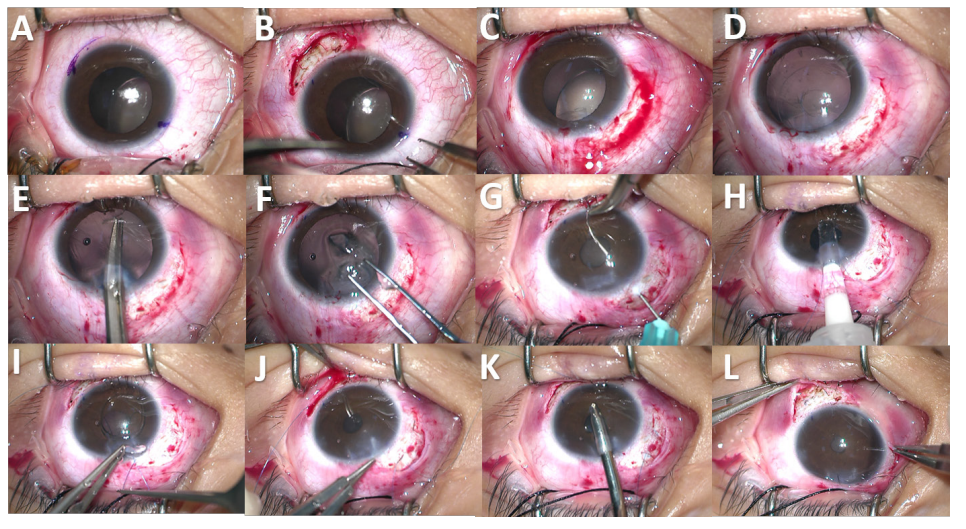
所有患者在全身麻醉下进行手术。开睑后,用 4-0 丝线固定上直肌便于术中暴露术野。改良无线结经巩膜缝线固定 PCIOL 手术:1) 巩膜刻槽:在 4 点和 10 点钟方位做以穹隆为基底的结膜瓣至角膜缘后 4 mm,暴露巩膜面并烧灼止血 ( 图1A),于角膜缘后 2 mm 做两道垂直于角膜缘、宽度为 3 mm 的平行巩膜凹槽,深度约为巩膜厚度的1/3~1/2 ( 图 1B);2) 摘除晶状体:于 12 点处做 3 mm 透明角膜隧道切口,行 4.5~5.5 mm 连续环形撕囊,根据晶状体脱位程度在前囊口植入 2~4 个弹性虹膜拉钩 (611.74,Alcon Grieshaber AG, 美 国 ) 使晶状体居中 ( 图 1C、D),水分离、水分层后清除晶状体皮质和核,晶状体和玻璃体前界间注入粘弹剂形成保护垫,用晶状体调位钩离断残留晶状体悬韧带,部分较为坚韧的悬韧带用囊膜剪剪断,取出全部晶状体囊膜 ( 图 1E、F),如有玻璃体溢出行前段玻璃体切割术;3) 植入 IOL:前房内注入卡巴胆碱缩瞳后,用 30 G 破囊针分别从 10 点、4 点角膜缘后 2 mm 对称的巩膜凹槽处穿入,经虹膜后方到前房,分别使用带 8-0 聚丙烯缝线 (8-0 普理灵缝线,美国强生公司 ) 的缝针从周边角膜切口穿入并对接入破囊针尖内,通过破囊针头将缝线分别从对称的巩膜凹槽处引出,2 条线尾从角膜主切口引出 ( 图 1G)。从主切口通过推注器推入 IOL,置于虹膜前,将 8-0 聚丙烯缝线对称捆绑打结于 IOL 两个襻的对称点 ( 图 1H、I),将 IOL 缓慢放入虹膜后,双手同时拉紧两侧缝线,调整 IOL 位置使 IOL光学面位正位,“Z”形来回缝合 5 次,缝线掩埋在巩膜凹槽内 ( 图 1J)。吸除粘弹剂后,水密主切口,必要时使用 10-0 尼龙线缝合主切口,使用止血器烧灼闭合结膜切口。指测眼压正常,包眼 ( 图1K、L)。
传统巩膜缝线固定 PCIOL 手术:1) 制作巩膜瓣,在 4 点和 10 点钟方位做和改良组相同的结膜瓣后,于角膜缘后 3 mm 做以角膜缘为基底的三角形板层巩膜瓣;2) 摘除晶状体,同改良组相同步骤;3) 与角膜缘后 2 mm 巩膜瓣下做引线,对称捆绑IOL 襻并植入后房,方法同改良组,依次打结固定8-0 聚丙烯缝线于两侧巩膜瓣下,10-0 尼龙线缝合巩膜瓣,吸除粘弹剂后,水密主切口,必要时使用10-0 尼龙线缝合主切口,使用止血器烧灼闭合结膜切口。指测眼压正常包眼。

1.3 观察指标

    术前搜集并记录所有患者的人口统计学信息。所有患者在手术前 1 周内行术前常规全身检查及眼部检查。
    术前及术后 1 年检查并记录患者主觉验光获得球镜度 (diopter sphere,DS)、柱镜度 (diopter sylinder,DC)、等效球镜 (sphere equivalent,SE) 和最佳矫正视力 (best corrected visual acuity, BCVA);
20230227153554_5809.png
图 1 改良无线结经巩膜缝线固定 IOL 手术方法示意图
Figure1 Schematic diagram of modified knotless transscleral suture fixation IOL
A:标记并暴露巩膜面;B:制作巩膜凹槽;C-D:撕囊后用虹膜拉钩固定晶状体并清除晶状体皮质和核;E-F:离断晶状体悬韧带并取出全部晶状体囊膜;G:预置固定 IOL 的缝线;H-I:前房内推入 IOL 并用预置的缝线固定 IOL 双襻;J:摆正 IOL 位置后,“Z”形穿梭缝合使缝线固定于双侧巩膜凹槽内;K-L:吸除前房内粘弹剂后封闭切口
A:Expose and mark the sclera; B:Make the scleral grooves; C-D:After capsulorhexis, fix the lens capsular with iris retractors and remove the len's cortex and nucleus; E-F:Disconnect the len's zonules and remove the whole lens capsular; G:Preset the sutures for IOL fixation; H-I:Put the IOL into the anterior chamber and fix the two loops of IOL with preset sutures; J:Correct the IOL position,and perform transscleral "Z" suture to fix the sutures in bilateral scleral grooves; K-L:Remove the viscoelastic in the anterior chamber and close the incision
使用非接触眼压计测量眼压 (intraocular pressure, IOP);使用角膜内皮镜检查获得角膜内皮细胞计数;使用 IOL Master 测量眼轴长度 (Axial Length, AL);使用前置镜或直接检眼镜散瞳观察眼底情况;使用光学相干断层成像观察有无黄斑水肿。使用 Pentacam(70100, Oculus Optikger?te GmbH, 德国 ) 获得垂直及水平方向 IOL 图片,并使用 Matlab(MATLAB 9.11,MathWorks.Inc, 美国 ) 计算 IOL 倾斜与偏心。

1.4 统计学处理

使用 SPSS 22.0 统计学软件分析数据。正态分布的计量资料采用均数 ± 标准差 (x±s) 描述,组间比较采用成组 t 检验,组内手术治疗比较采用配对 t 检验;非正态分布的计量资料采用中位数{M(P25,P75)} 表示,组间比较采用 Mann-Whitney U检验,组内手术治疗比较采用 Wilcoxon 配对秩和检验;计数资料采用频数、率、百分比描述,组间比较使用 χ2 检验或 Fisher 精确概率法。P<0.05 为差异有统计学意义。

2 结果

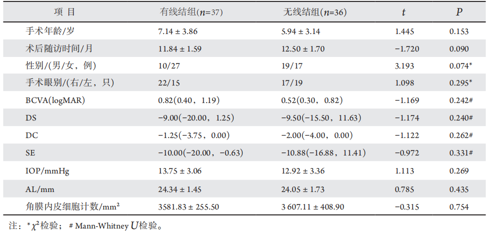
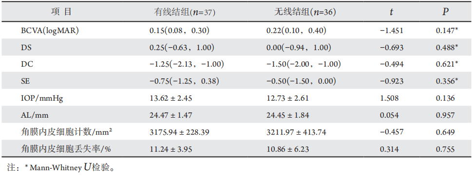
两组患者的性别、年龄、眼别比较差异均无统计学意义(均P>0.05)。见表1。术前和术后1年,两组患者术眼的 BCVA(logMAR)、DS、DC、SE、IOP、AL、角膜内皮细胞计数及角膜内皮丢失率在组间比较差异均无统计学意义(均P>0.05),见表1和表2。
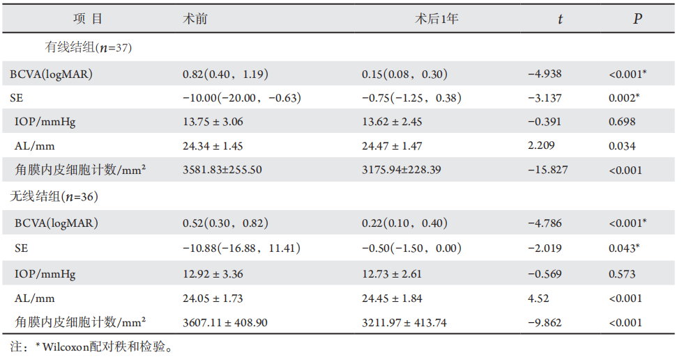
两组患者术后 1 年的 BCVA(logMAR)、等效球镜度数与术前比较均有明显改善 ( 均 P<0.05),AL较术前增加 (P=0.034),IOP 无明显变化 (P>0.05),见表 3。
术后1年并发症方面,两组患者均未出现高眼压、黄斑水肿、玻璃体积血,两组患者的垂直及水平方向IOL倾斜与偏心比较差异无统计学意义(均P>0.05)。而传统有线结组有5例(13.51%)患者出现缝线暴露,5例(13.51%)出现结膜充血,1例(2.70%)出现巩膜溃疡,而改良无线结组无缝线暴露、结膜充血及巩膜溃疡,两组间在缝线暴露及结膜充血方面有显著差异(均P=0.022),见表4。

表1 无线结组与有线结组患者术前基本资料
Table1 Preoperative basic data of patients in knotless group and knoed group

20230227154054_8299.png

表2 术后1年两组间患者视力、屈光结果和眼部参数分析
Table2 Analysis of postoperative 1 year visual acuity、refractive results and ocular parameters between the two groups

20230227154128_5571.png

表3 两组患者术前与术后1年视力和眼部参数的比较
Table3 Compared preoperative and postoperative 1 year visual acuity and ocular parameters in two groups respectively

20230227154229_3708.png

表4 无线结组与有线结组患者术后并发症的比较[例(%)]
Table4 Comparison of postoperative complications between knotless group and knotted group[n(%)]

20230227154358_6881.png

3 讨论

目前临床上治疗先天性晶状体脱位所采用的PCIOL手术方式主要包括两种:巩膜瓣下的线结埋藏和无线结“Z”形缝线固定。鉴于前者常出现并线结相关并发症,后者自问世以来获得更多临床医师的推崇及推广[14]。但研究发现,采用无线结“Z”形10-0缝线固定PCIOL在术后10年缝线断裂发生率较高[15]。因此,针对上述问题,在既往研究基础上笔者将8-0聚丙烯缝线应用在无线结“Z”形缝线固定PCIOL术上。结果显示,与传统有线结组相比,改良无线结组在术后1年屈光结果、眼部生物学参数方面均比较差异无统计学意义,而术后1年缝线暴露发生率则显著降低。
晶状体位置异常导致的严重屈光不正是引起CEL患者视力异常的主要原因之一,而大部分CEL患者仍处于视觉发育敏感期,对于带镜治疗效果不理想的患者,及时手术治疗有助于改善患儿的视觉质量。在本研究中,无线结组及有线结组的术后1年BCVA 较术前均显著提高,而两组间术后BCVA无显著差异,提示CEL患者通过手术可有效改善术前的视力不佳,其中改良无线结手术的临床视力改善程度与传统有线结手术效果相当。考虑到大部分CEL患者是儿童,手术时眼球尚未停止发育,本研究观察了术后1年时两组患者眼部生物学参数变化的情况。在术后1年,两组患者的 IOP、AL、角膜内皮细胞计数比较差异无统计学意义,这说明改良无线结术式对眼部生物学参数的影响与传统有线结术式是一致的。值得注意的是,两组患者术后1年的眼轴均较术前增加,考虑到两组患者手术年龄尚在学龄期,该结果可能与眼球发育有关。尽管眼轴发生变化,但两组间的DS、DC、SE均相近,提示术后1年改良无线结术式所带来的屈光误差与传统有线结术式相类似,证实了该术式的有效性。除此之外,角膜内皮细胞在角膜正常生理功能中承担重要作用,如维持角膜透明,角膜内皮细胞计数是评判内眼手术安全性的指标之一,Bourne等[27]研究表明白内障超声乳化术后1年角膜内皮细胞平均减少10%。本研究结果显示,两组患者术后1年的角膜内皮细胞丢失率无差异,并且与白内障术后角膜内皮细胞的损失接近,这也说明了改良无线结术式未加重对角膜内皮细胞的影响,更进一步验证了该术式的安全性。
此外,由于CEL患者多为儿童,而儿童的巩膜较成年人更薄,使缝线相关问题更不容忽视[11-13],改良无线结术式的初衷亦是为了探讨是否能在保证手术安全性及有效性的同时,减少传统有线结术式所带来的缝线相关并发症。本研究结果显示,有线结组中术后1年内共有 5例出现缝线暴露,且线结周围均可见结膜充血、其中1例出现线结周围巩膜溃疡形成,该组缝线暴露发生率与既往研究相一致[13];而无线结组中则未出现缝线暴露、缝线周围结膜充血及巩膜溃疡。同时,两组患者在垂直及水平方向上IOL倾斜度与偏心值无差别,且改良无线结术式未增加术后高眼压、黄斑水肿、玻璃体积血的发生率。因此,本研究提示改良无线结术式在保证术后长期安全性的同时,可有效减少术后缝线暴露的发生,这也意味着可降低患者未来因缝线暴露而引起的结膜持续炎症、巩膜溃疡甚至眼内感染等的风险。且由于低龄患儿合作欠佳,儿童CEL患者术后缝线暴露的处理一直是临床难点,而改良无线结术式可让患儿避免再次行全身麻醉手术以处理暴露的线结。同时,巩膜凹槽可掩埋缝线,不仅减少缝线相关并发症,也有增强美观的效果。因此,对于儿童患者居多的CEL,改良无线结 IOL 巩膜层间固定手术更有其临床优势。
综上所述,本研究介绍了改良无线结巩膜缝线固定PCIOL的术式,与传统巩膜缝线固定PCIOL手术相比,在保证安全性及有效性的同时,能有效减少缝线相关并发症,而且手术安全性更高,因此改良无线结经巩膜缝线固定IOL手术是值得推广的,亦为CEL患者手术方式的选择提供新的选择。但是,本研究仍存在局限性,即该研究为单中心回顾性研究,未来有待前瞻性的多中心研究,以获得更多可靠的循证医学证据。

开放获取声明

    本文适用于知识共享许可协议(Creative Commons),允许第三方用户按照署名(BY)-非商业性使用(NC)-禁止演绎(ND)(CCBY-NC-ND)的方式共享,即允许第三方对本刊发表的文章进行复制、发行、展览、表演、放映、广播或通过信息网络向公众传播,但在这些过程中必须保留作者署名、仅限于非商业性目的、不得进行演绎创作。详情请访问:https://creativecommons.org/licenses/by-nc-nd/4.0/
1、Burian HM, Allen L. Histologic study of the chamber angel of patients with Marfan's syndrome. A discussion of the cases of Theobald, Reeh and Lehman, and Sadi de Buen and Velazquez[ J]. Arch Ophthalmol Chic Ill 1960, 1961, 65: 323-33.Burian HM, Allen L. Histologic study of the chamber angel of patients with Marfan's syndrome. A discussion of the cases of Theobald, Reeh and Lehman, and Sadi de Buen and Velazquez[ J]. Arch Ophthalmol Chic Ill 1960, 1961, 65: 323-33.
2、Nelson LB, Maumenee IH. Ectopia lentis[ J]. Surv Ophthalmol, 1982, 27(3): 143-160.Nelson LB, Maumenee IH. Ectopia lentis[ J]. Surv Ophthalmol, 1982, 27(3): 143-160.
3、Maumenee IH. The eye in the Marfan syndrome[ J]. Trans Am Ophthalmol Soc, 1981, 79: 684-733.Maumenee IH. The eye in the Marfan syndrome[ J]. Trans Am Ophthalmol Soc, 1981, 79: 684-733.
4、Dagi LR, Walton DS. Anterior axial lens subluxation, progressive myopia, and angle-closure glaucoma: recognition and treatment of atypical presentation of ectopia lentis[ J]. J AAPOS, 2006, 10(4): 345- 350.Dagi LR, Walton DS. Anterior axial lens subluxation, progressive myopia, and angle-closure glaucoma: recognition and treatment of atypical presentation of ectopia lentis[ J]. J AAPOS, 2006, 10(4): 345- 350.
5、Dureau P. Pathophysiology of zonular diseases. Curr Opin Ophthalmol, 2008, 19(1): 27-30.Dureau P. Pathophysiology of zonular diseases. Curr Opin Ophthalmol, 2008, 19(1): 27-30.
6、Jin G M, Fan M, Cao QZ, et al. Trends and characteristics of congenital ectopia lentis in China[ J]. Int J Ophthalmol, 2018, 11(9): 1545-1549.Jin G M, Fan M, Cao QZ, et al. Trends and characteristics of congenital ectopia lentis in China[ J]. Int J Ophthalmol, 2018, 11(9): 1545-1549.
7、McAllister AS, Hirst LW. Visual outcomes and complications of scleral- fixated posterior chamber intraocular lenses[ J]. J Cataract Refract Surg, 2011, 37(7): 1263-1269.McAllister AS, Hirst LW. Visual outcomes and complications of scleral- fixated posterior chamber intraocular lenses[ J]. J Cataract Refract Surg, 2011, 37(7): 1263-1269.
8、Hannush SB. Sutured posterior chamber intraocular lenses: indications and procedure[ J]. Curr Opin Ophthalmol, 2000, 11(4): 233-240.Hannush SB. Sutured posterior chamber intraocular lenses: indications and procedure[ J]. Curr Opin Ophthalmol, 2000, 11(4): 233-240.
9、Wagoner MD, Cox TA, Ariyasu RG, et al. Intraocular lens implantation in the absence of capsular support: a report by the American Academy of Ophthalmology[ J]. Ophthalmology, 2003, 110(4): 840-859.Wagoner MD, Cox TA, Ariyasu RG, et al. Intraocular lens implantation in the absence of capsular support: a report by the American Academy of Ophthalmology[ J]. Ophthalmology, 2003, 110(4): 840-859.
10、Jacob S, Kumar DA, Rao NK. Scleral fixation of intraocular lenses[ J]. Curr Opin Ophthalmol, 2020, 31(1): 50-60.Jacob S, Kumar DA, Rao NK. Scleral fixation of intraocular lenses[ J]. Curr Opin Ophthalmol, 2020, 31(1): 50-60.
11、Solomon K, Gussler JR, Gussler C, et al. Incidence and management of complications of transsclerally sutured posterior chamber lenses[ J]. J Cataract Refract Surg, 1993, 19(4): 488-493.Solomon K, Gussler JR, Gussler C, et al. Incidence and management of complications of transsclerally sutured posterior chamber lenses[ J]. J Cataract Refract Surg, 1993, 19(4): 488-493.
12、Uthoff D, Teichmann KD. Secondary implantation of scleral-fixated intraocular lenses[ J]. J Cataract Refract Surg, 1998, 24(7): 945-950.Uthoff D, Teichmann KD. Secondary implantation of scleral-fixated intraocular lenses[ J]. J Cataract Refract Surg, 1998, 24(7): 945-950.
13、Holland EJ, Daya SM, Evangelista A, et al. Penetrating keratoplasty and transscleral fixation of posterior chamber lens[ J]. Am J Ophthalmol, 1992, 114(2): 182-187.Holland EJ, Daya SM, Evangelista A, et al. Penetrating keratoplasty and transscleral fixation of posterior chamber lens[ J]. Am J Ophthalmol, 1992, 114(2): 182-187.
14、Szurman P, Petermeier K, Aisenbrey S, et al. Z-suture: a new knotless technique for transscleral suture xation of intraocular implants[ J]. Br J Ophthalmol, 2010, 94(2): 167-169.Szurman P, Petermeier K, Aisenbrey S, et al. Z-suture: a new knotless technique for transscleral suture xation of intraocular implants[ J]. Br J Ophthalmol, 2010, 94(2): 167-169.
15、Dimopoulos S, Dimopoulos V, Blumenstock G, et al. Long-term outcome of scleral-fixated posterior chamber intraocular lens implantation with the knotless Z-suture technique[ J]. J Cataract Refract Surg, 2018, 44(2): 182-185.Dimopoulos S, Dimopoulos V, Blumenstock G, et al. Long-term outcome of scleral-fixated posterior chamber intraocular lens implantation with the knotless Z-suture technique[ J]. J Cataract Refract Surg, 2018, 44(2): 182-185.
16、McAllister AS, Hirst LW. Visual outcomes and complications of scleral- fixated posterior chamber intraocular lenses[ J]. J Cataract Refract Surg, 2011, 37(7): 1263-1269.McAllister AS, Hirst LW. Visual outcomes and complications of scleral- fixated posterior chamber intraocular lenses[ J]. J Cataract Refract Surg, 2011, 37(7): 1263-1269.
17、Por YM, Lavin MJ. Techniques of intraocular lens suspension in the absence of capsular/zonular support[ J]. Surv Ophthalmol, 2005, 50(5): 429-462.Por YM, Lavin MJ. Techniques of intraocular lens suspension in the absence of capsular/zonular support[ J]. Surv Ophthalmol, 2005, 50(5): 429-462.
18、Khan MA, Gupta OP, Smith RG, et al. Scleral fixation of intraocular lenses using Gore-Tex suture: clinical outcomes and safety profile[ J]. Br J Ophthalmol, 2016, 100(5): 638-643.Khan MA, Gupta OP, Smith RG, et al. Scleral fixation of intraocular lenses using Gore-Tex suture: clinical outcomes and safety profile[ J]. Br J Ophthalmol, 2016, 100(5): 638-643.
19、Gimbel HV, Condon GP, Kohnen T, et al. Late in-the-bag intraocular lens dislocation: incidence, prevention, and management[ J]. J Cataract Refract Surg, 2005, 31(11): 2193-2204.Gimbel HV, Condon GP, Kohnen T, et al. Late in-the-bag intraocular lens dislocation: incidence, prevention, and management[ J]. J Cataract Refract Surg, 2005, 31(11): 2193-2204.
20、Wallmann AC, Monson BK, Adelberg DA. Transscleral fixation of a foldable posterior chamber intraocular lens[ J]. J Cataract Refract Surg, 2015, 41(9): 1804-1809.Wallmann AC, Monson BK, Adelberg DA. Transscleral fixation of a foldable posterior chamber intraocular lens[ J]. J Cataract Refract Surg, 2015, 41(9): 1804-1809.
21、Khan MA, Samara WA, Gerstenblith AT,et al. Combined pars Plana vitrectomy and scleral fixation of an intraocular lens using gore-tex suture: one-year outcomes[ J]. Retina, 2018, 38(7): 1377-1384.Khan MA, Samara WA, Gerstenblith AT,et al. Combined pars Plana vitrectomy and scleral fixation of an intraocular lens using gore-tex suture: one-year outcomes[ J]. Retina, 2018, 38(7): 1377-1384.
22、Chee SP, Chan NSW. Suture snare technique for scleral fixation of intraocular lenses and capsular tension devices[ J]. Br J Ophthalmol, 2018, 102(10): 1317-1319.Chee SP, Chan NSW. Suture snare technique for scleral fixation of intraocular lenses and capsular tension devices[ J]. Br J Ophthalmol, 2018, 102(10): 1317-1319.
23、John T, Tighe S, Hashem O, et al. New use of 8-0 polypropylene suture for four-point scleral fixation of secondary intraocular lenses[ J]. J Cataract Refract Surg, 2018, 44(12): 1421-1425.John T, Tighe S, Hashem O, et al. New use of 8-0 polypropylene suture for four-point scleral fixation of secondary intraocular lenses[ J]. J Cataract Refract Surg, 2018, 44(12): 1421-1425.
24、Zhao P, Ou Z, Zhang Q, et al. Adjustable buckle-slide suture: a novel surgical technique for transscleral fixation of intraocular lenses[ J]. Retina, 2019, 39(Suppl 1): S24-S29.Zhao P, Ou Z, Zhang Q, et al. Adjustable buckle-slide suture: a novel surgical technique for transscleral fixation of intraocular lenses[ J]. Retina, 2019, 39(Suppl 1): S24-S29.
25、Rezar-Dreindl S, Stifter E, Neumayer T, et al. Visual outcome and surgical results in children with Marfan syndrome[ J]. Clin Exp Ophthalmol, 2019, 47(9): 1138-1145.Rezar-Dreindl S, Stifter E, Neumayer T, et al. Visual outcome and surgical results in children with Marfan syndrome[ J]. Clin Exp Ophthalmol, 2019, 47(9): 1138-1145.
26、Sridhar J, Chang JS. Marfan's syndrome with ectopia lentis[ J]. N Engl J Med, 2017, 377(11): 1076.Sridhar J, Chang JS. Marfan's syndrome with ectopia lentis[ J]. N Engl J Med, 2017, 377(11): 1076.
27、Bourne RR, Minassian DC, Dart JK, et al. Eect of cataract surgery on the corneal endothelium: modern phacoemulsication compared with extracapsular cataract surgery[ J]. Ophthalmology, 2004, 111(4): 679- 685.Bourne RR, Minassian DC, Dart JK, et al. Eect of cataract surgery on the corneal endothelium: modern phacoemulsication compared with extracapsular cataract surgery[ J]. Ophthalmology, 2004, 111(4): 679- 685.
1、国家自然科学基金 (81900841);广东省基础与应用基础研究基金 (2021A1515011673; 2022A1515011181)。
This work was supported by the National Natural Science Foundation of China (81900841), and Guangdong Basic and Applied Basic Research Foundation (2021A1515011673; 2022A1515011181).()
上一篇
下一篇
其他期刊
  • 眼科学报

    主管:中华人民共和国教育部
    主办:中山大学
    承办:中山大学中山眼科中心
    主编:林浩添
    主管:中华人民共和国教育部
    主办:中山大学
    浏览
  • Eye Science

    主管:中华人民共和国教育部
    主办:中山大学
    承办:中山大学中山眼科中心
    主编:林浩添
    主管:中华人民共和国教育部
    主办:中山大学
    浏览
推荐阅读
出版者信息
目录