您的位置: 首页 > 2022年11月 第37卷 第11期 > 文字全文
2023年7月 第38卷 第7期11
目录

经鼻内镜鼻泪管-泪囊切除术的应用解剖

Applied anatomy of transnasal endoscopic resection of nasolacrimal duct and lacrimal sac

来源期刊: 眼科学报 | 2022年11月 第37卷 第11期 856-863 发布时间: 收稿时间:2022/12/6 14:31:49 阅读量:6726
作者:
关键词:
鼻泪管泪囊泪道系统应用解剖内镜手术
nasolacrimal duct lacrimal sac lacrimal drainage system applied anatomy endoscopic surgery
DOI:
10.3978/j.issn.1000-4432.2022.11.06
收稿时间:
 
修订日期:
 
接收日期:
 
目的:通过在血管灌注尸头标本上模拟经鼻内镜下鼻泪管-泪囊切除术探讨该术式的基本操作以及在经鼻内镜下鼻泪管-泪囊和其周围结构的解剖关系,以期为临床开展该术式提供解剖学依据。方法:采用5个动、静脉双灌注成人尸头标本(共10侧)进行解剖学研究。在标本上进行经鼻内镜下暴露全程鼻泪管及泪囊,观察鼻泪管-泪囊切除过程的解剖标志和毗邻关系。结果:10侧泪囊-鼻泪管的解剖显示全部泪囊均位于鼻丘的前方稍外侧,泪囊顶基本和鼻丘顶平齐,泪囊体大部分位于中鼻甲腋窝水平线上方。泪囊窝后内壁由泪骨构成,泪骨后内方与鼻丘气房相邻。泪囊底向下移行为膜性鼻泪管进入骨性鼻泪管,鼻泪管在鼻腔外侧壁的投影位于钩突垂直部前缘前方约3~7mm。鼻泪管下鼻道开口距离下鼻甲前端的距离为(16±3)mm。泪囊长度为(13.8±1.8)mm,鼻泪管长度为(23.2±3.6)mm。结论:经鼻内镜入路可充分暴露和切除全程鼻泪管和泪囊。本解剖研究展示的基本操作过程和解剖标志可为临床开展经鼻内镜鼻泪管-泪囊切除术提供解剖学参考。
Objective: By simulating transnasal endoscopic resection of nasolacrimal duct and lacrimal sac on cadaveric specimens with vascular perfusion, the basic process of this procedure and the anatomical relationship between nasolacrimal duct and lacrimal sac were explored, providing anatomical basis for clinical application. Methods: Five adult cadaver head specimens (10 sides in total) were used for anatomical study. The nasolacrimal duct and lacrimal sac were exposed under transnasal endoscopy, and the anatomical landmarks and their relationship with adjacent areas during the resection of nasolacrimal duct and lacrimal sac were observed. Results: Ten sides of nasolacrimal duct and lacrimal sac showed that all lacrimal sacs were located anterior and slightly lateral to the nasal mound. The roof of lacrimal sac was almost the same height as the roof of nasal mound. The body of lacrimal sac was almost located above the horizontal line of the middle turbinate axilla. The posterior inner wall of the lacrimal fossa was composed of lacrimal bone. The posterior inner side of the lacrimal bone was adjacent to the agger nasi cell. The bottom of the lacrimal sac moved downward as the membranous part of the nasolacrimal duct located in the bony part of the nasolacrimal duct. The projection of the nasolacrimal duct on the lateral nasal wall was located about 3–7 mm in front of the anterior edge of the vertical part of the uncinate process. The distance between the inferior meatus opening of the nasolacrimal duct and the anterior end of the inferior turbinate was (16±3) mm. The length of the lacrimal sac was (13.8±1.8) mm, and the length of the nasolacrimal duct was (23.2±3.6) mm. Conclusion: The transnasal endoscopic approach can fully expose and resect the nasolacrimal duct and lacrimal sac. The basic operation process and anatomical landmarks demonstrated in this anatomical study provide an anatomical reference for the clinical development of transnasal endoscopic resection of nasolacrimal duct and lacrimal sac.
      泪囊位于眼眶前内侧的泪囊窝内,内侧连接泪总管,上方为盲袋状,下方移行为鼻泪管,开口于下鼻道[1]。由于泪囊和鼻泪管均和鼻腔外侧壁相毗邻,随着内窥镜技术的发展和应用,临床上已开始逐渐采用经鼻内镜入路开展泪囊和鼻泪管的手术,其中以内镜下泪囊鼻腔吻合术治疗慢性泪囊炎最为广泛[2]。在此基础上,对于部分原发于泪囊和鼻泪管的肿瘤,近年一些临床学者也开始采用经鼻内镜入路进行泪囊和鼻泪管切除的临床探索[3-7]。然而,目前仍缺乏针对经鼻内镜鼻泪管泪囊切除术的应用解剖研究。本研究采用新鲜尸头标本,通过模拟经鼻内镜鼻泪管-泪囊切除术进行解剖,探讨此术式的基本操作步骤,以及在经鼻内镜下鼻泪管-泪囊和其周围结构的解剖关系,以期为临床开展该术式提供解剖学依据。

1 材料与方法

1.1 材料

      采用5具成人新鲜尸头标本(经颈总动脉和颈内静脉分别灌注红色和蓝色乳胶溶液,由河南黄河科技学院解剖教研室提供),所有尸头在解剖前均行高分辨计算机断层扫描(computed tomography,CT),未发现明显外伤及鼻腔鼻窦病变。标本采用70%酒精浸泡保存。实验条件包括采用0°、直径4mm、长18cm的硬性鼻内窥镜、高清摄像头和显示器(德国艾克松内窥镜有限公司),并采用系统自带摄像系统对解剖过程进行图片和视频采集;内镜手术器械包括咬骨钳、探针、剥离子、剪刀等(杭州科锐特医疗设备有限公司);动力系统采用高速磨钻(美国美敦力公司)。游标卡尺进行内镜下测量,所有径线每个尸头平均测量3次,结果取平均值。本解剖研究已获得暨南大学附属祈福医院医学伦理委员会的审核批准。

1.2 解剖方法

      模拟鼻内镜下鼻泪管前径路切除全程鼻泪管及泪囊的尸头解剖。1)研究鼻腔内解剖结构标志。2)剥离子探梨状孔缘,确定在下鼻甲头端和鼻内孔之间切口位置,于中鼻甲头端根部开始,斜向前上至鼻丘隆起上缘,然后转向前下至下鼻甲头端作弧形切口,切开黏膜暴露下鼻甲鼻腔外侧壁骨性附着处;剥离子离断下鼻甲鼻腔外侧壁附着处;以下鼻甲鼻腔外侧壁附着处标志,用骨凿解剖鼻泪管;用电钻将骨性鼻泪管及上颌骨额突磨平,充分暴露膜性鼻泪管及泪囊、并对鼻泪管的长度进行测量;分离泪囊周围组织,切除泪囊及鼻泪管;复位鼻腔外侧壁黏膜。

2 结果

2.1 鼻腔内解剖结构

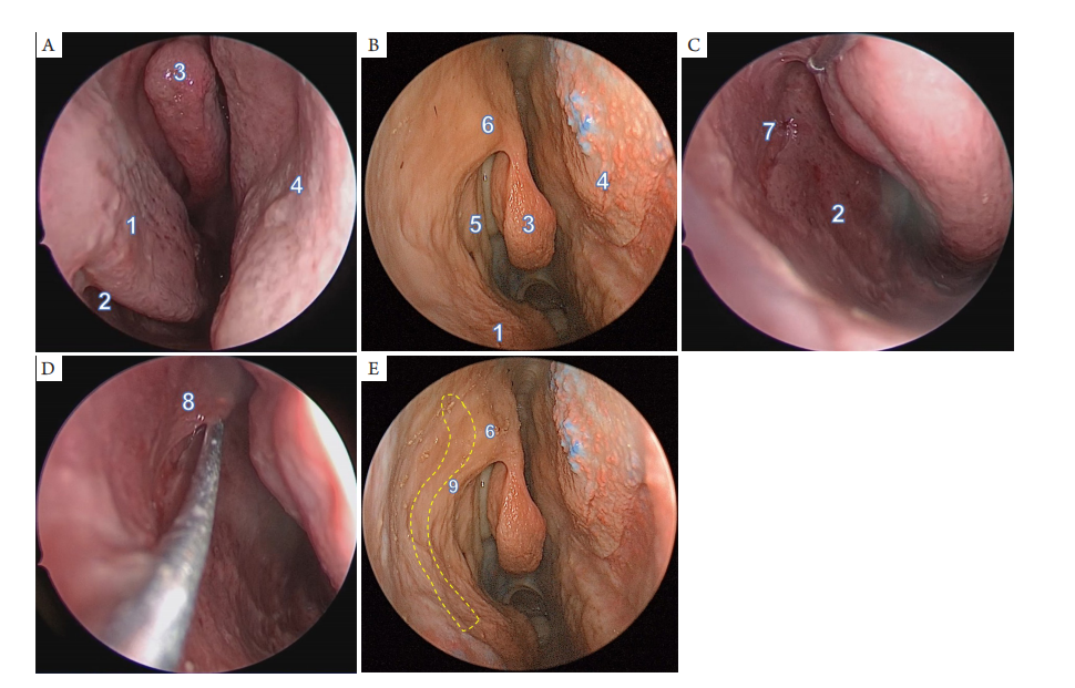
      鼻腔内部解剖标志包括下鼻甲、鼻中隔、中鼻甲、钩突和鼻丘(图1)。0°鼻内镜紧贴鼻阈从标本右侧鼻孔进入鼻腔,首先见到的是下鼻甲头端,继续向深部插入,可见鼻中隔和中鼻甲、钩突、鼻丘(图1A,1B)。用探针抬起下鼻甲,进入下鼻道可见鼻泪管开口于下鼻道外侧壁(图1C,1D)。泪囊在鼻腔外侧壁的投影位置位于鼻丘的前方,泪囊向前下斜行移行为鼻泪管,投影于上颌骨额突稍前方(图1E)。

2.2 经鼻内镜切除鼻泪管 - 泪囊的解剖观察

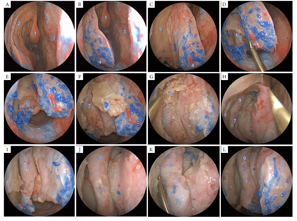
      模拟经鼻内镜右侧鼻泪管-泪囊切除术,在鼻腔外侧壁做弧形切口,向后分离黏膜瓣后,在下鼻甲头端处首先显露下鼻甲骨(图2A-2C)。以鼻泪管开口为解剖标志,咬除鼻泪管开口前方的下鼻甲骨质,见膜性鼻泪管下部位于骨性鼻泪管内。鼻泪管在下鼻道开口和下鼻道黏膜相互移行,形成Hasner瓣,遮盖鼻泪管开口。沿着鼻泪管开口处向上,探针在探及骨性鼻泪管的走向后,继而向上凿除骨性鼻泪管内侧壁骨质,电钻磨除骨管周围骨质,可见膜性鼻泪管完全位于骨性鼻泪管内,骨性鼻泪管的前方骨质致密,后方相对较为菲薄(图2D-2F)。沿着鼻泪管继续向上,部分标本可见厚实的上颌骨额突,继而用电钻磨除上颌骨额突骨质,即可暴露泪囊内侧壁。探针探及泪囊后,沿着泪囊继续磨除周围骨质,即可充分显露泪囊,可见泪囊位于泪囊窝内,前内方为较厚的上颌骨额突,后方为菲薄的泪骨(图2G-2L)。探针向下游离泪囊和鼻泪管全程,即可完整暴露泪囊和鼻泪管。
20230203162252_7525.png

图1 鼻内镜观察和鼻泪管相关的鼻腔外侧结构
Figure 1 Endoscopic views of nasolacrimal duct related structures on lateral nasal wall
(A,B)鼻内镜显示右侧鼻腔视野;(C,D)探针抬起下鼻甲,显露右侧鼻泪管在下鼻道外侧壁的开口;(E)鼻泪管-泪囊在鼻腔外侧壁的投影(黄色虚线)。1:下鼻甲;2:下鼻道;3:中鼻甲;4:鼻中隔;5:钩突;6:鼻丘;7:鼻泪管开口;8:Hasner瓣;9:上颌骨额突。
(A,B) Endoscopic views of the right side of nasal cavity; (C,D) Exposure of the opening of the right nasolacrimal duct on the later wall of inferior meatus; (E) The projection of the nasolacrimal duct and lacrimal sac on the lateral nasal wall (Yellow dash line). 1: inferior turbinate; 2: inferior meatus; 3: middle turbinate; 4: nasal septum; 5: uncinate process; 6: nasal mound; 7: opening of nasolacrimal duct; 8: Hasner valve; 9: frontal process of maxilla.

20230203162708_8058.png

图2 鼻内镜下显露鼻泪管-泪囊
Figure 2 Exposure of nasolacrimal duct and lacrimal sac under nasal endoscopy
(A-C)右侧鼻腔外侧做弧形纵向切口,于中鼻甲头端附着点起始,向前向上至鼻丘上方水平,弧形转至向前下方至下鼻甲头端前方,剥离子于骨面向后分离黏膜瓣,显示下鼻甲骨头端。(D-F)分离黏膜瓣于下鼻甲腋处的连接,定位鼻泪管开口,去除鼻泪管开口前方的下鼻甲头端骨质,并向上切除鼻泪管表面的鼻腔外侧壁骨质。(G-I)显露鼻泪管后,以鼻泪管为标志向上切除上颌骨额突的骨质,显露泪囊内侧壁;从下向上游离鼻泪管。( J-L)离断膜性鼻泪管周围连接组织,充分游离鼻泪管和泪囊。1:中鼻甲;2:鼻腔外侧壁黏膜瓣;3:鼻中隔;4:筛前动脉鼻外侧支;5:鼻腔外侧壁骨质;6:骨性鼻泪管下鼻道开口;7:膜性鼻泪管;8:泪囊内侧壁骨质;9:泪囊。
(A-C) An arc-shaped longitudinal incision was made on the lateral wall of the right nasal cavity, starting from the attachment point of the anterior end of the middle turbinate, forward and upward to the level above the nasal mound, and then turning forward and downward to the front of the anterior end of the inferior turbinate. Then elevator was used to separate the mucosal flap from the bony surface, and the head of the bony inferior turbinate was seen. (D-F) The connection of the mucosal flap at the axilla of the inferior turbinate was separated, and the opening of the nasolacrimal duct was located. Then the anterior part of the bony inferior turbinate was removed in front of the opening of the nasolacrimal duct, and the bony lateral nasal wall on the surface of the nasolacrimal duct was removed upward. (G-I) After the nasolacrimal duct was exposed, the bone of the frontal process of maxilla was removed upward with the nasolacrimal duct as a landmark, and then the medial wall of the lacrimal sac was exposed. The nasolacrimal duct was freed from the bottom up. ( J-L) After dissecting the connecting tissue around the membranous part of nasolacrimal duct, the nasolacrimal duct and lacrimal sac were fully freed. 1: middle turbinate; 2: mucosal flap on the nasal lateral wall; 3: nasal septum; 4: lateral nasal branch of anterior ethmoidal artery; 5: bony lateral nasal wall; 6: the inferior meatus opening of the bony part of nasolacrimal duct; 7: membrane part of nasolacrimal duct; 8: The inner bony wall of lacrimal sac; 9: lacrimal sac.

2.3 鼻泪管 - 泪囊和周围结构的解剖毗邻关系

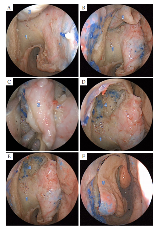
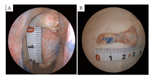
      10侧泪囊-鼻泪管的解剖显示全部泪囊均位于鼻丘的前方稍外侧,泪囊顶基本和鼻丘顶平齐,泪囊体大部分位于中鼻甲腋窝水平线上方。泪囊窝后内壁由泪骨构成,10侧标本中,有9侧显示泪骨后内方与鼻丘气房相邻,毗邻的范围和鼻丘气房大小有关,鼻丘气房越大,泪囊和鼻腔气房的关系越紧密。泪囊窝外侧壁无骨性结构,泪总管在此处和泪囊相连接(图3)。泪囊底向下移行为膜性鼻泪管进入骨性鼻泪管中走行,鼻泪管在鼻腔外侧壁的投影位于钩突垂直部前缘前方约3~7mm。鼻泪管向下开口于下鼻道外侧壁,在鼻泪管开口表面可见Hasner瓣(图1,2)。以此瓣为标志,鼻泪管下鼻道开口距离下鼻甲前端的距离为(16±3)mm。经测量,泪囊长度为(13.8±1.8)mm,鼻泪管长度为(23.2±3.6)mm(图4)。
20230203162857_4493.png

图3 经鼻内镜切除鼻泪管-泪囊
Figure 3 Transnasal endoscopic resection of nasolacrimal duct and lacrimal sac
(A-D)离断右侧鼻泪管开口和下鼻道黏膜的连接,从下向上将膜性鼻泪管从骨管中掀起,显露上方泪囊的外侧部分,从前向后分离泪囊和眶内侧组织之间的连接,完整切除泪囊和鼻泪管。(E)切除鼻泪管和泪囊后,内镜显示泪囊窝、骨性鼻泪管结构。(F)复位鼻腔外侧壁黏膜瓣。1:骨性鼻泪管;2:膜性鼻泪管;3:泪囊;4:泪囊窝;5:鼻腔外侧壁黏膜瓣。
(A-D) After dissecting the connection between the opening of the right nasolacrimal duct and the mucosa of the inferior meatus, the membranous part of the nasolacrimal duct was lifted from the bony canal from the bottom up. The lateral part of the upper lacrimal sac was revealed. Then the lacrimal sac and the medial orbital tissue was separated from anteriorly to posteriorly, and finally the lacrimal sac and nasolacrimal duct were resected completely. (E) Endoscopic views showing structures of the lacrimal fossa, the bony part of nasolacrimal duct after removal of nasolacrimal duct and lacrimal sac. (F) Reposition of the mucosal flap on the nasal lateral wall. 1: bony part of nasolacrimal duct; 2: membrane part of nasolacrimal duct; 3: lacrimal sac; 4: lacrimal fossa; 5: mucosal flap on the nasal lateral wall.

20230203163047_0529.png

图4 骨性鼻泪管(A)和膜性鼻泪管及泪囊(B)的测量
Figure 4 Measurement of the bony part of nasolacrimal duct (A) and the membrane part of nasolacrimal duct and the lacrimal sac (B)

3 讨论

      随着内镜软硬件技术的发展,内镜技术越来越多的被应用到眼鼻相关外科手术,最早采用经鼻内镜手术的眼部疾病是慢性泪囊炎。McDonogh等[8]在1989年率先报道了经鼻内镜的鼻腔泪囊造口术,获得了满意的疗效。随着临床技术的日臻成熟,目前该术式已成为临床上治疗慢性泪囊炎的主要手术方法[2]。除了慢性泪囊炎,原发于或可能侵犯泪囊和鼻泪管的疾病还包括多种良恶性肿瘤,如内翻性乳头状瘤、泪囊癌及一些特异性感染疾病等[9-14]。近年眼科或耳鼻咽喉科医师也开始在临床上积极探索经鼻内镜的方式进行手术切除泪囊和鼻泪管病变[15]。Villaret等[3]最早报道了1例采用经鼻内镜入路切除鼻泪管嗜酸性细胞癌的病例。新近Curragh等采用经鼻内镜下鼻甲摇门法的方式暴露和切除鼻泪管[4],以及经鼻内镜下泪前隐窝入路进行泪囊癌切除术[5]。Chang等[7]则在影像导航下进行泪囊肿瘤切除的经验探索。这些零散的临床报道提示经鼻内镜进行鼻泪管-泪囊切除术具有较好的可行性和应用前景。但目前尚无有关于经鼻内镜鼻泪管泪囊切除术应用解剖的研究。为探讨该内镜入路下的解剖基础,本研究采用血管灌注的新鲜尸头标本进行手术模拟。解剖结果展示了经鼻内镜鼻泪管泪囊切除术的基本操作步骤。以中鼻甲、鼻丘、钩突、下鼻甲头端、下鼻道的鼻泪管开口(Hasner瓣)等为解剖标志,能准确定位鼻泪管-泪囊在鼻腔外侧壁的投影;并且在解剖标志的引导下,从下向上逐步开放骨性鼻泪管,能够有效和安全地暴露膜性鼻泪管和泪囊,从而给予完整切除。采用本解剖研究的鼻腔外侧壁切口所分离的黏膜瓣,在切除鼻泪管-泪囊后可完全覆盖鼻腔外侧壁的手术创面,可能更有助于减少鼻腔功能的破坏和术后伤口的恢复。泪囊肿瘤治疗原则首选手术治疗,手术的切除范围通常包括括泪囊、鼻泪管和上下泪小管,完整切除泪道对彻底切除病变防止复发甚为重要,而病变的充分暴露和清晰的视野是手术成功的关键[11,16]。早年学者[17-18]多采用鼻侧切开术,切除泪道同时切除骨性泪囊窝和鼻泪管口,但该方法损伤大、愈合慢、术后颜面部瘢痕明显。鉴于内镜的技术优点,近年临床上已开始探索应用内镜进行泪道肿瘤的手术。然而,由于泪囊解剖位置的特殊性,骨性鼻泪管和泪囊窝内壁有坚硬的上颌骨额突和泪骨,因而经鼻内镜下泪囊、鼻泪管的病变切除仍具有较大的挑战性。本解剖研究显示采用经鼻内镜入路切除鼻泪管-泪囊具有较高的可行性,该入路显示鼻内镜能提供清晰的直观视野,使操作更为准确精细,更有助于泪道病变的暴露和彻底切除;并且由于面部无瘢痕,创面可能愈合更快,并发症也更少。本组解剖发现,要达到准确无误、操作省力省时、手术成功率高,关键在于准确定位鼻泪管-泪囊在鼻腔外侧壁的投影。既往文献[19]记载“泪囊位于中鼻甲在鼻腔外侧壁的附着”,但Wormald等[20]认为这是不正确的。Wormald等通过测量发现,泪囊在鼻腔外侧壁的投影定位比一般文献记载要高的多,与本研究结果一致,即泪囊的大部分位于中鼻甲腋窝的前外上方,小部分位于中鼻甲腋的前外方。为确定泪囊鼻腔外侧壁的准确位置。该研究以中鼻甲为基准点,测量发现泪囊后界(泪后嵴后缘)至中鼻甲前缘的距离为0~4.46mm,泪囊前界至中鼻甲附着处前缘的距离为(11.31±1.84)mm,中鼻甲前缘覆盖泪囊窝后界的占20%,不覆盖的占80%。张速勤等[21]研究显示泪囊顶、底与中鼻甲腋平面的垂直距离分别为(0.69±0.15)cm、(0.34±0.18)cm。本研究解剖发现全部标本的泪囊均位于鼻丘的前方稍外侧,泪囊顶基本和鼻丘顶平齐,泪囊体大部分位于中鼻甲腋窝水平线上方。因此,本研究采用的鼻腔外侧壁切口(中鼻甲头端根部开始,斜向前上至鼻丘隆起上缘,然后转向前下至下鼻甲头端的弧形切口),可完全暴露泪囊及鼻泪管,从而更有助于彻底切除病变部位。本研究也发现经鼻内镜入路进行鼻泪管-泪囊切除具有一定局限性。经鼻内镜入路对泪囊周围的眼眶内结构暴露范围有限,对切除泪囊恶性肿瘤侵及眶内结构的复杂病变具有明显的局限性。对于此类病变,需联合鼻外入路,以实现对最大程度的病变暴露。本研究存在一些不足。虽然对鼻泪管-泪囊及其周围解剖结构进行了内镜下测量,并得到了初步的测量结果,但需要强调的是,本研究只采用了5个(共10侧)新鲜尸头标本,数量可能不足以体现采集数据的整体分布,因此后续仍需更多尸头标本进行解剖测量,才能更准确地了解鼻泪管-泪囊的相关解剖参数。此外,由于前期无法获得足够的正常人的眼眶CT影像,且本研究的主要目的并非解剖测量,因此暂未对眼眶CT影像进行相关解剖结构测量分析。在后续研究中将完善此部分内容,以提供更完整和准确的鼻泪管-泪囊和其周围解剖结构参数。总之,经鼻内镜入路可以充分暴露全程鼻泪管和泪囊,从而对涉及该区域的病变进行彻底切除。本解剖研究展示的基本操作过程和解剖标志可为临床医师开展经鼻内镜鼻泪管-泪囊切除术提供解剖学参考。

开放获取声明

    本文适用于知识共享许可协议 (Creative Commons),允许第三方用户按照署名(BY)-非商业性使用(NC)-禁止演绎(ND)(CC BY-NC-ND)的方式共享,即允许第三方对本刊发表的文章进行复制、发行、展览、表演、放映、广播或通过信息网络向公众传播,但在这些过程中必须保留作者署名、仅限于非商业性目的、不得进行演绎创作。详情请访问:https://creativecommons.org/licenses/by-nc-nd/4.0/
1、Rajak SN, Psaltis AJ. Anatomical considerations in endoscopic lacrimal surgery[ J]. Ann Anat, 2019, 224: 28-32.Rajak SN, Psaltis AJ. Anatomical considerations in endoscopic lacrimal surgery[ J]. Ann Anat, 2019, 224: 28-32.
2、中华医学会眼科学分会眼整形眼眶病学组. 中国内镜泪囊鼻腔吻合术治疗慢性泪囊炎专家共识(2020年)[ J]. 中华眼科杂志,2020, 56(11): 820-823.
Ophthalmology Orbital Disease Group of Chinese Medical Association Ophthalmology Branch. Expert consensus on the treatment of chronic dacryocystitis with endoscopic dacryocystorhinostomy in China[ J]. Chinese Journal of Ophthalmology, 2020, 56(11): 820-823.
中华医学会眼科学分会眼整形眼眶病学组. 中国内镜泪囊鼻腔吻合术治疗慢性泪囊炎专家共识(2020年)[ J]. 中华眼科杂志,2020, 56(11): 820-823.
Ophthalmology Orbital Disease Group of Chinese Medical Association Ophthalmology Branch. Expert consensus on the treatment of chronic dacryocystitis with endoscopic dacryocystorhinostomy in China[ J]. Chinese Journal of Ophthalmology, 2020, 56(11): 820-823.
3、Villaret AB, Lombardi D, Schreiber A, et al. Oncocytic carcinoma of the nasolacrimal duct treated by transnasal endoscopic resection[ J]. Head Neck, 2013, 35(1): E24-E27.Villaret AB, Lombardi D, Schreiber A, et al. Oncocytic carcinoma of the nasolacrimal duct treated by transnasal endoscopic resection[ J]. Head Neck, 2013, 35(1): E24-E27.
4、Curragh DS, James C, Selva D. Swinging inferior turbinate approach to the nasolacrimal duct[ J]. Orbit, 2020, 39(2): 112-117.Curragh DS, James C, Selva D. Swinging inferior turbinate approach to the nasolacrimal duct[ J]. Orbit, 2020, 39(2): 112-117.
5、Curragh DS, Psaltis AJ, Tan NC, et al. Prelacrimal approach for nasolacrimal duct excision in the management of lacrimal system tumours[ J]. Orbit, 2019, 38(4): 308-312.Curragh DS, Psaltis AJ, Tan NC, et al. Prelacrimal approach for nasolacrimal duct excision in the management of lacrimal system tumours[ J]. Orbit, 2019, 38(4): 308-312.
6、Rajesh Raju G, Sandeep S. Lacrimal Sac Rhinosporidiosis and surgical management by transnasal endoscopic excision: A case series[ J]. Laryngoscope, 2018, 128(12): 2693-2696.Rajesh Raju G, Sandeep S. Lacrimal Sac Rhinosporidiosis and surgical management by transnasal endoscopic excision: A case series[ J]. Laryngoscope, 2018, 128(12): 2693-2696.
7、Chang CH, Ku WN, Kung WH, et al. Navigation-assisted endoscopic surgery of lacrimal sac tumor[ J]. Taiwan J Ophthalmol, 2020, 10(2): 141-143.Chang CH, Ku WN, Kung WH, et al. Navigation-assisted endoscopic surgery of lacrimal sac tumor[ J]. Taiwan J Ophthalmol, 2020, 10(2): 141-143.
8、McDonogh M, Meiring JH. Endoscopic transnasal dacryocystorhinostomy[ J]. J Laryngol Otol, 1989, 103(6): 585-587.McDonogh M, Meiring JH. Endoscopic transnasal dacryocystorhinostomy[ J]. J Laryngol Otol, 1989, 103(6): 585-587.
9、Alam MS, Mukherjee B, Krishnakumar S. Clinical profile and management outcomes of lacrimal drainage system malignancies[ J]. Orbit, 2022, 41(4): 429-436.Alam MS, Mukherjee B, Krishnakumar S. Clinical profile and management outcomes of lacrimal drainage system malignancies[ J]. Orbit, 2022, 41(4): 429-436.
10、Krishna Y, Coupland SE. Lacrimal sac tumors—A review[ J]. Asia Pac J Ophthalmol (Phila), 2017, 6(2): 173-178.Krishna Y, Coupland SE. Lacrimal sac tumors—A review[ J]. Asia Pac J Ophthalmol (Phila), 2017, 6(2): 173-178.
11、Singh S, Ali MJ. Primary malignant epithelial tumors of the lacrimal drainage system: A major review[ J]. Orbit, 2021, 40(3): 179-192.Singh S, Ali MJ. Primary malignant epithelial tumors of the lacrimal drainage system: A major review[ J]. Orbit, 2021, 40(3): 179-192.
12、蒋永强, 王彬, 李晓华, 等. 泪囊原发性恶性肿瘤22例临床病理学分析[ J]. 中华眼外伤职业眼病杂志, 2019, 41(2): 81-84. JIANG Yongqiang, WANG Bin, LI Xiaohua, et al. Clinicopathological analysis of 22 cases with primary malignant tumors of the lacrimal sac[ J]. Chinese Journal of Ocular Trauma and Occupational Eye Disease, 2019, 41(2): 81-84.蒋永强, 王彬, 李晓华, 等. 泪囊原发性恶性肿瘤22例临床病理学分析[ J]. 中华眼外伤职业眼病杂志, 2019, 41(2): 81-84. JIANG Yongqiang, WANG Bin, LI Xiaohua, et al. Clinicopathological analysis of 22 cases with primary malignant tumors of the lacrimal sac[ J]. Chinese Journal of Ocular Trauma and Occupational Eye Disease, 2019, 41(2): 81-84.
13、赵云, 惠靖雯, 杨丽红, 等. 原发性泪道肿物64例临床组织病理学分析. 中华眼科杂志, 2020, (05): 364-369.
ZHAO Yun, HUI Jingwen, YANG Lihong , et al. Clinical and pathological analysis of 64 patients with primary neoplasms of the lacrimal drainage system[ J]. Chinese Journal of Ophthalmology, 2020, 56(5): 364-369.
赵云, 惠靖雯, 杨丽红, 等. 原发性泪道肿物64例临床组织病理学分析. 中华眼科杂志, 2020, (05): 364-369.
ZHAO Yun, HUI Jingwen, YANG Lihong , et al. Clinical and pathological analysis of 64 patients with primary neoplasms of the lacrimal drainage system[ J]. Chinese Journal of Ophthalmology, 2020, 56(5): 364-369.
14、Heindl LM, Jünemann AG, Kruse FE, et al. Tumors of the lacrimal drainage system[ J]. Orbit, 2010, 29(5): 298-306.Heindl LM, Jünemann AG, Kruse FE, et al. Tumors of the lacrimal drainage system[ J]. Orbit, 2010, 29(5): 298-306.
15、Reshef ER , Bleier BS, Freitag SK . The endoscopic transnasal approach to orbital tumors: A review[ J]. Semin Ophthalmol, 2021, 36(4): 232-240.Reshef ER , Bleier BS, Freitag SK . The endoscopic transnasal approach to orbital tumors: A review[ J]. Semin Ophthalmol, 2021, 36(4): 232-240.
16、Ramberg I, Toft PB, Heegaard S. Carcinomas of the lacrimal drainage system[ J]. Surv Ophthalmol, 2020, 65(6): 691-707.Ramberg I, Toft PB, Heegaard S. Carcinomas of the lacrimal drainage system[ J]. Surv Ophthalmol, 2020, 65(6): 691-707.
17、Parmar DN, Rose GE. Management of lacrimal sac tumours[ J]. Eye (Lond), 2003, 17(5): 599-606.Parmar DN, Rose GE. Management of lacrimal sac tumours[ J]. Eye (Lond), 2003, 17(5): 599-606.
18、El-Sawy T, Frank SJ, Hanna E, et al. Multidisciplinary management of lacrimal sac/nasolacrimal duct carcinomas[ J]. Ophthalmic Plast Reconstr Surg, 2013, 29(6): 454-457.El-Sawy T, Frank SJ, Hanna E, et al. Multidisciplinary management of lacrimal sac/nasolacrimal duct carcinomas[ J]. Ophthalmic Plast Reconstr Surg, 2013, 29(6): 454-457.
19、Rebeiz EE, Shapshay SM, Bowlds JH, et al. Anatomic guidelines for dacryocystorhinostomy[ J]. Laryngoscope, 1992, 102(10): 1181-1184.Rebeiz EE, Shapshay SM, Bowlds JH, et al. Anatomic guidelines for dacryocystorhinostomy[ J]. Laryngoscope, 1992, 102(10): 1181-1184.
20、Wormald PJ, Kew J, Van Hasselt A. Intranasal anatomy of the nasolacrimal sac in endoscopic dacryocystorhinostomy[ J]. Otolaryngol Head Neck Surg, 2000, 123(3): 307-310.Wormald PJ, Kew J, Van Hasselt A. Intranasal anatomy of the nasolacrimal sac in endoscopic dacryocystorhinostomy[ J]. Otolaryngol Head Neck Surg, 2000, 123(3): 307-310.
21、张速勤, 贾沛靓, 唐海红, 等. 泪囊鼻内解剖研究及临床应用. 中华耳鼻咽喉头颈外科杂志, 2006, 41(7): 506-509.
ZHANG Suqin, JIA Peiliang, TANG Haihong, et al. Endonasal anatomy of lacrimal sac and its clinical significance in dacryocystorhinostomy[ J]. Chinese Journal of Otorhinolaryngology Head and Neck Surgery, 2006, 41(7): 506-509.
张速勤, 贾沛靓, 唐海红, 等. 泪囊鼻内解剖研究及临床应用. 中华耳鼻咽喉头颈外科杂志, 2006, 41(7): 506-509.
ZHANG Suqin, JIA Peiliang, TANG Haihong, et al. Endonasal anatomy of lacrimal sac and its clinical significance in dacryocystorhinostomy[ J]. Chinese Journal of Otorhinolaryngology Head and Neck Surgery, 2006, 41(7): 506-509.
1、石丽洪,刘成芳,张卫国等.基于BOPPPS的线上、线下混合式教学在系统解剖学中的应用[J].基础医学教育,2023,25(10):899-903.
2、聂时淮,郭丽旭,刘咏等.DR-泪道造影术在泪道阻塞性疾病中的临床运用价值[J].实用医学杂志,2023,39(22):2969-2973.
上一篇
下一篇
其他期刊
  • 眼科学报

    主管:中华人民共和国教育部
    主办:中山大学
    承办:中山大学中山眼科中心
    主编:林浩添
    主管:中华人民共和国教育部
    主办:中山大学
    浏览
  • Eye Science

    主管:中华人民共和国教育部
    主办:中山大学
    承办:中山大学中山眼科中心
    主编:林浩添
    主管:中华人民共和国教育部
    主办:中山大学
    浏览
推荐阅读
出版者信息
目录