Abstract: Effective and safe electrical stimulation of the retinal ganglion cells is at the heart of retinal prosthesis design. However, the effectiveness and safety demand of the electrical stimulation is often at odds against each other. Besides, the nerve fiber layer above retinal ganglion cells limits the spatial resolution of stimulation. Also, current retinal prosthesis still cannot selectively activate the ON or OFF visual pathways, thus cannot relay the correct luminance information to the brain. With decades of development, the stimulation protocol for retinal implants began to tackle these problems. We believe that a novel design of electrical stimulation scheme, combined with gene therapy technique, can improve the selectivity and spatial resolution of retinal implants and further lower the damage caused by electric stimulation.
Abstract: Optical coherence tomography (OCT) is a widely used non-invasive medical imaging technology that has revolutionized clinical care in ophthalmology. New developments, such as OCT angiography (OCTA) are expected to contribute even further to the widespread use of OCT-based imaging devices in the diagnosis and monitoring of patients with ophthalmic diseases. In recent years, many of the disadvantages such as limited field of view and imaging artefacts have been substantially reduced. Similar to the progress achieved in the assessment of retinal disorders, OCT is expected to change the approach to patients seen in the neuro-ophthalmology clinic. In this article, we review the technical features of OCT and OCT-based imaging techniques, highlighting the specific factors that should be taken into account when interpreting OCT in the field of neuro-ophthalmology.
Abstract: Optical coherence tomography (OCT) provides a non-invasive analysis of the retina in vivo. Lesions which compress the anterior visual pathway can cause anterograde and retrograde neuro-degeneration. Retrograde structural changes to the retina can be detected by OCT. Analyzing patterns of change on OCT can guide diagnostic and treatment decisions for lesions compressing the optic nerve and chiasm to minimize loss of visual function. From our review of current literature, it is clear that thinning of both the retinal nerve fiber and ganglion cell layers (GCLs) can indicate compression. These parameters correlate with visual function loss as detected by perimetry. Furthermore, these measurements have shown to be the most reliable biomarkers to date in predicting visual recovery after treatment of these compressive lesions.
Abstract: Hereditary, metabolic and toxic optic neuropathies cause bilateral, central vision loss and therefore can result in severe impairment in visual function. Accurate, early diagnosis is critical, as nutritional and toxic optic neuropathies may be reversible if identified early, and diagnosis of hereditary optic neuropathies can prevent unnecessary invasive workup, provide prognostic information, and allow for effective genetic counseling. Optical coherence tomography (OCT) is a valuable tool that aids in the diagnosis and prognostication of optic neuropathies as it allows for quantification of changes in the retinal ganglion cells (RGCs) and retinal nerve fiber layer (RNFL) over time. We review the characteristic clinical presentations of hereditary, metabolic and toxic optic neuropathies, with an emphasis on OCT findings.
Abstract: Pediatric uveitis is an inflammatory ocular disease that can lead to sight-threatening complications. Pediatric patients have distinct challenges in the diagnosis and management of uveitis, secondary to difficulties in performing ophthalmic examinations in young children, delayed diagnosis due to lack of adherence with recommended screening schedules, medication side-effects, and increased burden of disease into adulthood. Measurement of outcomes in pediatric uveitis has traditionally relied upon the ophthalmic examination and general quality of life (QOL) measures. However, the ophthalmic examination does not take into account the impact of uveitis on a child’s QOL and general QOL measures do not adequately assess the specific effects of vision. Several vision-related quality of life (VR-QOL) instruments have been used to measure outcomes in both adults and children including: the National Eye Institute Visual Function Questionnaire (NEI VFQ-25), Vision-related Quality of Life of Children and Young People (VQoL_CYP), the Children’s Visual Function Questionnaire (CVFQ), and the Effect of Youngsters’ Eyesight on Quality of Life (EYE-Q). However, the NEI VFQ-25 is not a valid or applicable measure in children, and the VQoL_CYP and CVFQ are not uveitis specific and may not characterize disease specific burdens. The EYE-Q is the only uveitis-specific pediatric questionnaire that measures visual functioning and VR-QOL in 5–18 years old children and adolescents with uveitis. It has been shown to be a valid and reliable assessment tool in several cohorts of children with uveitis. A comprehensive assessment of the impact of uveitis on a child that includes a vision-specific measure, such as the EYE-Q, allows for better understanding of the true burden of uveitis in children. For this review, we describe traditional outcome measures in uveitis studies, general QOL measures and vision-specific measures in adults and in children.
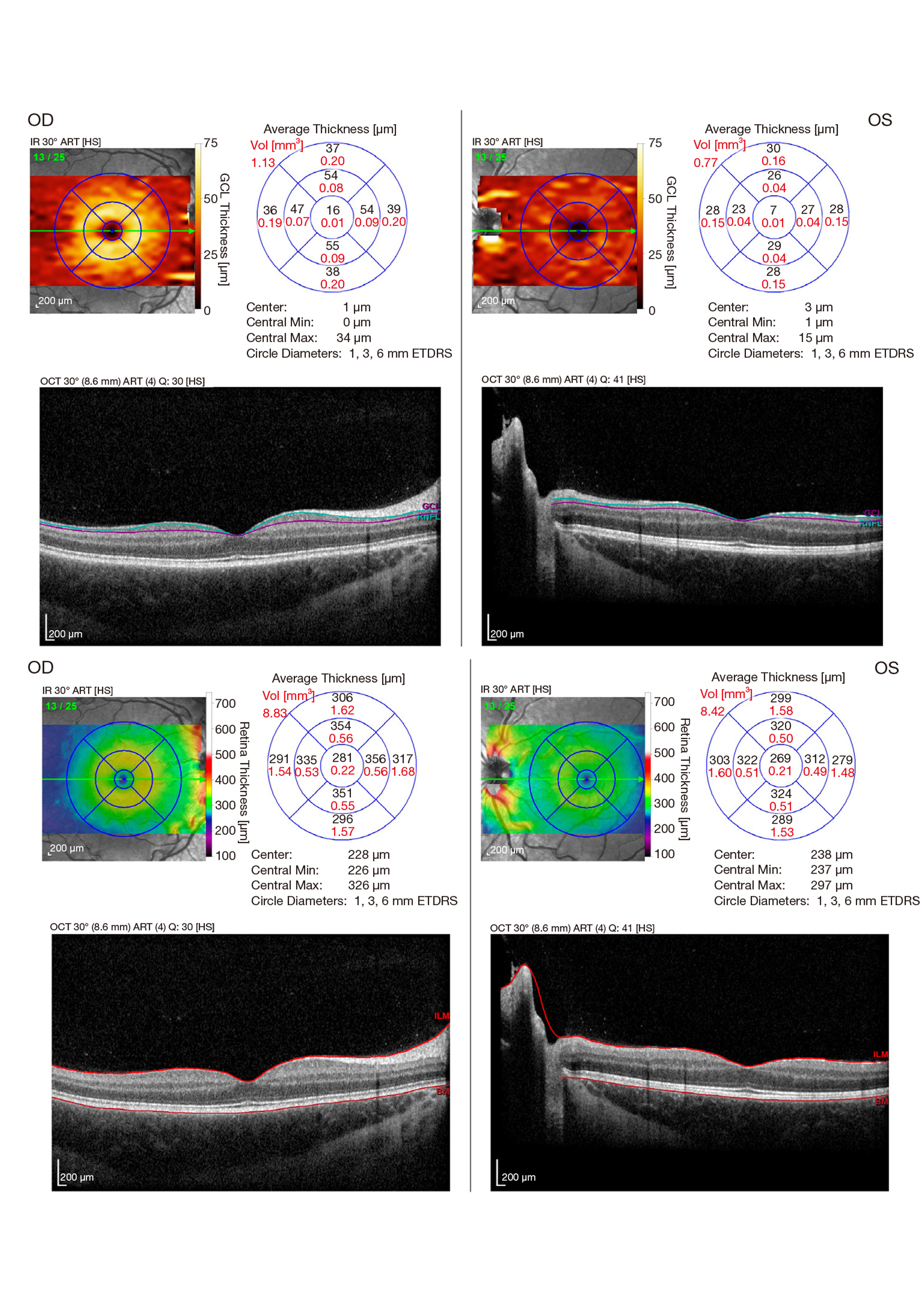
Abstract: Optical coherence tomography (OCT) is a technology that is widely used to assess structural abnormalities in the retina for a variety of pediatric conditions. The introduction of this instrument has allowed for widespread access to minimally invasive standardized, reproducible quantified structural assessments of the optic nerve and retina. This has had important implications in pediatric optic neuropathies, populations in whom monitoring of disease activity is essential to making treatment decisions. OCT has had particular relevance for inflammatory optic neuropathies, as onset of an inflammatory optic neuropathy may herald the onset of a chronic inflammatory disorder of the central nervous system (CNS) such as multiple sclerosis, neuromyelitis optica spectrum disorder (aquaporin 4 antibody positive), and myelin oligodendrocyte glycoprotein (MOG) associated disorders. This paper will focus on the application of OCT technology to this group of disorders in pediatrics. After reviewing pediatric-specific anatomic and practical issues pertinent to OCT, we will review knowledge related to the use of OCT in inflammatory pediatric optic neuropathies, with a focus on structural outcomes and their correlation with functional outcome metrics.
Abstract: Artificial intelligence (AI) methods have become a focus of intense interest within the eye care community. This parallels a wider interest in AI, which has started impacting many facets of society. However, understanding across the community has not kept pace with technical developments. What is AI, and how does it relate to other terms like machine learning or deep learning? How is AI currently used within eye care, and how might it be used in the future? This review paper provides an overview of these concepts for eye care specialists. We explain core concepts in AI, describe how these methods have been applied in ophthalmology, and consider future directions and challenges. We walk through the steps needed to develop an AI system for eye disease, and discuss the challenges in validating and deploying such technology. We argue that among medical fields, ophthalmology may be uniquely positioned to benefit from the thoughtful deployment of AI to improve patient care.