您的位置: 首页 > 2023年3月 第38卷 第3期 > 文字全文
2023年7月 第38卷 第7期11
目录

枕极脑梗死病变导致不典型视野缺损一例

Atypical visual field defect caused by occipital tip cerebral infarction: a case report

来源期刊: 眼科学报 | 2023年3月 第38卷 第3期 287-292 发布时间:2023-03-01 收稿时间:2023/3/24 15:56:11 阅读量:7734
作者:
关键词:
后视路视皮质枕叶梗死视野缺损同向偏盲 神经系统症状
posterior visual pathway visual cortex infarction of occipital lobe visual field defect homonymous hemianopsia neurological symptoms
DOI:
10.12419/j.issn.1000-4432.2023.03.13
收稿时间:
 
修订日期:
 
接收日期:
 
后视路病变是视交叉以后的视觉通路其本身或毗邻结构发生病变,引起视觉功能改变的一类疾病。神经眼科医生比较熟悉枕叶病变引起的对称性同侧偏盲,但枕极(纹状皮质的最后部分)的病变产生中心性对称性同向盲点,此类视野改变容易被忽略或误诊。该文报道一例老年男性患者,因双眼视觉清晰度下降、视物变形就诊。眼科检查:最佳矫正视力:右眼0.8,左眼1.0,FM-100检查提示重度色觉异常,颅脑磁共振成像(magnetic resonance imaging,MRI)提示双侧枕叶脑梗死(右侧枕极部,左侧纹状皮质前部),24-2 Humphrey视野检查可见双眼同向暗点趋势(不典型),10-2 Humphrey视野检查可见双眼中心视野同向偏盲(暗点),故而确诊。后视路病变可引起多种特征性的视野改变,可伴有高级视功能异常及其他神经系统症状和体征,是神经眼科的重要组成部分。该例枕极脑梗死病变产生对称性同向性盲点伴色觉改变患者的诊治过程,提示需关注后视路病变视野改变的多样性及其他视觉功能异常,提高早期诊断率,改善患者预后。
The disease of the posterior visual pathway is a kind of lesion in which the visual pathway itselfor its adjacent structure changes after optic chiasma causes pathological changes, resulting in changes in visual function. Neuro-ophthalmologists are familiar with symmetrical ipsilateral hemianopia caused by occipital lobe lesions, but occipital tip (the last part of the striatal cortex) lesions produce central symmetrical homonymous scotomas, which can easily be overlooked or misdiagnosed. This article reported a case of an olderly male patient treated with decreased binocular visual clarity and distortion. Ophthalmology examination: best corrected visual acuity: 0.8 in the right eye, 1.0 in the left eye; FM-100 examination indicated severe dyschromatopsia; cranial magnetic resonance imaging: infarction of bilateral occipital lobe (right portion of the occipital tip and left anterior portion of striate cortex); 24-2 Humphrey field examination showed a tendency of homonymous scotoma in bilateral eyes (atypical); 10-2 Humphrey field examination showed homonymous hemianopia (scotoma) in the central visual field. These results confirm a diagnosis of the disease of the posterior visual pathway. As an important part of neuro-ophthalmology, the posterior visual pathway can cause various characteristic visual field defects, which can be accompanied by advanced visual dysfunction and other neurological symptoms and signs. The diagnosis and treatment process of this case of occipital tip cerebral infarction with symmetrical homonymous blind spot accompanied by color vision changes suggests that attention should be paid to the diversity of visual field changes and other visual functional abnormalities in the posterior visual pathway lesions, so as to improve the early diagnosis rate and prognosis of the patient s.
    后视路是指视交叉至大脑枕叶视觉皮质的传递视觉信息的神经通路,包括视束、外侧膝状体、视放射、枕叶视皮质,这些结构本身或其毗邻结构发生诸如血管异常、肿瘤、外伤等病变时,会引起视力下降、视野缺损等视功能改变的表现[1-2]。在成人群体中,多见如脑梗死、颅内出血等血管性原因(70%~89%),病变部位以枕叶多见(69.6%)[3–5];而在儿童群体中,神经系统肿瘤是最常见的病因(39%)[6]。后视路病变的临床症状多不典型,往往伴有神经系统症状与体征[2],容易被眼科医生忽略而造成误诊和漏诊。本文报道一例双侧枕叶脑梗死的病例,患者常规眼科检查表现不典型但主观视觉异常明显,既往被误诊为玻璃体浑浊,最终完善中心视野检查,以典型的同向偏盲表现确诊。

1 临床资料

1.1 病史与主诉

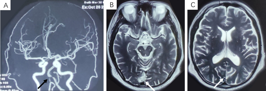
    患者,男性,64岁,因双眼视觉清晰度下降1年半于2022年2月11日就诊。2020年7月患者突发双眼一过性黑朦,缓解后出现双眼眼前大片水墨样遮挡,以视野上方为重,间断伴有闪光感,持续数分钟缓解;因眼前遮挡持续不缓解,先后至多家医院眼科就诊,均诊断为双眼玻璃体浑浊,未予特殊诊治。2020年8月,患者出现口眼歪斜症状3次,每次持续十余分钟自行缓解;遂就诊于神经内科,经头颅磁共振血管造影(magnetic resonance angiography,MRA) 检查提示:基底动脉起始部狭窄(图1A),经头颅磁共振成像(magnetic resonance imaging, MRI)检查提示:双侧枕叶梗死(图1B)。后于神经内科行基底动脉支架植入术,治疗后口眼歪斜症状缓解,视功能无明显变化。至2022年2月(起病以来第18个月),患者诉眼前水墨样遮挡逐渐呈小片状弥漫至全视野范围,伴视物显小,以双眼视觉清晰度下降前来解放军总医院第三医学中心眼科医学部求诊。

1.2 体格检查及辅助检查

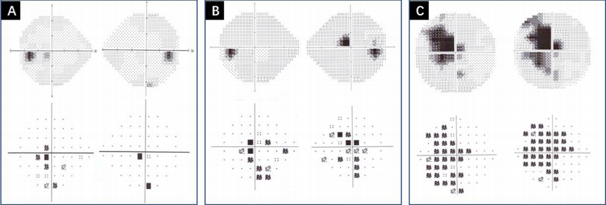
    患者全身情况可。眼科检查:右眼矫正视力0.8,左眼矫正视力1.0,双眼前节未见异常,玻璃体絮状浑浊,眼底检查见左眼黄斑鼻侧少量陈旧性黄白色病,余未见明显异常(图2A)。双眼各频率对比敏感度在正常范围。FM-100孟塞尔色棋检查提示双眼色觉障碍(总分:右眼316,左眼296)。眼科B超检查提示双眼玻璃体浑浊,左眼玻璃体后脱离。闪光视觉诱发电位 (flash visual evoked potential,FVEP) 检查提示:双眼P2波峰时均延迟,右眼P2波峰值较左眼降低,左眼波峰值可(图2B)。黄斑及视盘相干光断层成像 (Optical coherence tomography, OCT) 检查结果示双眼视盘视网膜神经纤维层 (retinal nerve fiber layer, RNFL)、黄斑区节细胞未见明显异常(图2C、D)。24-2Humphrey自动视野检查提示:右眼中心暗点,双眼点状视野缺损(图3A)。

1.3 诊 断

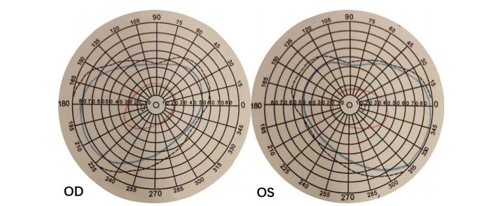
    结合患者临床表现与眼科检查,入院诊断考虑为双眼玻璃体浑浊,枕叶陈旧性脑梗死;但患者诉既往5年前即诊断双眼玻璃体浑浊,本次起病眼前遮挡呈弥漫小片状,相对固定,无漂浮感,伴视物显小。复查头颅MRI检查提示:双侧枕叶梗死,右侧枕极部,左侧纹状皮质上前部,与既往头颅MRI检查对比未见明显变化(图4)。因患者既往视野检查中心绝对暗点有明显象限分界表现,复查24-2视野检查提示右眼中心暗点(图3B),完善10-2Humphrey 视野检查提示:双眼左侧视野同向偏盲(图3C),完善Goldmann动态视野检查提示:右眼颞侧新月状视野缺损(图5)。至此,结合患者视觉改变特点及神经影像表现,诊断为双眼同侧偏盲、右眼颞侧新月状视野缺损、双侧枕叶脑梗死。

2 讨 论

    不同于视交叉前视神经损伤所致的单眼视觉改变,单侧的后视路病变所造成的视觉改变通常是病变对侧的双眼同向偏盲[7],这种特征性的视觉改变是由于单侧的后视路汇集双眼视觉神经纤维,而双眼的视觉神经纤维在视觉通路中又按照一定的拓扑结构排列,最终汇总至视皮质[8]。后视路病变患者由于存在视野缺损,往往在学习及工作等方面受限,会出现阅读障碍及偏盲侧视觉忽视症状等,严重影响患者的生活质量[9]

20230324155939_0186.png

图1 头颅MRA和MRI检查结果
Figure 1 Cranial vascular MRA and MRI images
(A)头颅血管MRA检查:基底动脉起始部狭窄(黑色箭头);(B、C)头颅MRI检查:双侧枕叶可见T2高信号,符合枕叶梗死改变(白色箭头) 。
(A)Cranial vascular MRA shows the stenosis of basilar artery initial portion(black arrow); (B, C)Cranial MRI shows that T2 hyperintensity is seen in both occipital lobes and consistent with occipital infarction (white arrow).

20230324160133_1395.png

图2 双眼彩色眼底照相、F-VEP及OCT检查结果
Figure 2 Fundus color photo, F-VEP and OCT images
(A)彩色眼底照相:双眼视盘界清,色可;(B)FVEP结果示双眼P2波峰时均延迟,右眼P2波峰值较左眼降低,左眼波峰值可;(C、D)OCT检查结果示双眼视盘RNFL、黄斑区节细胞未见明显异常。
(A)Fundus color photo: Clear boundary of optic disc with good color in bilateral eyes; (B)The results of F-VEP showed that the peak of P2 wavewas delayed in bilateral eyes, and the peak of P2 wave in right eye was lower than that in left eye; (C, D) OCT examination showed no obvious abnormalities in RNFL of optic disc and ganglion cells of macular area.

20230324160331_6713.png

图3 双眼24-2及10-2 Humphrey自动视野检查
Figure 3 24-2 and 10-2 program Humphrey automatic visual field examination of bilateral eyes
24-2双眼Humphrey自动视野检查(上方:灰度图;下方:模式偏差图):(A) 左眼中心暗点(同侧性暗点趋势,2020-09-01);(B)右眼中心暗点(偏鼻上,双眼数个暗点,趋势不明确,2022-02-14); 10-2 Humphrey自动视野检查:(C)左侧同向偏盲(同向暗点,2022-02-14)。
24-2 program Humphrey automatic visual field examination(upward: grey-scale plot; downward: pattern deviation plot): A: central field scotoma in left eye(homonymous scotoma tendency, September 1, 2020), B: central scotoma in right eye (in superior nasal visual field, and several scotomas in bilateral eyes with unclear tendency, February 14, 2022); 10-2 program Humphrey automatic visual field examination: C: left homonymous hemianopia; (homonymous scotoma, February 14, 2022).

20230324160630_7887.png

图4 头颅MRI检查
Figure 4 Cranial magnetic resonance imaging
头颅MRI(2022-0218)提示:双侧枕叶梗死(箭头)。
Cranial magnetic resonance imaging(February 18, 2022): Infarction of bilateral occipital lobe(arrow).

20230324160724_8229.png

图5 Goldmann动态视野检查
Figure 5 Goldmann kinetic perimeter
Goldmann动态视野检查提示:右眼颞侧新月状视野缺损。
Goldmann kinetic perimeter: Visual field defect of temporal crescent of right eye.

    同向偏盲的类型可因后视路病变部位不同而存在差异,且不同脑叶损伤后也会出现特征性的神经症状,这些特征性的眼部及神经症状可以辅助定位诊断[1,10]。视束病变可产生完全或不全性的同向偏盲,由于支配瞳孔对光反射的传入神经纤维在视束中走行,如单纯视束病变可导致病变对侧眼出现相对性瞳孔传入障碍(relative efferent pupil defect,RAPD),还会导致偏盲性瞳孔反应(Wernicke瞳孔) [11]。而外侧膝状体处脉络丛血管的异常会导致特征性的扇形盲及扇形回避盲。视放射腹侧束纤维行经颞叶,形成Meyer环,投射到枕叶距状裂下唇,支配上半视野区域;背侧束直接向后延伸,穿过后部顶叶至枕叶,投射到枕叶距状裂上唇,支配下半视野区域;外侧束纤维代表同侧视网膜周边区域,投射到枕叶上极。视放射神经纤维越接近枕叶皮质,其视网膜皮质映射关系越明显[12]。由于视放射的前部构成了内囊的大部,此处存在大量的大脑上下行神经纤维,此处病变可导致“三偏综合征”,即同向偏盲、偏瘫及偏身感觉障碍。颞叶视放射损伤所致的同向偏盲通常是不对称、不完全性的,可局限于上象限,而且可以伴随多种形式的神经系统表现,包括失语、记忆障碍、复杂性癫痫发作及幻听和幻视等。顶叶视放射损伤所致的同向偏盲通常是不全性的,且通常局限于下象限,由于顶叶为大脑皮质的主要感觉区,其损伤常会导致感觉障碍。初级视皮质也称纹状皮层;其最前部接受视放射外侧束的神经纤维,支配周边视野,因双眼最外侧30°视野范围与对侧眼的鼻侧视野不相重叠,故此处损伤可导致病变对侧眼颞侧新月状视野缺损,这是后视路病变引起单眼视野缺损的唯一形式。此病例Goldmann动态视野检查提示右眼颞侧新月状视野缺损,考虑与左侧距状裂前部梗死有关;上部初级视皮质接受来自视放射背侧束的神经纤维,支配下部视野;下部接受来自视放射腹侧束的神经纤维,支配上部视野;枕极部支配中央区域视野。枕叶病变所致的同向偏盲通常会伴有黄斑回避,即中央视野保留的现象,对此的解释有:表征黄斑处视野的枕极部脑组织接受大脑中动脉及大脑后动脉的双重血供,一支血管闭塞后仍有血供,故而单支血管闭塞后黄斑处视野仍得以保留[13]。双侧枕叶病变会导致皮质盲,部分患者可能无明显视觉缺失感,因幻觉存在而否认视觉异常,这种现象称为Anton综合征[14]。枕极(纹状皮质的最后部分)的病变通常产生对称性同向性盲点,此类视野改变容易被忽略造成漏诊或误诊;本例患者双侧枕叶梗死表现,Humphrey自动视野24-2程序检查仅表现为右眼象限性中心暗点,边界较为明确,未见明显同侧偏盲改变,应用10-2程序检查,可见明显同侧性偏盲(暗点),结合患者临床表现及相关检查结果,考虑右侧枕极部梗死灶为主要责任病灶。
    初级视皮层(V1)接受来自外侧膝状体、视放射的信息,然后通过腹侧传导通路及背侧传导通路将信息逐步整合并传至纹外皮层进行高级加工[15-16]。背侧通路包括枕叶到顶叶的一系列脑区(V1/V2-MT-MST/VIP)[17-18],主要处理运动和深度相关的视知觉信息;腹侧通路包括枕叶到颞叶的一系列脑区(V1/V2-V4-TEO/IT),主要处理形状和颜色相关信息。但目前研究表明,背侧及腹侧通路仅对视觉加工过程中主要参与脑区的粗略分类,人体大脑对视觉信息的处理遵循分布、层级、网格化等组织原则,不仅信息处理通路内部存在双向投射,各通路间也存在广泛的投射联系。随着视觉信息的层级传递,视觉信息的处理也逐渐从一般到复杂、具体到抽象,如初级视觉皮层可区分物体的朝向和运动状态,纹外皮层则可经此而区分出观察者自身的运动状态,故初级视皮层损伤亦会导致诸如中枢性色盲、面容失认、视觉共济失调等高级视觉功能异常的症状[19-20]。本例患者双眼最佳矫正视力尚可,主诉视物显小,检查提示双眼色觉障碍,患者此形觉与色觉障碍提示枕叶梗死导致了初级视觉腹侧传导通路的异常,启发眼科临床医生对于脑叶梗死患者的视功能评估,不应局限于中心视力这单一指标,还应进行如色觉、形觉、亮度、对比敏感度、深度视觉、立体视觉等高级视觉功能的评估。
    Zhang[21]等报道,在后视路病变发生后的1个月内,50%患者的同向偏盲症状可自发缓解,至损伤发生后6个月,视野改变基本稳定[22]。目前,针对后视路病变患者的治疗方法较为有限,主要包括重复经颅磁刺激(repetitive transcranial magnetic stimulation, rTMS)等[23]。康复性方法主要基于刺激毗邻偏盲半侧视野的过渡区以拓宽视野,包括刺激相对暗点激活、训练替代通路激活、代偿性眼球运动训练等[24]
    本文介绍一例枕极脑梗死病变产生对称性同向性盲点伴色觉改变的患者,旨在向临床医生展示此类视野的特点及枕叶损伤可能导致除视野以外其他高级视功能的异常,提高对本类疾病的认识,有助于早期明确诊断及治疗。

开放获取声明

本文适用于知识共享许可协议 (Creative Commons),允许第三方用户按照署名(BY)-非商业性使用(NC)-禁止演绎(ND)(CCBY-NC-ND)的方式共享,即允许第三方对本刊发表的文章进行复制、发行、展览、表演、放映、广播或通过信息网络向公众传播,但在这些过程中必须保留作者署名、仅限于非商业性目的、不得进行演绎创作。详情请访问:https://creativecommons.org/licenses/by-nc-nd/4.0/。
1、Moss HE. Chiasmal and postchiasmal disease[ J]. Continuum (Minneap Minn), 2019, 25(5): 1310-1328.Moss HE. Chiasmal and postchiasmal disease[ J]. Continuum (Minneap Minn), 2019, 25(5): 1310-1328.
2、Pula JH, Yuen CA. Eyes and stroke: the visual aspects of cerebrovascular disease[ J]. Stroke Vasc Neurol, 2017, 2(4): 210-220.Pula JH, Yuen CA. Eyes and stroke: the visual aspects of cerebrovascular disease[ J]. Stroke Vasc Neurol, 2017, 2(4): 210-220.
3、Trobe JD, Lorber ML, Schlezinger N S. Isolated homonymous hemianopia:a review of 104 cases[ J]. Arch Ophthalmol, 1973, 89(5): 377-381.Trobe JD, Lorber ML, Schlezinger N S. Isolated homonymous hemianopia:a review of 104 cases[ J]. Arch Ophthalmol, 1973, 89(5): 377-381.
4、Zhang X, Kedar S, Lynn M J, et al. Homonymous hemianopias: clinical-anatomic correlations in 904 cases[ J]. Neurology, 2006, 66(6): 906- 910.Zhang X, Kedar S, Lynn M J, et al. Homonymous hemianopias: clinical-anatomic correlations in 904 cases[ J]. Neurology, 2006, 66(6): 906- 910.
5、Pambakian AL, Kennard C. Can visual function be restored in patients with homonymous hemianopia? [ J]. Br J Ophthalmol, 1997, 81(4): 324-328.Pambakian AL, Kennard C. Can visual function be restored in patients with homonymous hemianopia? [ J]. Br J Ophthalmol, 1997, 81(4): 324-328.
6、Liu GT, Galetta S L. Homonymous hemifield loss in childhood[ J]. Neurology, 1997, 49(6): 1748-1749.Liu GT, Galetta S L. Homonymous hemifield loss in childhood[ J]. Neurology, 1997, 49(6): 1748-1749.
7、Fraser JA, Newman NJ, Biousse V. Disorders of the optic tract, radiation, and occipital lobe[ J]. Handb Clin Neurol, 2011, 102: 205- 221.Fraser JA, Newman NJ, Biousse V. Disorders of the optic tract, radiation, and occipital lobe[ J]. Handb Clin Neurol, 2011, 102: 205- 221.
8、Li N, Gu Y. The visual pathway for binocular integration[ J]. Neurosci Bull, 2020, 36(9): 1089-1091.Li N, Gu Y. The visual pathway for binocular integration[ J]. Neurosci Bull, 2020, 36(9): 1089-1091.
9、Schaller-Paule MA, Friedauer L, You SJ. Sudden onset homonymous quadrantanopia[ J]. BMJ, 2020, 371: m3338.Schaller-Paule MA, Friedauer L, You SJ. Sudden onset homonymous quadrantanopia[ J]. BMJ, 2020, 371: m3338.
10、Short RA, Graff-Radford N R. Localization of Hemiachromatopsia[ J]. Neurocase, 2001, 7(4): 331-337.Short RA, Graff-Radford N R. Localization of Hemiachromatopsia[ J]. Neurocase, 2001, 7(4): 331-337.
11、Kardon R, Kawasaki A, Miller NR. Origin of the relative afferent pupillary defect in optic tract lesions[ J]. Ophthalmology, 2006, 113(8): 1345-1353.Kardon R, Kawasaki A, Miller NR. Origin of the relative afferent pupillary defect in optic tract lesions[ J]. Ophthalmology, 2006, 113(8): 1345-1353.
12、Mehra D, Moshirfar M. Neuroanatomy, Optic Tract. Treasure Island (FL): StatPearls Publishing, 2022.Mehra D, Moshirfar M. Neuroanatomy, Optic Tract. Treasure Island (FL): StatPearls Publishing, 2022.
13、Horton JC, Economides JR, Adams DL. The mechanism of macular sparing[ J]. Annu Rev Vis Sci, 2021, 7: 155-179.Horton JC, Economides JR, Adams DL. The mechanism of macular sparing[ J]. Annu Rev Vis Sci, 2021, 7: 155-179.
14、Kartsounis LD, James-Galton M, Plant GT. Anton syndrome, with vivid visual hallucinations, associated with radiation induced leucoencephalopathy[ J]. J Neurol Neurosurg Psychiatry, 2009, 80(8): 937-938.Kartsounis LD, James-Galton M, Plant GT. Anton syndrome, with vivid visual hallucinations, associated with radiation induced leucoencephalopathy[ J]. J Neurol Neurosurg Psychiatry, 2009, 80(8): 937-938.
15、Zachariou V, Klatzky R, Behrmann M. Ventral and dorsal visual stream contributions to the perception of object shape and object location[ J]. J Cogn Neurosci, 2014, 26(1): 189-209.Zachariou V, Klatzky R, Behrmann M. Ventral and dorsal visual stream contributions to the perception of object shape and object location[ J]. J Cogn Neurosci, 2014, 26(1): 189-209.
16、Murray MM, Thelen A, Thut G, et al. The multisensory function of the human primary visual cortex[ J]. Neuropsychologia, 2016, 83: 161-169.Murray MM, Thelen A, Thut G, et al. The multisensory function of the human primary visual cortex[ J]. Neuropsychologia, 2016, 83: 161-169.
17、Kwan WC, Chang CK, Yu HH, et al. Visual cortical area MT is required for development of the dorsal stream and associated visuomotor behaviors[ J]. J Neurosci, 2021, 41(39): 8197-8209.Kwan WC, Chang CK, Yu HH, et al. Visual cortical area MT is required for development of the dorsal stream and associated visuomotor behaviors[ J]. J Neurosci, 2021, 41(39): 8197-8209.
18、Galletti C, Fattori P. The dorsal visual stream revisited: stable circuits or dynamic pathways?[ J]. Cortex, 2018, 98: 203-217.Galletti C, Fattori P. The dorsal visual stream revisited: stable circuits or dynamic pathways?[ J]. Cortex, 2018, 98: 203-217.
19、Morenas-Rodríguez E, Camps-Renom P, Pérez-Cordón A, et al. Visual hallucinations in patients with acute stroke: a prospective exploratory study[ J]. Eur J Neurol, 2017, 24(5): 734-740.Morenas-Rodríguez E, Camps-Renom P, Pérez-Cordón A, et al. Visual hallucinations in patients with acute stroke: a prospective exploratory study[ J]. Eur J Neurol, 2017, 24(5): 734-740.
20、R afique SA , Richards JR , Steeves JKE. Altered white matter connectivity associated with visual hallucinations following occipital stroke[ J]. Brain Behav, 2018, 8(6): e01010.R afique SA , Richards JR , Steeves JKE. Altered white matter connectivity associated with visual hallucinations following occipital stroke[ J]. Brain Behav, 2018, 8(6): e01010.
21、Zhang X, Kedar S, Lynn M J, et al. Natural history of homonymous hemianopia[ J]. Neurology, 2006, 66(6): 901-905.Zhang X, Kedar S, Lynn M J, et al. Natural history of homonymous hemianopia[ J]. Neurology, 2006, 66(6): 901-905.
22、de Haan GA, Heutink J, Melis-Dankers BJ, et al. Spontaneous recovery and treatment effects in patients with homonymous visual field defects: a meta-analysis of existing literature in terms of the ICF framework[ J]. Surv Ophthalmol, 2014, 59(1): 77-96.de Haan GA, Heutink J, Melis-Dankers BJ, et al. Spontaneous recovery and treatment effects in patients with homonymous visual field defects: a meta-analysis of existing literature in terms of the ICF framework[ J]. Surv Ophthalmol, 2014, 59(1): 77-96.
23、Koch G, Bonnì S, Giacobbe V, et al. θ-burst stimulation of the left hemisphere accelerates recovery of hemispatial neglect[ J]. Neurology, 2012, 78(1): 24-30.Koch G, Bonnì S, Giacobbe V, et al. θ-burst stimulation of the left hemisphere accelerates recovery of hemispatial neglect[ J]. Neurology, 2012, 78(1): 24-30.
24、Fahrenthold BK, Cavanaugh MR, Jang S, et al. Optic tract shrinkage limits visual restoration aer occipital stroke[ J]. Stroke, 2021, 52(11): 3642-3650.Fahrenthold BK, Cavanaugh MR, Jang S, et al. Optic tract shrinkage limits visual restoration aer occipital stroke[ J]. Stroke, 2021, 52(11): 3642-3650.
1、国家自然科学基金青年项目(82101110)。
This work was supported by the National Natural Science Foundation of China (82101110).()
上一篇
下一篇
其他期刊
  • 眼科学报

    主管:中华人民共和国教育部
    主办:中山大学
    承办:中山大学中山眼科中心
    主编:林浩添
    主管:中华人民共和国教育部
    主办:中山大学
    浏览
  • Eye Science

    主管:中华人民共和国教育部
    主办:中山大学
    承办:中山大学中山眼科中心
    主编:林浩添
    主管:中华人民共和国教育部
    主办:中山大学
    浏览
推荐阅读
出版者信息
目录