您的位置: 首页 > 2023年4月 第38卷 第4期 > 文字全文
2023年7月 第38卷 第7期11
目录

结膜松弛症动物模型的构建和评估

Construction and evaluation of animal models of conjunctivochalasis

来源期刊: 眼科学报 | 2023年4月 第38卷 第4期 305-311 发布时间:2023-04-18 收稿时间:2023/4/19 10:31:42 阅读量:4471
作者:
关键词:
结膜松弛症动物模型基质金属蛋白酶-1基质金属蛋白酶-3
conjunctivochalasis animal model matrix metalloproteinase-1 matrix metalloproteinase-3
DOI:
10.12419/j.issn.1000-4432.2023.04.01
收稿时间:
 
修订日期:
 
接收日期:
 
目的:通过球结膜下注射基质金属蛋白酶(metalloproteinase,MMP)-1、3构建结膜松弛症的动物模型。法:20只新西兰大白兔随机分为4组:对照组无干预措施;伪造模组球结膜下注射生理盐水;MMP-1组球结膜下注射MMP-1;MMP-3组球结膜下注射MMP-3。8周后观察眼前节照相和眼前节光学相干检查、泪液蕨类试验及结膜组织光镜检查结果。结果:对照组和伪造模组未见结膜皱褶形成,泪液羊齿状结晶样结构完整,光镜下未见胶原和弹力纤维破坏;MMP-1组可见结膜皱褶形成,泪液羊齿状结晶样结构破坏,光镜下见胶原和弹力纤维破坏;MMP-3组未见结膜皱褶形成,泪液羊齿状结晶样结构部分破坏,光镜下见胶原和弹力纤维部分破坏。结论:球结膜下注射MMP-1可构建结膜松弛症1~2级的动物模型。
Objective:To establish an animal model of conjunctivochalasis by subconjunctival injection of matrix metalloproteinase 1 and 3. Methods:New Zealand white rabbits were randomly divided into four groups, the control group had no intervention measures; Pseudo model group was injected susbconjunctival with normal saline; MMP-1 group was injected subconjunctival with matrix metalloproteinase 1; MMP-3 group was injected subconjunctival with matrix metalloproteinase 3. Afer 8 weeks, the results of anterior segment photography and optical coherence test, tear fern test and conjunctival tissue light microscopy were observed. Results:No conjunctival folds were observed in the control group and Pseudo model group, the lacrimal fern-shaped crystalline structures were intact, and no destruction of collagen and elastic fbers was found under the light microscope. In MMP-1 group, conjunctival folds were formed, lacrimal ferns crystalline structures were damaged, and collagen and elastic fbers were damaged under light microscope. No conjunctival folds were found in MMP-3 group, and the lacrimal ferns crystalline structure was partially destroyed, while the collagen and elastic fibers were partially destroyed under light microscope. Conclusion:Subconjunctival injection of matrix metalloproteinase-1 can construct the animal model of conjunctivochalasis of 1-2 grades.
结膜松弛症(conjunctivochalasis,CCH)是过多的、松弛的、非水肿性的球结膜堆积在眼球与下睑缘、内、外眦部之间引起的疾病[1]。目前该病尚缺乏一个公认的疾病动物模型,该动物模型的构建对探索结膜松弛症的发生发展规律、有效防治方法具有重要的科学意义和临床意义。
在前期建模的初步试验中,本课题组尝试了基质金属蛋白酶(metalloproteinase,MMP)-3球结膜下注射、肿瘤坏死因子-α(Tumour necrosis factor alpha,TNF-α)溶液点眼、D-半乳糖皮下注射三种方法[2]。结果示MMP-3球结膜下注射的动物的下球结膜在外力作用下形成皱褶,并见疏松和排列紊乱的胶原纤维、碎片状的弹力纤维、成纤维细胞的局灶性坏死。表明球结膜下注射MMPs可能是构建CCH动物模型的可行方法。
在此结果的基础上,课题组通过球结膜下注射MMP-1、MMP-3的方法来构建疾病动物模型;通过眼前节照相和光学相干成像检查、泪液蕨类试验、结膜组织光镜检查结果来评估造模效果。现将实验结果报告如下。

1 材料与方法

1.1 实验材料

1.1.1 实验仪器
眼前节照相仪,产自芬兰Smartscope M5 OPTOMED公司;Cirrus 4000 HD OCT光学相干断层扫描仪,产自德国CARL ZEISS集团。
1.1.2 实验试剂
重组人MMP-1,购自美国PerproTech公司;重组人MMP-3,购自美国PerproTech公司;弹力纤维染色试剂盒,购于北京索莱宝科技有限公司;盐酸丙美卡因滴眼液,产自爱尔康(中国)眼科产品有限公司;盐酸左氧氟沙星滴眼液,产自山东博士伦福瑞达制药有限公司。
1.1.3 实验试剂的配置
10 μg重组人MMP-1(现配现用)+3.33 mL灭菌注射用水,配成浓度为3 000 μg/L;10 μg重组人MMP-3(现配现用)+3.33 mL灭菌注射用水,配成浓度为3 000 μg/L。
1.1.4 实验动物
20只普通级12~14周雄性新西兰白兔,体重2.0~2.5 kg,来自上海市松江区松联实验动物厂。动物许可证号:SCXK(沪)2017-0008。
饲养环境:单笼常规饲养,自由饮水和进食,使用全价颗粒饲料喂饲;室温控制在25 ℃左右;湿度为40%~70%;通风:每小时15次空气交换;每天12 h光照,工作照度150~300 lx,动物照度100~200 lx;噪声≤60 dB。
本动物实验的所有过程均遵循“视觉和眼科学研究学会(Association for research in vision and ophthalmology,ARVO)关于在眼科和视觉研究中使用动物的声明”以及中华人民共和国科学技术部制定的“关于善待实验动物的指导意见”。

1.2 实验方法

1.2.1 实验动物的分组
对20只新西兰白兔称重,体重相近的4只兔配成1个区组,给予相应编号。应用SPSS21.0统计软件得出随机分配序列,将20只实验兔随机分配至空白对照组、伪造模组、MMP-1组和MMP-3组,每组5只。
1.2.2 干预措施
空白对照组:无任何干预措施。伪造模组:给予灭菌注射用水兔眼球结膜下注射,每侧兔眼0.3 mL,2侧/次,每周1次,共8周。MMP-1组:给予MMP-1灭菌注射用水溶液兔眼球结膜下注射,每侧兔眼0.3 ml L,2侧/次,每周1次,共8周。MMP-3组:给予MMP-3灭菌注射用水溶液兔眼球结膜下注射,每侧兔眼0.3 L,2侧/次,每周1次,共8周。
1.2.3 球结膜下注射药物方法
固定器固定实验动物兔,注射前使用盐酸丙美卡因滴眼液点兔眼行表面麻醉。1 mL注射器抽取配制好的MMP-3溶液,针头剖面向上,由外眦向内眦方向进针,以5 °的角度刺入结膜,待针头有突破感后停止进针,回抽无血液后注入药物。注射后球结膜肿胀,一般3~5 d完全消退,无需特殊处理。注射结束后给予盐酸左氧氟沙星滴眼液点兔眼预防眼部感染(每日3次,共3 d)。

1.3 观测指标

1.3.1 眼前节照相
造模前及造模后8周分别使用便携式眼前节照相仪为实验动物兔双眼照相并保留照片,观察球结膜有无皱褶形成。
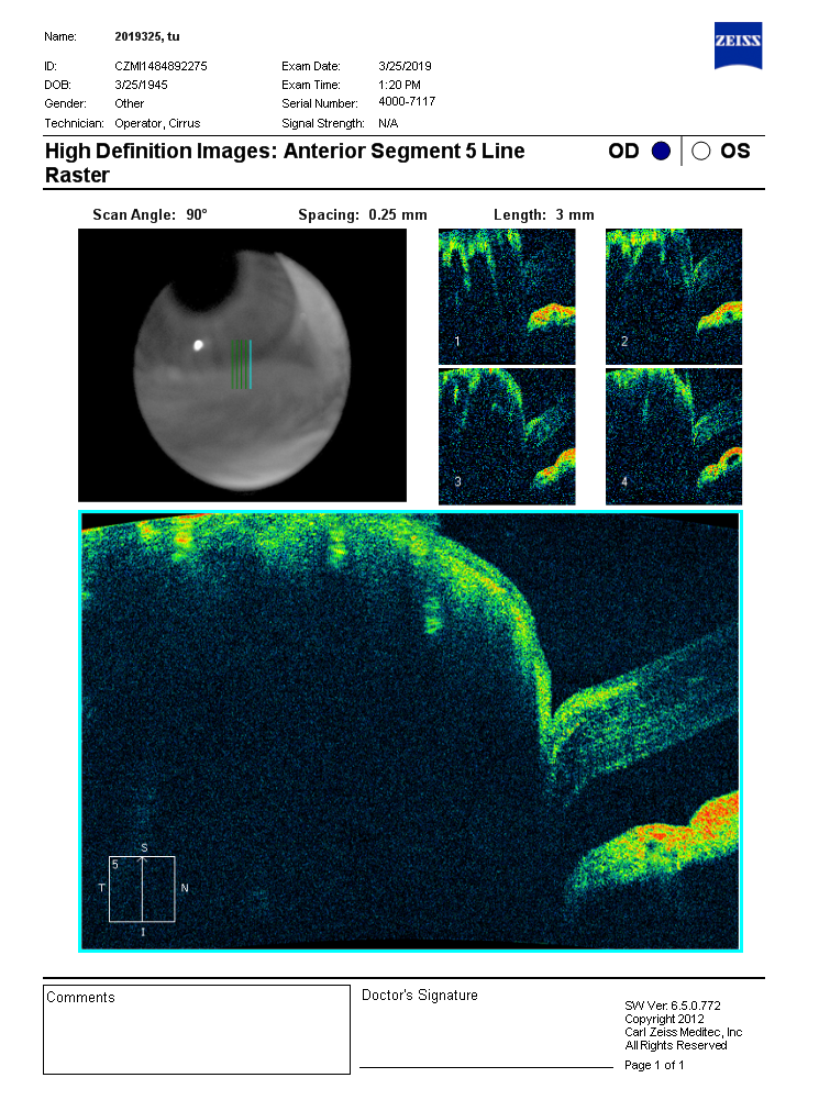
1.3.2 眼前节光学相干断层扫描(AS-OCT)
根据来自角膜缘鼻侧、颞侧以及角膜中央垂直于下睑缘的直线相交于下睑缘的位置可以得到鼻侧点、中央点和颞侧点。OCT检查得到的图像即位于此三点处,见图1。
20230419094857_6811.png
图1 兔右眼颞侧点结膜囊OCT检测示意图
Figure 1 OCT detection of conjunctival sac in temporal point of rabbit right eye
(A)鼻侧点,角膜缘鼻侧垂线和下睑缘的交点处; (B)中央点,角膜中央垂线和下睑缘的交点处; (C)颞侧点,角膜缘颞侧垂线和下睑缘的交点处。
(A) The nasal point, the intersection of the nasal vertical line of the limbal cornea and the lower eyelid margin ;(B) the central point, the intersection of the central vertical line of the cornea and the lower eyelid margin; (C) the temporal point, the intersection of the temporal vertical line of the limbal cornea and the lower eyelid margin.
1.3.3 泪液蕨类试验(tear reining test, TFT)
使用10 mL的玻璃毛细管在下睑结膜囊泪阜处通过虹吸方法吸取泪液标本(未使用表面麻醉药物或其他化学性药物),尽量不接触眼表。将泪液标本吹入载玻片,在25 ℃室温下(空调控制温度与湿度)干燥10~20 min,置于双目光学显微镜下观察(放大倍数为100)评级。
1.3.4 球结膜组织光镜检查
所有实验动物静脉注射戊巴比妥钠处死。完整摘取兔眼,摘除过程中从上下结膜穹隆处离断结膜,以保留完整的球结膜组织。去除玻璃体和晶状体后将眼球固定于10%的中性甲醛溶液中。
组织标本取自实验动物下球结膜的中央,制备组织石蜡切片。分别行苏木素-伊红(Hematoxylin-eosin,HE)染色、改良Gomori醛品红染色、Masson三色染色。显微镜下观察上述染色的组织标本并拍照。

2 结果

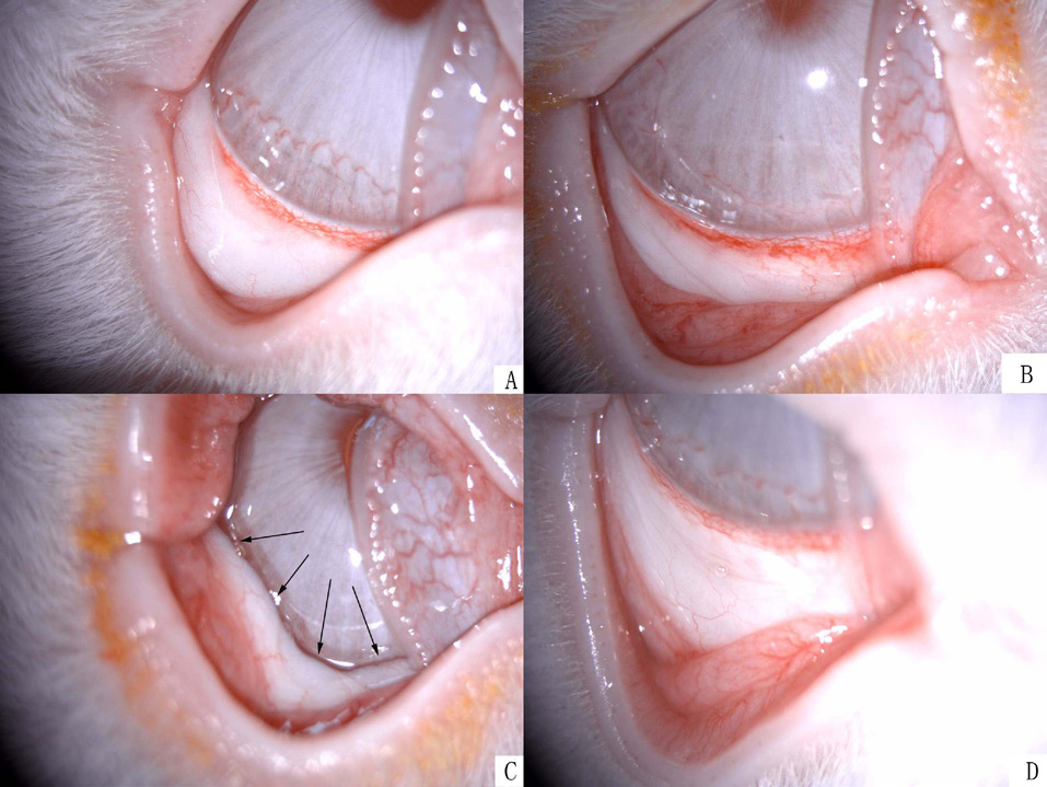
2.1 兔眼前节照片

对照组(A组)、伪造模组(B组)及MMP-3组(D组)未见结膜皱褶形成,MMP-1组(C组)可见结膜皱褶形成。见图2。
20230419095254_1843.png
图2 兔眼前节照片
Figure 2 Photo of rabbit’s anterior segment
(A)对照组;(B)伪造模组;(C)MMP-1组;(D)MMP-3组。其中图A、B、D均未见结膜皱褶形成,图C黑色箭头所指为球结膜皱褶。
(A) Control group; (B) Pseudo Model group; (C) MMP-1 group; (D) MMP-3 group. No conjunctival folds were observed in Figures A, B, and D, while the black arrow in Figure C indicates bulbar conjunctival folds.

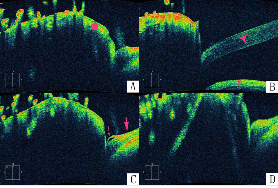
2.2 兔结膜囊OCT照片

对照组( A组)、伪造模组( B组)及MMP-3组( D组)未见结膜皱褶形成,MMP-1组(C组)可见结膜皱褶形成。见图3。
20230419095326_1219.png
图3 兔结膜囊OCT照片
Figure 3 OCT photo of rabbit conjunctival sac
(A)对照组,红色圆点处为下眼睑;(B)伪造模组,红色三角处为角膜,红色五角处为虹膜;(C)MMP-1组;(D)MMP-3组。其中图A、B、D均未见结膜皱褶,图C红色细箭头所指为结膜皱褶,红色粗箭头所指为球结膜和其下巩膜。
(A) Control group, the red dot was the lower eyelid; (B) Pseudo Model group: the red triangle representing the cornea and the red pentagon representing the iris; (C) MMP-1 group; (D) MMP-3 group. In FIG. A, B and D, conjunctival folds were not observed. In FIG. C, the red thin arrow pointed to conjunctival folds, while the red thick arrow pointed to bulbar conjunctiva and its lower sclera.

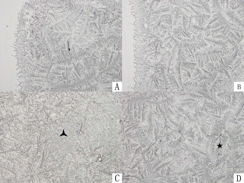
2.3 泪液蕨类实验

对照组(A组)和伪造模组(B组)泪液羊齿状结晶样结构完整,MMP-1组(C组)可见泪液羊齿状结晶样结构破坏,MMP-3组(D组)见泪液羊齿状结晶样结构部分破坏。见图4。
20230419095615_5153.png
图4 泪液蕨类实验(×100)
Figure 4 Tear reining test
(A)对照组,细箭头所指为完整的泪液羊齿状结晶;(B)伪造模组,泪液羊齿状结晶完整;(C)MMP-1组,黑色三角处为泪液羊齿状结晶样结构完全破坏;(D)MMP-3组,黑色五角处为泪液羊齿状结晶样结构部分破坏。
(A) Control group, the fine arrow indicated complete lacrimal ferns; (B) Pseudo Model group, complete lacrimal ferns; (C) MMP-1 group, the black triangle showed that the lacrimal fernion crystalline structure was completely destroyed; (D) MMP-3 group, the black pentagon showed partial destruction of lacrimal ferns crystalline structure.

2.4 球结膜组织H E染色

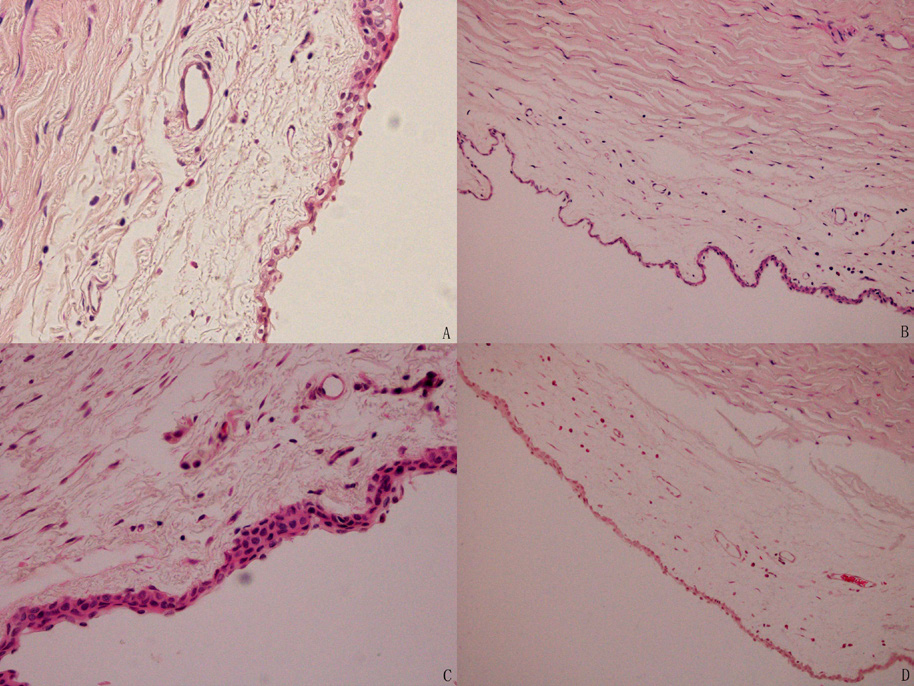
对照组( A组)、伪造模组( B组)、MMP-1组(C组)及MMP-3组( D组)之间未见明显区别。见图5。
/20230419095602_9684.png
图5 球结膜组织HE染色(×100)
Figure 5 HE staining of bulbar conjunctival tissue
(A)对照组;(B)伪造模组;(C)MMP-1组;(D)MMP-3组。图中可见结膜组织经HE染色后,细胞核呈蓝色,细胞质、肌纤维、胶原纤维等呈深浅不等的红色。结膜上皮层由2~4层的上皮细胞组成。
(A) Control group; (B) Pseudo Model group; (C) MMP-1 group; (D) MMP-3 group. In the figure, it can be seen that after HE staining, the nucleus of the conjunctival tissue is blue, while the cytoplasm, muscle fibers, collagen fibers, etc. are red in varying shades. The conjunctival epithelium consists of two to four layers of epithelial cells.

2.5 球结膜组织Masson染色

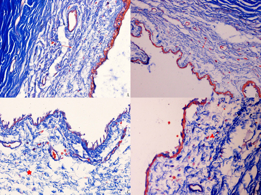
对照组(A组)和伪造模组(B组)光镜下未见胶原纤维破坏,MMP-1组(C组)光镜下见胶原纤维破坏,MMP-3组(D组)光镜下见胶原纤维部分破坏。见图6。
20230419095956_3610.png
图6 球结膜组织Masson染色(×200)
Figure 6 MASSON staining of bulbar conjunctival tissue
(A)对照组;(B)伪造模组;(C)MMP-1组;(D)MMP-3组。图A、B红色细箭头所指处为胶原纤维致密并成平行排列;图C红色五角星处示胶原纤维结合疏松,失去平行排列结构;图D红色三角星左侧区域胶原纤维结合疏松,右侧区域尚可见平行排列结构。
(A) Control group; (B) Pseudo Model group; (C) MMP-1 group; (D) MMP-3 group. The red arrows in Figures A and B indicate dense and parallel arrangement of collagen fibers; The red pentagram in Figures C shows that the collagen fibers are loosely combined and lose their parallel arrangement structure; The collagen fibers in the left area of the red triangle star in Figure D are loosely bound, while a parallel arrangement structure can still be seen in the right area.

2.6 球结膜组织弹力纤维染色

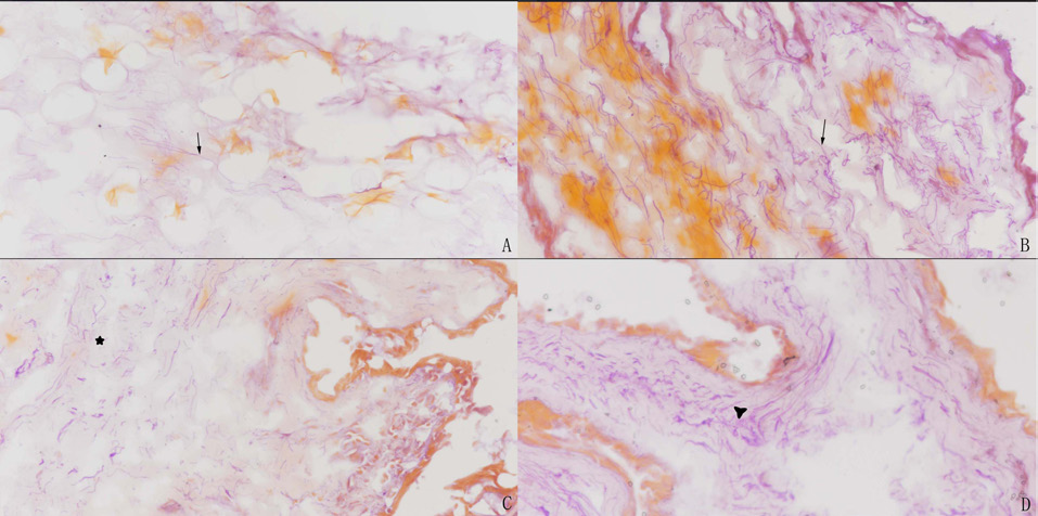
对照组(A组)和伪造模组(B组)光镜下未见弹力纤维破坏,MMP-1组(C组)光镜下见弹力纤维破坏,MMP-3组(D组)光镜下见弹力纤维部分破坏。见图7。
20230419100317_7674.png
图7 球结膜组织弹力纤维染色(×200)
Figure 7 Dyeing of elastic fibers in bulbar conjunctival tissue
(A)对照组;(B)伪造模组;(C)MMP-1组;(D)MMP-3组。图A、B黑色细箭头示弹力纤维染成紫色,纤细、完整,走行自然;图C黑色五角星示弹力纤维破裂成细小段,成簇分布;图D黑色三角星左侧区域见弹力纤维碎片,右侧区域尚可见完整弹力纤维。
(A) Control group; (B) Pseudo Model group; (C) MMP-1 group; (D) MMP-3 group. In Figure A and B, the black arrows show that the elastic fibers are dyed purple, slim, complete and walk naturally. In Figure C, the black five-pointed star shows that the elastic fibers are broken into small segments and distributed in clusters. In Figure D, the elastic fiber fragments can be seen in the left area of the black triangle star, and the intact elastic fiber can be seen in the right area.

3 讨论

到目前为止,构建CCH疾病动物模型的问题尚没有明确答案。该病发病相关的因素包括:老龄化[3]、基因突变[4]、下睑缘张力增高[5]、细胞凋亡[6]、紫外线辐射[7]、氧化应激[8]、屈光不正和眼轴长度[9]、眼泪的渗透压、热休克蛋白[10]、佩戴角膜接触镜(干燥和摩擦引起结膜炎症)[11]、自身免疫性甲状腺疾病(机体过度表达炎症因子)[12]等。上述病因诱使炎症因子增多,导致MMP与基质金属蛋白酶组织抑制因子(tissue inhibitor of metalloproteinase,TIMP)的平衡失调,或者直接影响MMP活性(譬如老龄化[13]),引起弹力纤维的降解变性。结膜固有层中弹力纤维明显减少可引起球结膜弹性下降、张力降低。进而球结膜与眼球的附着力下降,在下睑缘的反复挤压下形成皱褶[14]。结膜皱褶突出于眼球表面曲面,影响眼泪的流动和泪河的形成[15]。引起结膜松弛症患者的泪液排泄延缓[16]。泪液清除延迟导致泪液中炎症因子大量堆积,形成恶性循环,致结膜松弛不断进展。
根据前述炎症因子-MMP-弹力纤维降解机制,我们提出假设:可以通过球结膜下注射MMP来直接破坏动物结膜基质层的弹力纤维,从而得到CCH的动物模型。
同样,评价疾病动物模型是否成功的问题也没有明确的答案。CCH的临床表现为松弛结膜形成的皱褶影响泪河以及泪膜的稳定性,导致患者出现眼部干涩、泪、烧灼感、异物感等不适症状。相关症状与体征可归结为2大类:一类为眼部不适症状,一类为特征性的体征。由此,我们提出以特征性的体征和眼部不适症状作为CCH造模动物是否成功的标准。
松弛结膜形成的皱褶为CCH特征性的临床体征,而结膜皱褶可以通过仪器直接观测并留取影像。本研究中我们采取了2种仪器来观测。眼前节照相仪直接对实验对象-兔眼进行拍照和观察,可以直接观察到有无结膜皱褶的形成。眼前节光学相干断层扫描(AS-OCT)则能对实验动物的结膜囊成像,所提供的结膜囊横截面图可以直接观察到结膜细小皱褶的形成。
症状是患者对自身主观不适的一种描述,动物是无法发声和沟通的。评价实验动物是否有眼部不适症状是一个难题。患者眼部不适症状是由松弛结膜形成的皱褶影响泪河以及泪膜的稳定性所造成的,症状的发生和泪河、泪膜的关系密切。由此,我们可以反过来通过检测造模动物泪河、泪膜的改变来推测其主观不适症状。这样动物主观不适症状评估的问题就被转变成泪河和泪膜的客观检测问题。
临床上常用的泪河、泪膜的客观检测指标有泪膜破裂时间、泪液分泌实验、泪液蕨类实验。泪液分泌实验主要检测的是泪腺及副泪腺的分泌功能,与CCH的泪河以及泪膜的不稳定性机制无关,不适合作为检测指标。泪膜破裂时间反映的是泪膜的稳定性,适合作为检测指标。它的测量有裂隙灯下荧光素染色和干眼综合仪这两种方法,检测过程中均需主动眨眼。由于实验动物无法配合该动作,检测难以完成。文献资料显示,泪液中盐类和高分子物质(包括蛋白和黏蛋白)是影响泪液蕨样结晶的决定因素[17] ,TFT试验间接反映了泪液黏蛋白的功能,黏蛋白是泪膜的重要组成部分。结膜松弛症患者的泪液中羊齿状结晶明显减少,出现黏蛋白异常[18-19]。且该实验方法易于操作,重复性好,故其适合作为检测指标。
临床结膜松弛症的诊断和治疗效果的判定均无需结膜的活组织检查(活检),故结膜组织的光镜检测结果不适合作为造模是否成功的标准,仅可作为评估标准的辅助和参考。
对照组和伪造模组的实验结果说明无干预措施及生理盐水球结膜下注射不会对结膜的解剖结构及泪膜性状造成改变,同时表明其他两组动物结膜改变的原因来自实验药物。
MMP-3组造成泪液蕨类试验结果部分改变,光镜下见胶原和弹力纤维部分破坏,但未观察到结膜皱褶形成,该方法构建CCH动物模型失败。MMP-1组观察到结膜皱褶形成和泪液蕨类试验结果改变,这些改变符合前述结膜松弛症模型的评估标准。光镜下见胶原和弹力纤维破坏,也符合临床对CCH的认识[20]。总之,球结膜下注射MMP-1可构建结膜松弛症的动物模型。
临床上根据CCH的结膜皱褶的严重程度将CCH患者分为4级:1级为细小单层皱褶未超过泪河高度;2级为明显多层皱褶超过泪河高度;3级为皱褶骑跨或覆盖下睑缘;4级为松弛结膜皱褶影响眼睑闭合。本次动物实验结果按此原则可分类为1~2级[21],而实验设计模型结果为3~4级。两者之间差异的原因推测如下:1)球结膜下注射MMP后药物会被吸收入血,在组织中的浓度按照半衰期的规律而衰减,不能长时间在组织中维持一个较高的有效浓度;2)球结膜下注射MMP是通过MMP直接破坏球结膜下的细胞外基质,这是一个外源性MMP造成组织损伤的过程,和结膜松弛症的炎症因子-MMPs-细胞外基质降解机制不完全相符。
未来我们将进一步完善CCH的动物模型:1)尝试构建缓释给药系统,系统在组织中均匀缓慢的释出药物,从而在组织中保持一个能发挥细胞外基质降解的有效浓度。如将MMP和透明质酸共同注入球结膜下,以使药物缓慢吸收入血;2)尝试球结膜下注射致炎因子(如脂多糖等),诱发组织无菌性炎症,结膜组织自身产生高浓度的内源性MMP来降解细胞外基质。

利益冲突

所有作者均声明不存在利益冲突

开放获取声明

本文适用于知识共享许可协议(Creative Commons),允许第三方用户按照署名(BY)-非商业性使用(NC)-禁止演绎(ND)(CC BY-NC-ND)的方式共享,即允许第三方对本刊发表的文章进行复制、发行、展览、表演、放映、广播或通过信息网络向公众传播,但在这些过程中必须保留作者署名、仅限于非商业性目的、不得进行演绎创作。详情请访问:https://creativecommons.org/licenses/by-nc-nd/4.0/。
1、Zhang X, Li Q, Zou H, et al. Assessing the severity of conjunctivochalasis in a senile population: a community-based epidemiology study in Shanghai, China[ J]. BMC Public Health, 2011, 11: 198.Zhang X, Li Q, Zou H, et al. Assessing the severity of conjunctivochalasis in a senile population: a community-based epidemiology study in Shanghai, China[ J]. BMC Public Health, 2011, 11: 198.
2、Gan JY, Li QS, Zhou HM, et al. A preliminar y study on the establishment of an animal model of conjunctivochalasis[ J]. Int J Ophthalmol, 2018, 11(6): 899-904.Gan JY, Li QS, Zhou HM, et al. A preliminar y study on the establishment of an animal model of conjunctivochalasis[ J]. Int J Ophthalmol, 2018, 11(6): 899-904.
3、Mimura T, Yamagami S, Usui T, et al. Changes of conjunctivochalasis with age in a hospital-based study[ J]. Am J Ophthalmol, 2009, 147(1): 171-177.e1. 45Mimura T, Yamagami S, Usui T, et al. Changes of conjunctivochalasis with age in a hospital-based study[ J]. Am J Ophthalmol, 2009, 147(1): 171-177.e1. 45
4、Kantaputra PN, Kaewgahya M, Wiwatwongwana A, et al.Cutis laxa with pulmonary emphysema, conjunctivochalasis, nasolacrimal duct obstruction, abnormal hair, and a novel FBLN5 mutation[ J]. Am J Med Genet A, 2014, 164A(9):2370-2377.Kantaputra PN, Kaewgahya M, Wiwatwongwana A, et al.Cutis laxa with pulmonary emphysema, conjunctivochalasis, nasolacrimal duct obstruction, abnormal hair, and a novel FBLN5 mutation[ J]. Am J Med Genet A, 2014, 164A(9):2370-2377.
5、严雅静, 张兴儒, 项敏泓, 等. 结膜松弛症下睑缘位置及张力观察[ J]. 国际眼科杂志, 2009, 9(3): 495-497.
Yan YJ, Zhang XR, Xiang MH, et al. The observation of lower eyelid location and tension of conjunctivochalasis [ J]. Int J Ophthalmol, 2009, 9 (3): 495-497.
严雅静, 张兴儒, 项敏泓, 等. 结膜松弛症下睑缘位置及张力观察[ J]. 国际眼科杂志, 2009, 9(3): 495-497.
Yan YJ, Zhang XR, Xiang MH, et al. The observation of lower eyelid location and tension of conjunctivochalasis [ J]. Int J Ophthalmol, 2009, 9 (3): 495-497.
6、张兴儒,项敏泓,吴庆庆,等. 结膜松弛症患者泪液蛋白质组学研究[ J]. 中华眼科志,2009,45(2):135-140.
Zhang XR, Xiang MH, Wu QQ, et al. The tear proteomics analysis of conjunctivochalasis[ J]. Chin J Ophthalmol, 2009, 45(2): 135-140.
张兴儒,项敏泓,吴庆庆,等. 结膜松弛症患者泪液蛋白质组学研究[ J]. 中华眼科志,2009,45(2):135-140.
Zhang XR, Xiang MH, Wu QQ, et al. The tear proteomics analysis of conjunctivochalasis[ J]. Chin J Ophthalmol, 2009, 45(2): 135-140.
7、Mimura T, Mori M, Obata H, et al. Conjunctivochalasis: associations with pinguecula in a hospital-based study[ J]. Acta Ophthalmol, 2012, 90(8): 773-782.Mimura T, Mori M, Obata H, et al. Conjunctivochalasis: associations with pinguecula in a hospital-based study[ J]. Acta Ophthalmol, 2012, 90(8): 773-782.
8、Ward SK, Wakamatsu TH, Dogru M, et al. e role of oxidative stress and inammation in conjunctivochalasis[ J]. Invest Ophthalmol Vis Sci, 2010, 51(4):1994-2002.Ward SK, Wakamatsu TH, Dogru M, et al. e role of oxidative stress and inammation in conjunctivochalasis[ J]. Invest Ophthalmol Vis Sci, 2010, 51(4):1994-2002.
9、Mimura T, Usui T, Yamagami S, et al. Relationship between conjunctivochalasis and refractive error[ J]. Eye Contact Lens, 2011, 37(2):71-78.Mimura T, Usui T, Yamagami S, et al. Relationship between conjunctivochalasis and refractive error[ J]. Eye Contact Lens, 2011, 37(2):71-78.
10、刘晔翔, 李轶捷, 张兴儒, 等. 结膜松弛症球结膜组织中热休克蛋白的表达[ J]. 中华眼科杂志, 2010, 46(8):743-745.
Liu YX, Li YJ, Zhang XR, et al. Expression of heat shock protein in conjunctival tissue of conjunctivochalasis [ J]. Chin J Ophthalmol, 2010, 46(8):743-745.
刘晔翔, 李轶捷, 张兴儒, 等. 结膜松弛症球结膜组织中热休克蛋白的表达[ J]. 中华眼科杂志, 2010, 46(8):743-745.
Liu YX, Li YJ, Zhang XR, et al. Expression of heat shock protein in conjunctival tissue of conjunctivochalasis [ J]. Chin J Ophthalmol, 2010, 46(8):743-745.
11、Mimura T, Usui T, Yamamoto H, et al. Conjunctivochalasis and contact lenses[ J]. Am J Ophthalmol, 2009, 148(1):20-25.Mimura T, Usui T, Yamamoto H, et al. Conjunctivochalasis and contact lenses[ J]. Am J Ophthalmol, 2009, 148(1):20-25.
12、de Almeida SF, de Sousa LB, Vieira LA, et al. Clinic-cytologic study of conjunctivochalasis and its relation to thyroid autoimmune diseases: prospective cohort study[ J]. Cornea, 2006, 25(7):789-793.de Almeida SF, de Sousa LB, Vieira LA, et al. Clinic-cytologic study of conjunctivochalasis and its relation to thyroid autoimmune diseases: prospective cohort study[ J]. Cornea, 2006, 25(7):789-793.
13、Wang M, Kim SH, Monticone RE, et al. Matrix metalloproteinases promote arterial remodeling in aging, hypertension, and atherosclerosis[J]. Hypertension, 2015, 65(4): 698-703. Wang M, Kim SH, Monticone RE, et al. Matrix metalloproteinases promote arterial remodeling in aging, hypertension, and atherosclerosis[J]. Hypertension, 2015, 65(4): 698-703.
14、Otaka I, Kyu N. A new surgical technique for management of conjunctivochalasis[J]. Am J Ophthalmol, 2000, 129(3):385-387. Otaka I, Kyu N. A new surgical technique for management of conjunctivochalasis[J]. Am J Ophthalmol, 2000, 129(3):385-387.
15、张兴儒,刘晔翔,许琰,等. 结膜松弛症的泪液学观察[J]. 中国眼耳鼻喉科杂志, 2002, 2(6): 364-365, 374.
Zhang XR, Liu YX, Xu Y, et al. The observation on lacrimal fluid in conjunctivochalasis [J]. Chin J Ophthalmol Otolaryngology, 2002, 2(6): 364-365, 374.
张兴儒,刘晔翔,许琰,等. 结膜松弛症的泪液学观察[J]. 中国眼耳鼻喉科杂志, 2002, 2(6): 364-365, 374.
Zhang XR, Liu YX, Xu Y, et al. The observation on lacrimal fluid in conjunctivochalasis [J]. Chin J Ophthalmol Otolaryngology, 2002, 2(6): 364-365, 374.
16、李青松, 杨振燕, 张兴儒, 等. 结膜松弛症泪液排泄系统99mTc-SPECT动态显像的临床研究[J]. 同济大学学报(医学版), 2006, 27(4): 56-60.
Li QS, Yang ZY, Zhang XR, et al. Clinical study of 99mTc SPECT dynamic imaging of tear excretory system in conjunctivochalasis [J]. Journal of Tongji University (Medical Edition), 2006, 27 (4): 56-60
李青松, 杨振燕, 张兴儒, 等. 结膜松弛症泪液排泄系统99mTc-SPECT动态显像的临床研究[J]. 同济大学学报(医学版), 2006, 27(4): 56-60.
Li QS, Yang ZY, Zhang XR, et al. Clinical study of 99mTc SPECT dynamic imaging of tear excretory system in conjunctivochalasis [J]. Journal of Tongji University (Medical Edition), 2006, 27 (4): 56-60
17、Golding TR, Baker AT, Rechberger J, et al. X-ray and scanning electron microscopic analysis of the structural composition of tear ferns[J]. Cornea, 1994, 13(1):58-66. Golding TR, Baker AT, Rechberger J, et al. X-ray and scanning electron microscopic analysis of the structural composition of tear ferns[J]. Cornea, 1994, 13(1):58-66.
18、项敏泓, 张兴儒, 蔡瑞霞, 等. 结膜松弛症泪液中羊齿状结晶的观察[J]. 眼科, 2008, 17(1): 37-39.
Xiang MH, Zhang XR, Cai RX, et al. The observation of tear ferning in conjunctivochalasis [J]. Ophthalmology, 2008, 17(1): 37-39.
项敏泓, 张兴儒, 蔡瑞霞, 等. 结膜松弛症泪液中羊齿状结晶的观察[J]. 眼科, 2008, 17(1): 37-39.
Xiang MH, Zhang XR, Cai RX, et al. The observation of tear ferning in conjunctivochalasis [J]. Ophthalmology, 2008, 17(1): 37-39.
19、Zhang X,Li Q,Xiang M,et al.Analysis of tear mucin and goblet cells in patients with conjunctivochalasis[J].Spektrum Der Augen-heilkd,2010,24( 4) : 206-213.Zhang X,Li Q,Xiang M,et al.Analysis of tear mucin and goblet cells in patients with conjunctivochalasis[J].Spektrum Der Augen-heilkd,2010,24( 4) : 206-213.
20、李轶捷, 张兴儒, 项敏泓, 等. 结膜松弛症球结膜及筋膜组织的超微结构观察[J]. 中华实验眼科杂志, 2012, 30(7): 638-640.
Li YJ, Zhang XR, Xiang MH, et al. Ultrastructure change of conjunctiva and fascia tissue of conjunctivochalasis [J]. Chin J Exp Ophthalmol, 2012, 30(7): 638-640.
李轶捷, 张兴儒, 项敏泓, 等. 结膜松弛症球结膜及筋膜组织的超微结构观察[J]. 中华实验眼科杂志, 2012, 30(7): 638-640.
Li YJ, Zhang XR, Xiang MH, et al. Ultrastructure change of conjunctiva and fascia tissue of conjunctivochalasis [J]. Chin J Exp Ophthalmol, 2012, 30(7): 638-640.
21、Zhang XR, Zou HD, Li QS, et al. Comparison study of two diagnostic and grading systems for conjunctivochalasis[J]. Chin Med J (Engl), 2013, 126(16): 3118-3123. Zhang XR, Zou HD, Li QS, et al. Comparison study of two diagnostic and grading systems for conjunctivochalasis[J]. Chin Med J (Engl), 2013, 126(16): 3118-3123.
1、上海市普陀区卫生系统自主创新项目(PTKWWS201812);上海市卫生健康委员会科研课题(202040418)。This work was supported by the Health System Independent Innovation Project of Shanghai Putuo District (PTKWWS201812) and Research Project of Shanghai Municipal Health Commission(202040418).()
上一篇
下一篇
其他期刊
  • 眼科学报

    主管:中华人民共和国教育部
    主办:中山大学
    承办:中山大学中山眼科中心
    主编:林浩添
    主管:中华人民共和国教育部
    主办:中山大学
    浏览
  • Eye Science

    主管:中华人民共和国教育部
    主办:中山大学
    承办:中山大学中山眼科中心
    主编:林浩添
    主管:中华人民共和国教育部
    主办:中山大学
    浏览
推荐阅读
出版者信息
目录