您的位置: 首页 > 2021年12月 第36卷 第12期 > 文字全文
2023年7月 第38卷 第7期11
目录

特发性黄斑前膜在光学相干断层扫描上的形态学改变与视力的相关性

Correlation between morphological changes on optical coherence tomography and visual acuity in idiopathic macular epiretinal membranes

来源期刊: 眼科学报 | 2021年12月 第36卷 第12期 977-983 发布时间:2021-12 收稿时间:2023/5/9 10:15:08 阅读量:3834
作者:
关键词:
特发性黄斑前膜光学相干断层成像视网膜厚度囊样改变囊样水肿
idiopathic macular epiretinal membrane optical coherence tomography retina thickness cystic change cystic edma
DOI:
10.3978/j.issn.1000-4432.2021.11.03
收稿时间:
 
修订日期:
 
接收日期:
 
目的:探究特发性黄斑前膜(idiopathic macular epiretinal membrane,IMEM)患者在光学相干断层扫描(optical coherence tomography,OCT)的形态学改变与视力的相关性。方法:回顾性研究。基于OCT图像4级分级方案,纳入3级及以下的特发性黄斑前膜患者35例共37只眼进行OCT扫描,并借助第三方软件分析并获取每只眼在黄斑中心凹直径分别为1、3、6 mm共3个同心圆区域内视网膜各层的平均厚度值,并按照是否存在内核层(inner nuclear layer,INL)囊样改变分为两组。通过建立最小分辨角对数(minimum resolution angle in logarithmic,logMAR)最佳矫正视力(best corrected vision acuity,BCVA)与视网膜各层厚度值的多元线性回归方程,以及比较有INL囊样改变及无INL囊样改变两组的视力,分析视力与视网膜各层厚度值及INL囊样改变的关系。结果:多元线性回归分析显示,在直径1 mm的区域内,logMAR BCVA与神经纤维层(retina nerve fiber layer,RNFL)、INL的厚度值均呈正相关(均P<0.05),而在直径3 mm和6 mm的区域内,logMAR BCVA仅与INL的厚度值呈正相关(均P<0.05)。与无INL囊样改变组相比,有INL囊样改变组视力及INL厚度差异均有统计学意义(均P<0.05)。结论:特发性黄斑前膜患者当前的视力主要与RNFL和INL厚度值相关。发生在INL层的囊样改变可能很好的提示INL增厚,其与视力具有相关性,具有这种囊样改变的患者往往视力也较差。
Objective: To investigate the morphological changes detected by spectral domain optical coherence tomography (OCT) and their relevance to vision in patients with idiopathic macular epiretinal membranes (IMEM). Methods: This is a retrospective study. OCT recorded 37 eyes with stage 3 and below IMEM according to the 4-level grading system based on OCT images among 35 patients. We managed to obtain thickness of all retina layers in three concentric circular regions with a diameter of 1 mm, 3 mm and 6 mm across macular fovea with the help of a third-party software. Plus we divided all 37 eyes into two groups according to whether there were cystic changes in the inner nuclear layer (INL). Multiple linear regression analyses were performed between thickness of all retina layers and current minimum resolution angle in logarithmic (logMAR) best corrected visual acuity (BCVA). The difference of BCVA between groups with cystic changes and without was also compared. Thus, the relationships between visual acuity and thickness of retina layers, cystic changes in INL were analyzed. Results: Multiple linear regression analyses revealed that thickness of both retina nerve fiber layer (RNFL) and inner nuclear layer have positive correlation with logMAR BCVA in the region with a diameter of 1 mm (P<0.05), while in regions of a diameter of 3 mm and 6 mm, only INL thickness remained positively relevant (all P<0.05). There were significant differences in BCVA and thickness of INL between groups with INL cystic changes and without (all P<0.05). Conclusion: Current visual acuity among preoperative IMEM patients was mostly associated with thickness of RNFL and INL. Cystic change in INL layer may be a good indicator of INL thickening and was visually correlated. Those with this change tend to have worse vision.
黄斑前膜是一种发生在黄斑部玻璃体视网膜交界面的非血管性纤维细胞膜。特发性黄斑前膜是特指其中发病原因无法明确的类型。
临床发现很多特发性黄斑前膜患者存在不同程度的视物变形及视力下降的症状,但同时也观察到少部分患者没有症状或者症状十分轻微。从光学相干断层扫描(optical coherence tomography,OCT)图像的改变来看,后者在形态上往往受累程度较轻,提示患者的视功能损害程度与OCT改变存在一定的相关性。既往许多研究也证实了这一点。Joe等[1]发现视力与视网膜神经纤维层(retina nerve fiber layer,RNFL)、节细胞层(ganglion cell layer,GCL)、内核层(inner nuclear layer,INL)等的厚度相关,Cacciamani等[2]也有类似的发现,但后者发现外丛状层(outer plexiform layer,OPL)及外核层(outer nuclear layer,ONL)也具有相关性;且Cacciamani等[3]近来研究发现:前膜患者术后视力提高的程度与INL及OPL的厚度值降低程度呈正相关。与这截然不同的是,Okamoto等[4]却发现GCL、INL、和ONL + OPL等的厚度值与视力无关。国内宁玲[5]发现:黄斑前膜患者的术前最佳矫正视力(best corrected vision acuity,BCVA)与术前黄斑中心凹的厚度呈负相关(r=?0.638)。国内汪向利等[6]发现:黄斑前膜患者的视力与黄斑中心光感受器细胞层完整性和光感受器外节长度明显相关。这些研究的结果有冲突的地方,但似乎都有根有据,也合情合理。本研究旨在进一步明确黄斑前膜患者视网膜各层厚度值与视力的相关性以及相关性程度。此外,既往文献[7-9]报道:发生在黄斑区中心凹INL的囊样改变(囊样水肿)与视力具有相关性,本研究也一并进行了探讨及验证。

1 对象与方法

1.1 对象

选取自2017年10月至2018年11月在湘雅医院眼科门诊就诊,经过详细病史采集,裂隙灯检查,散瞳后前置镜检查及OCT(德国蔡司Cirrus 5000 HD-OCT)检查明确诊断为特发性黄斑前膜患者35例37只眼作为研究对象。其中男9例9眼,女26例28眼,患者年龄(64.1±7.0)岁。纳入标准:1)年龄>18岁,智力正常,自愿且可以配合验光及OCT检查。2)OCT图像明确显示视网膜表面有符合前膜的带状高反射信号,且黄斑前膜分级均在Govetto等[10]分级方案中三级及以下。3)所采集的眼部OCT图像位置均符合本研究标准,即采集的6 mm ×6 mm的区域均是以黄斑最中心凹为中心。排除标准:1)存在角膜斑翳/白斑,晶体明显混浊(晶状体混浊程度国际分级标准[11]3级及以上),玻璃体积血等屈光介质混浊明显影响视力的患者;2)继发性黄斑前膜(眼外伤、眼内炎症、眼内肿瘤或者眼内手术);3)高度近视(球镜度数>?6.0D或者眼轴>26 mm);4)合并糖尿病性视网膜病变、年龄相关性黄斑变性等视网膜疾病,青光眼及其他眼底疾病患者。

1.2 方法

1.2.1 数据采集
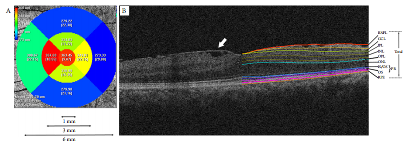
所有患者进行BCVA、裂隙灯、+90D前置镜(散瞳后)、OCT等检查。以黄斑中心凹为中心,应用Macular Cube 512×128模式对所有前膜眼进行扫描,以获取6 mm ×6 mm范围内黄斑区视网膜数据。将所获取的OCT源文件运用Iowa Reference Algorithm (Version 3.8)进行读取及运算,从而获取视网膜各层在直径分别为1、3、6 mm的3个同心圆,共9个亚分区(1个圆形区域,8个扇形区域)内的平均厚度值(图1)。
20230509092218_3506.png
图1 分区及视网膜分层示意图
Figure 1 Image of regions dividing and retina segmenting
(A) Iowa Reference Algorithm将OCT采集的区域自动分成9个亚分区并测量每个分区的平均厚度值。这9个分区同时构成了3个直径分别为1、3、6 mm的同心圆区域。(B)1例前膜患者黄斑区OCT图像及视网膜分层示意图。左半部分可以看到视网膜表面的前膜组织(白色箭头)。右半部分为同一只眼经软件自动分层后的各视网膜层次。IS/OS:光感受器的内节/外节交界处;OS:外节;RPE:色素细胞层。
(A) The Iowa Reference Algorithm automatically divided the area obtained by OCT into nine subfields and measured the mean thickness of each subfield. These nine subfields also constituted three concentric circular regions with a diameter of 1, 3 and 6 mm. (B) Image of OCT in macular of a IMEM patient and retina segmenting. The left part showed membrane tissue above the retina (white arrow). The right part displayed all the retina layers after segmentation by software in the same eye. IS/OS: photoreceptor inner/outer segments; OS: outer segment; RPE: retinal pigment epithelium.
1.2.2 根据中心凹INL是否有囊样改变分组
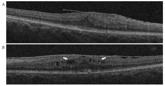
按OCT显示的中心凹处(1 mm)INL中是否存在肉眼可辨的囊样水肿改变将3 7只眼进一步分为两个亚组,即有囊样改变组,共11只;以及无囊样改变组,共26只眼(图2)。
20230509093449_8833.png
图2 INL层无囊样改变(A)及存在囊样改变(B)的黄斑前膜患者OCT图像,中心凹处INL层存在多个低信号的囊样改变区域(B;白色箭头)
Figure 2 OCT image of a IMEM patient without INL cysitc changes (A) and a IMEM patient with INL cysitc changes (B), there were many low signal cysitc areas (B; white arrows)

1.3 观察指标

1)BCVA,采用国际标准视力表检测所有观察眼的矫正视力,并转换为最小分辨角的对数值(minimum resolution angle in logarithmic,logMAR)以方便统计分析。2 )测量并分析在3个不同直径区域内RNFL、GCL、内丛状层(inner plexiform layer,IPL)、INL、OPL、ONL、光感受器层(photoreceptor,PR)的平均厚度。3)观察是否存在INL囊样改变,将37只眼分为有INL囊样改变组及非INL囊样改变组。

1.4 统计学处理

采用IBM SPSS Statistics 20.0软件进行数据分析。将所有观察眼的logMAR BCVA作为方程的因变量,以RNFL、GCL、IPL、INL、OPL、ONL、PR的厚度值作为自变量,分别在直径1、3、6 mm三个区域建立多因素线性回归方程。运用独立样本t检验比较INL囊样改变组和无INL囊样改变组的logMAR BCVA和1 mm区域内INL厚度值的差异。P<0.05为差异有统计学意义。

2 结果

2.1 视力与厚度值的多元线性回归分析

分别在直径1、3、6 mm区域,以logMAR BCVA作为因变量,以RNFL、GCL、IPL、INL、OPL、ONL、PR的厚度值作为可能的自变量,采用逐步策略,作多因素线性回归分析,建立的方程均具有统计学意义(均P<0.001)。
在直径1 mm区域,logMAR BCVA仅与RNFL和INL的厚度呈正相关,而与GCL、IPL、INL、OPL 、 P R等层次的厚度无关(表1) ,具体方程为:logMAR BCVA = 0.032 + 0.003 × RNFL + 0.003 × INL。
可以看到,方程的调整R2较为满意为0.53,即近53%的黄斑前膜人群的视力与厚度关系可以用该方程进行解释,且容忍度(tolerance)及方差膨胀因子(variance inflation factor,VIF)均接近于1,提示建立方程时引入的自变量之间共线性较弱,进一步提示所建立的方程具有很高的可信度。
在3 mm和6 mm区域以同样方式建立有效的多元线性回归分析(表2和表3),方程分别为:logMAR BCVA = ?0.407 + 0.013 × INL;logMAR BCVA = ?0.555 + 0.021 × INL。

表1 直径1 mm区域内logMAR BCVA与视网膜各层厚度的多元线性回归分析
Table 1 Multiple linear regression analysis of the correlation between logMAR BCVA and thickness of retinal layers in the region of a diameter of 1 mm

/20230509094125_3529.png

表2 直径3 mm区域内logMAR BCVA与视网膜各层厚度的多元线性回归分析
Table 2 Multiple linear regression analysis of the correlation between logMAR BCVA and thickness of retinal layers in the region of a diameter of 3 mm

20230509095806_8549.png

表3 直径6 mm区域内logMAR BCVA与视网膜各层厚度的多元线性回归分析
Table 3 Multiple linear regression analysis of the correlation between logMAR BCVA and thickness of retinal layers in the region of a diameter of 6 mm

20230509095838_7299.png

2.2 囊样改变组与非囊样改变组的比较

在37只前膜眼中,根据黄斑中心凹直径1 mm区域内INL层是否存在囊样(水肿)改变,将其分为有囊样改变组(11只眼)及非囊样改变组(26只眼),并比较两组之间的BCVA,以及1 mm范围内的INL厚度值。结果显示,两组差异均有统计学意义(P<0.05),其中囊样水肿组的BCVA较差,而INL厚度值更高(表4)。

表4 有INL囊样改变组与无囊样改变组INL厚度及logMAR BCVA的比较
Table 4 Comparison of INL thickness and logMAR BCVA between groups with INL cystic changes and without

20230509100001_2145.png

3 讨论

本研究确定了特发性黄斑前膜在OCT上的形态改变(厚度值及INL囊样改变)与视力损害的相关性,且将视网膜各层厚度这一指标进行了量化,并用方程这样一种直观的方式来体现哪层的厚度改变与视力损害最具相关性。
为进一步明确视力与视网膜厚度的关系,本研究分别在3个不同直径的同心圆区域建立了多元线性回归分析,得到的方程均具有统计学意义。且从调整R 2和容忍度,方差膨胀因子的数值来看,方程的可信度均较高。本研究发现:在中心凹处(1 mm)区域,logMAR BCVA与RNFL和INL的厚度值呈正相关,方程1显示,RNFL和INL的系数均为0.003,可以理解为RNFL和INL的厚度值每增加100 μm,黄斑前膜患者的BCVA(logMAR)会降低0.03。然而,在3 mm和6 mm区域,logMAR BCVA仅仅与INL的厚度相关,推测可能原因为RNFL的厚度值增加主要集中在1 mm区域,随着区域扩大,RNFL的平均厚度越来越被“稀释”,其具体数值越来越低,对视力的影响越来越小。由于logMAR BCVA的数值越小,BCVA值越大,即黄斑前膜患者的实际BCVA与RNFL及INL的厚度值大小呈负相关。这也许可以进一步解释具有INL囊样改变的黄斑前膜患者具有更高的INL厚度值,但BCVA值却较无囊样改变组更低。从建立的3个方程来看,每个具体的视力与厚度值可以一一对应,如果设定一个视力为损害的标准,甚至可以估算对应厚度的临界值,如假定该患者的视力<0.5,即定义为视力损害(小数视力),其对应的logMAR BCVA为0.3,按照方程1计算,对应的RNFL和INL值为89.3 μm。即中心凹处的RNFL和INL要至少增加到89.3 μm,前膜患者的视力才会有所明显下降。这也许可以解释临床中观察到一部分前膜患者视力损害非常轻微或者没有症状,推测黄斑前膜只有对视网膜损害达到一定程度时才会导致视力损伤。
关于RNFL及INL增厚导致黄斑前膜患者视力下降的具体病理生理学机制目前尚不能明确。在众多解释中,笔者倾向认可Reddy等[12]的观点,即来源于前膜本身的的牵拉会造成神经纤维层机械性延长,通过直接牵拉和/或微循环损伤介导,使得轴突损伤,导致视觉冲动传导受阻,从而造成视功能受损。至于INL增厚影响视力的原因,Okamoto等[4]推测,由于INL层主要是由水平细胞、双极细胞及无长突细胞等神经细胞构成,前膜的牵拉力量势必导致该层次的机械延长,结构的紊乱势必导致许多突触功能障碍。视觉冲动的传导因此会受到影响。另外,Ichikawa等[13-14]认为,INL的厚度值与黄斑前膜患者视物变形的程度呈正相关。视物变形在一定程度上也会影响视锐度,这也许也是一方面原因。
关于黄斑前膜患者发生的INL囊样改变,既往文献也有描述。Sigler等[9]发现4例前膜患者在术后1个月复诊时视力即有提升,然而术后3个月复诊时,由于发生了INL的囊样改变,视力随即下降,因而他们认为,这种囊样改变与视力具有相关性。本研究按是否存在INL囊样改变,将所有前膜眼分为有囊样改变眼和非囊样改变眼,并比较了两组间的BCVA和1 mm区域INL的厚度,最终发现这两个参数之间差异具有统计学意义。囊样改变组的平均logMAR视力明显高于非囊样改变组,囊样改变组的INL厚度也高于后者,即囊样改变与INL厚度值和BCVA具有相关性。它可能是INL增厚的一种原因,也可能是INL增厚的一种结局,提示囊样改变可能是一项反映INL增厚的良好指标。INL囊样改变提示INL厚度增加,这类患者视力可能更差,这与前面本研究多元分析多得到INL厚度与视力具有相关性的结论一致。关于INL囊样改变的病理机制尚不明确。Sigler[15]猜测这种改变可能是黄斑前膜对视网膜的机械牵拉继而引起该层细胞退行性改变及Muller功能障碍所导致。Abegg等[16]也认为,这种类似水肿的改变可能是INL层Muller细胞退行性改变继发液体吸收障碍所引起。关于这一方面有待进一步研究。
本研究主要描述了特发性黄斑前膜患者视网膜各层厚度的改变以及发生在INL的囊样改变,探讨了这两个参数与视力的相关性。通过建立多元回归分析方程,更加直观而且具体地阐述了视力与厚度的相关性以及相关性程度。此外,通过采用具备良好重复性的第三方自动分层软件[17-19],降低了来源于人工分层的误差,这一点有别于既往的类似研究[1-4]。本研究发现:特发性黄斑前膜患者视力与RNFL、INL的厚度值呈负相关。这对进一步理解黄斑前膜进展到视力损害阶段时发生的病理改变提供了更多的理论支持。发生在INL的囊样改变可以作为反映INL增厚的一种良好指标,其与视力也具有相关性,存在这种改变的患者,往往视力较差。对于存在囊样改变的患者,临床医师在术前谈话中可能需要留意,强调可能存在的不良预后,避免不必要的医疗风险。
本研究也有一些不足之处,包括样本量比较小。另外研究局限于术前的患者,缺乏术后分析,这使得手术预后评估方面显得不足。今后的研究应该纳入更多的术后患者,通过分析术后视力与术前OCT显示的相关视网膜结构(厚度值或者囊样改变等)参数的关系,尝试建立一个包含不同术前参数,甚至是类似本研究中线性方程一样的可量化的整体预估模型,以便通过术前的相关指标就可以预估IMEM患者术后视力恢复的情况。这不仅对于指导手术前谈话,降低医患纠纷风险具有重要参考价值,也为临床医师选择手术时机提供了理论依据。

开放获取声明

本文适用于知识共享许可协议(Creative Commons),允许第三方用户按照署名(BY)-非商业性使用(NC)-禁止演绎(ND)(CC BY-NC-ND)的方式共享,即允许第三方对本刊发表的文章进行复制、发行、展览、表演、放映、广播或通过信息网络向公众传播,但在这些过程中必须保留作者署名、仅限于非商业性目的、不得进行演绎创作。详情请访问:https://creativecommons.org/licenses/by-nc-nd/4.0/。
1、Joe SG, Lee KS, Lee JY, et al. Inner retinal layer thickness is the major determinant of visual acuity in patients with idiopathic epiretinal membrane[ J]. Acta Ophthalmol, 2013, 91(3): e242-e243.Joe SG, Lee KS, Lee JY, et al. Inner retinal layer thickness is the major determinant of visual acuity in patients with idiopathic epiretinal membrane[ J]. Acta Ophthalmol, 2013, 91(3): e242-e243.
2、Cacciamani A, Cosimi P, Di Nicola M, et al. Correlation between outer retinal thickening and retinal function impairment in patients with idiopathic epiretinal membranes[ J]. Retina, 2019, 39(2): 331-338.Cacciamani A, Cosimi P, Di Nicola M, et al. Correlation between outer retinal thickening and retinal function impairment in patients with idiopathic epiretinal membranes[ J]. Retina, 2019, 39(2): 331-338.
3、Cacciamani A, Cosimi P, Ripandelli G, et al. Epiretinal membrane surgery: structural retinal changes correlate with the improvement of visual function[ J]. J Clin Med, 2020, 10(1): 90.Cacciamani A, Cosimi P, Ripandelli G, et al. Epiretinal membrane surgery: structural retinal changes correlate with the improvement of visual function[ J]. J Clin Med, 2020, 10(1): 90.
4、Okamoto F, Sugiura Y, Okamoto Y, et al. Inner nuclear layer thickness as a prognostic factor for metamorphopsia after epiretinal membrane surgery[ J]. Retina, 2015, 35(10): 2107-2114.Okamoto F, Sugiura Y, Okamoto Y, et al. Inner nuclear layer thickness as a prognostic factor for metamorphopsia after epiretinal membrane surgery[ J]. Retina, 2015, 35(10): 2107-2114.
5、宁玲. 特发性黄斑前膜手术前后光学相干断层扫描形态变化与 视力的关系[ J]. 眼科新进展, 2017, 37(11): 1068-1070.
NING Ling. Correlation between morphological changes in the macula and visual acuity before and after idiopathic macular epiretinal surgery[ J]. Recent Advances in Ophthalmology, 2017, 37(11): 1068-1070.
宁玲. 特发性黄斑前膜手术前后光学相干断层扫描形态变化与 视力的关系[ J]. 眼科新进展, 2017, 37(11): 1068-1070.
NING Ling. Correlation between morphological changes in the macula and visual acuity before and after idiopathic macular epiretinal surgery[ J]. Recent Advances in Ophthalmology, 2017, 37(11): 1068-1070.
6、汪向利, 马建军. 特发性视网膜前膜术后视力恢复的两种预测 因素[ J]. 国际眼科杂志, 2018, 18(1): 166-168.
WANG Xiangli, MA Jianjun. Two structural predictors of visual outcome of idiopathic epiretinal membrane surgery[ J]. International Eye Science, 2018, 18(1): 166-168.
汪向利, 马建军. 特发性视网膜前膜术后视力恢复的两种预测 因素[ J]. 国际眼科杂志, 2018, 18(1): 166-168.
WANG Xiangli, MA Jianjun. Two structural predictors of visual outcome of idiopathic epiretinal membrane surgery[ J]. International Eye Science, 2018, 18(1): 166-168.
7、Chen SJ, Tsai FY, Liu HC, et al. Postoperative inner nuclear layer microcysts affecting long-term visual outcomes after epiretinal membrane surgery[ J]. Retina, 2016, 36(12): 2377-2383.Chen SJ, Tsai FY, Liu HC, et al. Postoperative inner nuclear layer microcysts affecting long-term visual outcomes after epiretinal membrane surgery[ J]. Retina, 2016, 36(12): 2377-2383.
8、Frisina R, Pinackatt SJ, Sartore M, et al. Cystoid macular edema after pars plana vitrectomy for idiopathic epiretinal membrane[ J]. Graefes Arch Clin Exp Ophthalmol, 2015, 253(1): 47-56.Frisina R, Pinackatt SJ, Sartore M, et al. Cystoid macular edema after pars plana vitrectomy for idiopathic epiretinal membrane[ J]. Graefes Arch Clin Exp Ophthalmol, 2015, 253(1): 47-56.
9、Sigler EJ, Randolph JC, Charles S. Delayed onset inner nuclear layer cystic changes following internal limiting membrane removal for epimacular membrane[ J]. Graefes Arch Clin Exp Ophthalmol, 2013, 251(7): 1679-1685.Sigler EJ, Randolph JC, Charles S. Delayed onset inner nuclear layer cystic changes following internal limiting membrane removal for epimacular membrane[ J]. Graefes Arch Clin Exp Ophthalmol, 2013, 251(7): 1679-1685.
10、Govetto A, Lalane RA 3rd, Sarraf D, et al. Insights into epiretinal membranes: presence of ectopic inner foveal layers and a new optical coherence tomography staging scheme[ J]. Am J Ophthalmol, 2017, 175: 99-113.Govetto A, Lalane RA 3rd, Sarraf D, et al. Insights into epiretinal membranes: presence of ectopic inner foveal layers and a new optical coherence tomography staging scheme[ J]. Am J Ophthalmol, 2017, 175: 99-113.
11、Chylack LT Jr, Wolfe JK , Singer DM, et al. The lens opacities classification system III. The longitudinal study of cataract study group[ J]. Arch Ophthalmol, 1993, 111(6): 831-836.Chylack LT Jr, Wolfe JK , Singer DM, et al. The lens opacities classification system III. The longitudinal study of cataract study group[ J]. Arch Ophthalmol, 1993, 111(6): 831-836.
12、Reddy RK, Lalezary M, Kim SJ, et al. Prospective Retinal and Optic Nerve Vitrectomy Evaluation (PROVE) study: findings at 3 months[ J]. Clin Ophthalmol, 2013, 7: 1761-1769.Reddy RK, Lalezary M, Kim SJ, et al. Prospective Retinal and Optic Nerve Vitrectomy Evaluation (PROVE) study: findings at 3 months[ J]. Clin Ophthalmol, 2013, 7: 1761-1769.
13、Ichikawa Y, Imamura Y, Ishida M. Inner nuclear layer thickness, a biomarker of metamorphopsia in epiretinal membrane, correlates with tangential retinal displacement[ J]. Am J Ophthalmol, 2018, 193: 20-27.Ichikawa Y, Imamura Y, Ishida M. Inner nuclear layer thickness, a biomarker of metamorphopsia in epiretinal membrane, correlates with tangential retinal displacement[ J]. Am J Ophthalmol, 2018, 193: 20-27.
14、Takabatake M, Higashide T, Udagawa S, et al. Postoperative changes and prognostic factors of visual acuity, metamorphopsia, and aniseikonia after vitrectomy for epiretinal membrane[ J]. Retina, 2018, 38(11): 2118-2127.Takabatake M, Higashide T, Udagawa S, et al. Postoperative changes and prognostic factors of visual acuity, metamorphopsia, and aniseikonia after vitrectomy for epiretinal membrane[ J]. Retina, 2018, 38(11): 2118-2127.
15、Sigler EJ. Microcysts in the inner nuclear layer, a nonspecific SD-OCT sign of cystoid macular edema[ J]. Invest Ophthalmol Vis Sci, 2014, 55(5): 3282-3284.Sigler EJ. Microcysts in the inner nuclear layer, a nonspecific SD-OCT sign of cystoid macular edema[ J]. Invest Ophthalmol Vis Sci, 2014, 55(5): 3282-3284.
16、Abegg M, Dysli M, Wolf S, et al. Microcystic macular edema: retrograde maculopathy caused by optic neuropathy[ J]. Ophthalmology, 2014, 121(1): 142-149.Abegg M, Dysli M, Wolf S, et al. Microcystic macular edema: retrograde maculopathy caused by optic neuropathy[ J]. Ophthalmology, 2014, 121(1): 142-149.
17、Song SJ, Lee MY, Smiddy WE. Ganglion cell layer thickness and visual improvement after epiretinal membrane surgery[ J]. Retina, 2016, 36(2): 305-310.Song SJ, Lee MY, Smiddy WE. Ganglion cell layer thickness and visual improvement after epiretinal membrane surgery[ J]. Retina, 2016, 36(2): 305-310.
18、Zou J, Tan W, Huang W, et al. Association between individual retinal layer thickness and visual acuity in patients with epiretinal membrane: a pilot study[ J]. PeerJ, 2020, 8: e9481Zou J, Tan W, Huang W, et al. Association between individual retinal layer thickness and visual acuity in patients with epiretinal membrane: a pilot study[ J]. PeerJ, 2020, 8: e9481
19、Chua J, Tham YC, Tan B, et al. Age-related changes of individual macular retinal layers among Asians[ J]. Sci Rep, 2019, 9(1): 20352.Chua J, Tham YC, Tan B, et al. Age-related changes of individual macular retinal layers among Asians[ J]. Sci Rep, 2019, 9(1): 20352.
上一篇
下一篇
其他期刊
  • 眼科学报

    主管:中华人民共和国教育部
    主办:中山大学
    承办:中山大学中山眼科中心
    主编:林浩添
    主管:中华人民共和国教育部
    主办:中山大学
    浏览
  • Eye Science

    主管:中华人民共和国教育部
    主办:中山大学
    承办:中山大学中山眼科中心
    主编:林浩添
    主管:中华人民共和国教育部
    主办:中山大学
    浏览
推荐阅读
出版者信息
目录