您的位置: 首页 > 2021年5月 第36卷 第5期 > 文字全文
2023年7月 第38卷 第7期11
目录

不同类型黄斑裂孔患病的相关因素

Related factors of various types of macular hole diseases

来源期刊: 眼科学报 | 2021年5月 第36卷 第5期 333-336 发布时间: 收稿时间:2023/5/17 11:22:55 阅读量:3526
作者:
关键词:
黄斑裂孔患病因素回顾性分析
macular hole disease factors retrospective analysis
DOI:
10.3978/j.issn.1000-4432.2021.04.09
收稿时间:
 
修订日期:
 
接收日期:
 
目的:观察并分析不同类型黄斑裂孔的患病相关影响因素。方法:回顾性分析2012年1月 至2018年12月在兰州大学第二医院住院治疗的1 150例(1 202只眼)全层黄斑裂孔患者的年龄、性别、民族、最佳矫正视力、黄斑裂孔类型以及是否继发孔源性视网膜脱离等临床资料。结果:黄斑裂孔患病人数随年龄增长呈趋势性分布,60~69岁年龄段患病人数最多;男女患病差异有统计学意义(P<0.05),眼别差异无统计学意义(P>0.05);特发性黄斑裂孔继发孔源性视网膜脱离的占比明显低于其他类型;特发性黄斑裂孔的入院最佳矫正视力较高,与高度近视黄斑裂孔相比,差异有统计学意义(P<0.05)。结论:黄斑裂孔多发于老年人,女性多见,高度近视黄斑裂孔容易继发孔源性视网膜脱离,可考虑尽早安排手术治疗;外伤性黄斑裂孔好发于中青年男性,常见于眼球钝挫伤,若未引发视网膜脱离等并发症,应适当观察而不急于手术。每1例黄斑裂孔都可能有其特殊性,需要认真分析病情综合判断,酌情选择观察或手术。
Objective: To observe and analyze the disease related factors of different types of macular hole. Methods: Clinical data including age, sex, ethnicity, best corrected visual acuity, macular hole type and whether accompanied by rhegmatogenous retinal detachment were retrospectively analyzed in 1 150 patients (1 202 eyes) hospitalized in our hospital from January 2012 to December 2018. Results: The number of patients with macular hole increased with age. There was statistically significant difference between male and female patients (P<0.05), and no statistically significant difference between eyes (P>0.05). The rate of rhegmatogenous retinal detachment caused by idiopathic macular hole was significantly lower than other types. The optimal corrected visual acuity of idiopathic macular hole was higher in admission, which was statistically different from that of high myopia macular hole and ametropia macular hole (P<0.05). Conclusion: Macular hole is more common in the elderly and more common in women. High myopia and ametropia macular hole are easily associated with rhegmatogenous retinal detachment, so surgical treatment should be arranged as soon as possible. Traumatic macular hole often occurs in young and middle-aged men and is often seen in blunt contusion of the eyeball. Each case of macular hole may have its particularity, which requires careful analysis of the condition, comprehensive analysis, and appropriate choice of observation or surgery.
黄斑裂孔是发生于黄斑中央或中心凹的神经知觉视网膜的解剖上不连续的状态[1],是指从视网膜内界膜到感光细胞层组织破裂的黄斑中心凹全层视网膜裂孔[2]。多数研究者[3-5]认为黄斑裂孔是由于黄斑中心凹发生病理性玻璃体视网膜牵拉而引起的。根据发病原因,目前临床上大致将其分为特发性和继发性两大类。继发性黄斑裂孔的原因主要有高度近视、外伤等。各类黄斑裂孔的病因、病程、治疗方案及预后均不尽相同,但在其诊断和治疗的过程中又存在共性的规律。本研究通过对特发性黄斑裂孔(idiopathic macular hole,IMH)、高度近视黄斑裂孔(屈光度≥-6.00D)、外伤性黄斑裂孔的发病特点和相关因素的总结和分析,进一步探讨黄斑裂孔的发病机制以及不同类型黄斑裂孔的差异,以期能为不同类型黄斑裂孔的个体化治疗方案提供一定的思路。

1 对象与方法

1.1 对象

本研究对2012年1月至2018年12月在兰州大学第二医院眼科住院治疗的全层黄斑裂孔患者1 150例(其中双眼患病52人,双眼均纳入研究)的临床资料及相干光断层扫描检查(optical coherence tomography,OCT)结果进行回顾性分析,准确记录患者入院的年龄、性别、民族、最佳矫正视力、黄斑裂孔类型及是否继发孔源性视网膜脱离等临床资料。

1.2 仪器与方法

使用国际标准对数视力表检查最佳矫正视力(best corrected visual acuity,BCVA),拓普康KR-800型电脑角膜验光仪[东京光学(东莞)科技有限公司]验光,裂隙灯显微镜检查眼前节,前置镜(90 D)检查眼底,使用Carl Zeiss公司CirrusTM HD-OCT 4000扫描仪测量黄斑裂孔大小。该仪器描绘分辨率:轴向5 μm,横向15 μm,具有黄斑中心凹自动居中Fovea FinderTM功能,自动寻找和定位黄斑中心凹的位置,确保了黄斑裂孔大小测量的精确性。采用高清5线高清扫描模式,以黄斑中心凹为中心的36°×30°范围进行扫描。

1.3 统计学处理

采用SPSS 16.0统计学软件进行数据分析,计量资料以均数±标准差(x±s)表示,采用应用χ2检验,计数资料以例(%)表示,采用F检验。P<0.05为差异有统计学意义。

2 结果

2.1 年龄

住院患者患病年龄最小为7岁,最大为83岁,患病人数随着年龄的增长呈现趋势性分布(Pearson2=3.232E2,P<0.001),其中以60~69岁年龄段患病人数最多,50~59岁次之,不同类型黄斑裂孔年龄分布详见表1。

表1 不同类型黄斑裂孔年龄分布情况
Table 1 Age distribution of macular holes of difffferent types

20230517111159_4186.png

2.2 性别

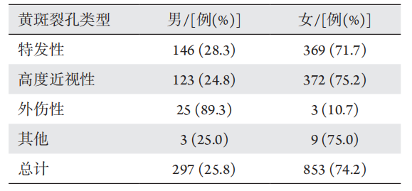
特发性、高度近视性和其他类型黄斑裂孔均为女性患者多,外伤性黄斑裂孔则为男性患者较多,男女患病差异有统计学意义( χ2=71.616,P<0.001;表2)。

表2 不同类型黄斑裂孔在不同性别的构成比
Table 2 Composition ratio of different types of macular holes in difffferent genders

20230517111325_9813.png

2.3 眼别

总计右眼590只眼,左眼560只眼,双眼52例,眼别差异无统计学意义(χ2=0.421,P=0.516),且不同类型黄斑裂孔患病眼别差异均无统计学意义。双眼患病患者共计52例,其中IMH患者8例,双眼患病占比为15.4%;高度近视性黄斑裂孔25例,占比48%,外伤性黄斑裂孔1例,占比2%,双眼患病率差异无统计学意义(χ2=5.649,P=0.171)。

2.4 民族

汉族共计1 055例(91.7%),回族63例(5.5%),藏族20例(1.7%),其他民族共计12例(1%),差异有统计学意义(χ2=44.623,P<0.001)。

2.5 继发孔源性视网膜脱离

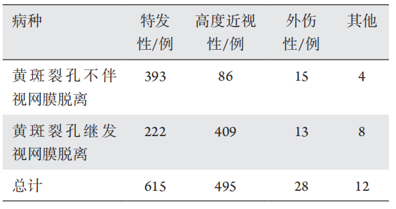
      IMH占比为7.3%,高度近视性为82.6%,外伤性为46.4%,其他类型为66.7%。IMH继发孔源性视网膜脱离的占比明显低于其他类型(表3)。

表3 不同类型黄斑裂孔继发孔源性视网膜脱离的比例
Table 3 Proportion of macular holes with retinal detachment of difffferent types

20230517111519_7003.png

2.6 视力

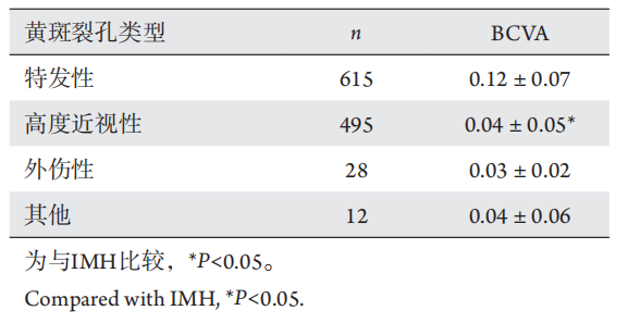
共纳入1 150例不同类型的黄斑裂孔患者的入院最佳矫正视力准确记录,统计各组入院患者BCVA,结果显示:IMH视力最佳(0.12±0.07),外伤性黄斑裂孔视力最差(0.03±0.02),完全随机分组F检验结果显示不同类型黄斑裂孔患者的入院视力差异有统计学意义(F=39.235,P<0.001),进一步做各组均数的两两比较,发现IMH与高度近视性黄斑裂孔的入院最佳矫正视力间,差异有统计学意义(P<0.05),其他任意两种类型黄斑裂孔差异均无统计学意义(P>0.05;表4)。

表4 不同类型黄斑裂孔入院视力差异
Table 4 Differences of visual acuity in different types of macular holes at admission

20230517111808_1381.png

3 讨论

随着眼科诊疗水平的日益提高,特别是OCT技术的广泛应用,黄斑裂孔的检出率大大提高。不同类型黄斑裂孔各有特点,兰州大学第二医院52.9%的IMH患者为60~69岁人群,其中81%为女性患者,无眼别差异,患病视力较其他类型稍高,该数据与国内外流行病学研究结果相符[6-7]。高度近视性黄斑裂孔容易继发视网膜脱离,是严重威胁高度近视患者视功能的常见并发症之一,这类黄斑裂孔可考虑尽早安排手术治疗。外伤性黄斑裂孔好发于中青年男性,其中眼球钝挫伤占82.1%,应重视详细的眼底检查,其发病率占闭合性眼外伤的1.4%[8],裂孔的形成可以是致伤原因的直接作用或外力的间接作用所致[9-10],外伤性黄斑裂孔有一定的自愈率,若不伴有视网膜脱离等并发症,确诊后不宜急于手术,应定期观察患眼黄斑OCT和视功能状况,如裂孔扩大或视力逐渐下降,则施行手术[11-12]。在其他类型黄斑裂孔中,多数与糖尿病视网膜病变(diabetic retinopathy,DR)和视网膜静脉阻塞(retinal vein occlusion,RVO)这两种眼病相关,DR继发性黄斑裂孔可发生于糖尿病黄斑水肿或牵拉性视网膜脱离,其形成原因可能与玻璃体视网膜牵拉、黄斑视网膜内渗出和中心凹萎缩变薄等多方面因素有关[13],手术中发现部分RVO继发性黄斑裂孔病例有玻璃体黄斑牵拉存在,但也考虑长期黄斑水肿和缺血对黄斑裂孔形成的影响。
综上所述,每一例黄斑裂孔都可能有其特殊性,需要认真分析病情,综合判断,酌情选择观察或手术治疗,本研究回顾性分析兰州大学第二医院黄斑裂孔患者的几项患病因素,以综合分析本地区各类型黄斑裂孔患者的患病特征,有助于探索各类型黄斑裂孔的形成机制从而指导临床治疗。
1、美国眼科学会. 眼科临床指南(第3版)[M]. 赵家良译. 北京: 人民 卫生出版社, 2013: 299.
American Academy of Ophthalmology. Clinical guidelines for ophthalmology (3rd edition)[M]. Translated by ZHAO Jialiang. Beijing: People’s Medical Publishing House, 2013: 299.
美国眼科学会. 眼科临床指南(第3版)[M]. 赵家良译. 北京: 人民 卫生出版社, 2013: 299.
American Academy of Ophthalmology. Clinical guidelines for ophthalmology (3rd edition)[M]. Translated by ZHAO Jialiang. Beijing: People’s Medical Publishing House, 2013: 299.
2、唐仕波, 李加青, 赖铭莹. 黄斑部疾病手术学[M]. 北京: 人民卫 生出版社, 2005: 212-229.
TANG SB, LI JQ, LAI MY. Surgical study of macular diseases[M]. Beijing: People’s Medical Publishing House, 2005: 212-229.
唐仕波, 李加青, 赖铭莹. 黄斑部疾病手术学[M]. 北京: 人民卫 生出版社, 2005: 212-229.
TANG SB, LI JQ, LAI MY. Surgical study of macular diseases[M]. Beijing: People’s Medical Publishing House, 2005: 212-229.
3、Gass JD. Idiopathic senile macular hole: its early stage and Pathogenesis[ J]. Arch Ophthalmol, 1988, 106: 629-639.Gass JD. Idiopathic senile macular hole: its early stage and Pathogenesis[ J]. Arch Ophthalmol, 1988, 106: 629-639.
4、Tognetto D, Grandin R . Sanguinetti G, et al. Internal limiting membrane removal during macular hole surgery: results of a multicenter retrospective study[ J]. Ophthalmology, 2006, 113: 1401-1410.Tognetto D, Grandin R . Sanguinetti G, et al. Internal limiting membrane removal during macular hole surgery: results of a multicenter retrospective study[ J]. Ophthalmology, 2006, 113: 1401-1410.
5、吕林. 进一步加强黄斑裂孔的临床研究, 努力提高黄斑裂孔的 治疗效果[ J]. 中华眼底病杂志, 2010, 11(26): 501-504.
Lü L. Clinical study on the treatment of macular hole[ J]. Chinese Journal of Ocular Fundus Diseases, 2010, 11(26): 501-504.
吕林. 进一步加强黄斑裂孔的临床研究, 努力提高黄斑裂孔的 治疗效果[ J]. 中华眼底病杂志, 2010, 11(26): 501-504.
Lü L. Clinical study on the treatment of macular hole[ J]. Chinese Journal of Ocular Fundus Diseases, 2010, 11(26): 501-504.
6、Wang S, Xu L, Jonas JB. Prevalence of full-thickness macular holes in urban and rural adult Chinese: the Beijing Eye Study[ J]. Am J Ophthalmol, 2006, 141(3): 589-591.Wang S, Xu L, Jonas JB. Prevalence of full-thickness macular holes in urban and rural adult Chinese: the Beijing Eye Study[ J]. Am J Ophthalmol, 2006, 141(3): 589-591.
7、The Eye Disease Case-Control Study Group. Risk factors for idiopathic macular holes[ J]. Am J Ophthalmol 1994, 118(6): 754-761.The Eye Disease Case-Control Study Group. Risk factors for idiopathic macular holes[ J]. Am J Ophthalmol 1994, 118(6): 754-761.
8、Lange C, Feltgen N, Junker B, et al. Resolving the clinical acuity categories “hand motion” and “counting fingers”, using the Freiburg Visual Acuity Test (FrACT)[ J]. Graefes Arch Clin Exp Ophthalmol, 2009, 247(1): 137-142.Lange C, Feltgen N, Junker B, et al. Resolving the clinical acuity categories “hand motion” and “counting fingers”, using the Freiburg Visual Acuity Test (FrACT)[ J]. Graefes Arch Clin Exp Ophthalmol, 2009, 247(1): 137-142.
9、Johnson RN, McDonald HR, Lewis H, et al. Traumatic macular hole: observations, pathogenesis, and results of vitrectomy surgery[ J]. Ophthalmology, 2002, 108: 853-857.Johnson RN, McDonald HR, Lewis H, et al. Traumatic macular hole: observations, pathogenesis, and results of vitrectomy surgery[ J]. Ophthalmology, 2002, 108: 853-857.
10、Yamashita T, Uemara A, Uchino E, et al. Spontaneous closure of traumatic macular hole[ J]. Am J Ophthalmol, 2002, 133: 230-235.Yamashita T, Uemara A, Uchino E, et al. Spontaneous closure of traumatic macular hole[ J]. Am J Ophthalmol, 2002, 133: 230-235.
11、Imai M, Ohshiro T, Gotoh T, et al. Spontaneous closure of stage 2 macular hole observed with optical coherence tomography[ J]. Am J Ophthamol, 2003, 136(1): 187-188.Imai M, Ohshiro T, Gotoh T, et al. Spontaneous closure of stage 2 macular hole observed with optical coherence tomography[ J]. Am J Ophthamol, 2003, 136(1): 187-188.
12、Sanjay S, Yeo TK, Au Eong KG. Spontaneous closure of traumatic macular hole[ J]. Saudi J Ophthalmol, 2012, 26(3): 343-345.Sanjay S, Yeo TK, Au Eong KG. Spontaneous closure of traumatic macular hole[ J]. Saudi J Ophthalmol, 2012, 26(3): 343-345.
13、Brazitikos PD, Stangos NT. Macular hole formation in diabetic retinopathy: the role of coexisting macular edema[ J]. Doc Ophthalmol, 1999, 97: 273-278.Brazitikos PD, Stangos NT. Macular hole formation in diabetic retinopathy: the role of coexisting macular edema[ J]. Doc Ophthalmol, 1999, 97: 273-278.
上一篇
下一篇
其他期刊
  • 眼科学报

    主管:中华人民共和国教育部
    主办:中山大学
    承办:中山大学中山眼科中心
    主编:林浩添
    主管:中华人民共和国教育部
    主办:中山大学
    浏览
  • Eye Science

    主管:中华人民共和国教育部
    主办:中山大学
    承办:中山大学中山眼科中心
    主编:林浩添
    主管:中华人民共和国教育部
    主办:中山大学
    浏览
推荐阅读
出版者信息
目录