您的位置: 首页 > 2021年6月 第36卷 第6期 > 文字全文
2023年7月 第38卷 第7期11
目录

温州青光眼进展研究报告之四:睡眠体位与其青光眼视神经损害及其进展的关系

No. 4 report of Wenzhou Glaucoma Progression Study: association between sleeping position and glaucomatous optic nerve damage and its progression

来源期刊: 眼科学报 | 2021年6月 第36卷 第6期 457-464 发布时间: 收稿时间:2023/5/25 17:38:11 阅读量:4548
作者:
关键词:
正常眼压性青光眼睡眠体位不对称视野损害
normal tension glaucoma sleeping position asymmetric visual field defect
DOI:
10.3978/j.issn.1000-4432.2021.06.13
收稿时间:
 
修订日期:
 
接收日期:
 
目的:探讨正常眼压性青光眼(normal tension glaucoma,NTG)患者睡眠体位与其双眼不对称损害的关系。方法:纳入2014年1月至2018年9月在温州青光眼进展研究(Wenzhou Glaucoma Progression Study,WGPS)项目中的NTG患者。眼部主要检查有视野和光学相干断层扫描(optical coherence tomography,OCT)。睡眠体位数据通过基线睡眠体位问卷获得。根据侧卧位睡眠偏好,将NTG患者眼部参数分为卧位高侧眼和卧位低侧眼进行讨论;根据双眼不对称损害,将患者眼部参数分为较好眼和较差眼讨论。双眼不对称损害定义为双眼视野平均偏差(mean deviation,MD)差值>6 dB或杯盘比差值>0.2。结果:共纳入158例NTG患者,最长随访时间为48个月,其中122例(77.22%)患者存在睡眠偏好;存在睡眠偏好的患者中,83例(68.03%)患者存在侧卧位偏好;存在侧卧位偏好的患者中,大多数患者偏好右侧卧位[右vs左:59 (71.1%) vs 24 (28.9%),P<0.001]。对存在侧卧位偏好的患者进行分析,发现卧位高侧眼与卧位低侧眼眼部参数之间,差异无统计学意义(P>0.05);卧位低侧眼的视野进展速率[视野指数(visual field index,VFI)、MD]慢于卧位高侧眼(0.48%±1.66%/年 vs ?0.45%±3.07%/年;0.54±0.96 dB/年 vs 0.2±1.15 dB/年),差异无统计学意义(P=0.086,P=0.308)。对同时存在侧卧位偏好及双眼不对称损害的患者进行分析,发现卧位高侧眼与卧位低侧眼的眼部参数之间,差异无统计学意义(P>0.05);卧位低侧较好眼的个数及占比高于卧位低侧较坏眼[23 (57.5%) vs 17(42.5%)],但差异无统计学意义(P=0.132);卧位低侧眼的视野进展速率(VFI、MD)也慢于卧位高侧眼(1.19%±1.65%/年 vs ?0.86%±3.65%/年;0.71±1.13 dB/年 vs0.13 dB/年),但差异无统计学意义(P=0.064,P=0.419)。结论:存在睡眠体位偏好的NTG患者中,约68%存在侧卧位偏好;存在侧卧位偏好的患者中,约70%偏好右侧卧位。但本研究并未发现睡眠体位与青光眼患者双眼不对称损害及其疾病进展存在相关性。
Objective: To investigate the association between lateral decubitus position (LDP) and asymmetric loss in normal tension glaucoma (NTG) patients. Methods: NTG patients were enrolled from Wenzhou Glaucoma Progression Study (WGPS) in Jan. 2014 to Sep. 2018. The main eye examinations included visual field test and optical coherence tomography (OCT). A questionnaire to determine the preferred sleeping position was administered to each patient in the baseline. According to the LDP, the eye parameters were divided into non-dependent eyes(higher lateral eyes) and dependent eyes (lower lateral position eyes) for discussion. According to the asymmetric damage, the ocular parameters of the patients were divided into the better eyes and the worse eyes for analysis.Asymmetric loss was defined as a difference in mean deviation (MD) between the 2 eyes of at least 6 dB or disc/cup >0.2. Results: One hundred and twenty-two patients (77.22%) had sleep preferences among the 158 NTG patients who was finally recruited and the longest follow up time was 48 months. Among the patients with sleep preference, 83 patients (68.03%) preferred the lateral decubitus position. Patients who had the lateral decubitus position mostly preferred the right lateral position [59 (71.1%) vs 24 (28.9%), P<0.001]. For patients who had the lateral decubitus position, the ocular parameters between the dependent eyes and the non-dependent eyes had no statistical difference(P>0.05); the rate of visual field progression in the dependent eyes was slower than that in non-dependent eyes, but there was no statistical difference between the two groups (0.48%±1.66%/year vs ?0.45%±3.07%/year; 0.54±0.96 dB/year vs 0.2±1.15 dB/year; P=0.086, P=0.308, respectively). For patients who had the lateral decubitus position and asymmetric damage, the ocular parameters between the dependent eyes and the non-dependent eyes had also no statistical difference (P>0.05); the number and ratio of the dependent-better eye and the dependent-worse eye were 48 and 41, respectively [23 (57.5%) vs 17 (42.5%), P=0.132]; the rate of visual field progression in the dependent eyes was also lower than that in non-dependent eyes, but there was no statistical difference between the two groups (1.19%±1.65%/year vs ?0.86%±3.65%/year; 0.71±1.13 dB/year vs 0.13 dB/year; P=0.064, P=0.419 respectively). Conclusion: About 68% of NTG patients with sleep preferences preferred the lateral decubitus position; and about 70% of patients with the lateral decubitus position preferred the right side sleeping. However, this study did not find a correlation between lateral decubitus position and asymmetric visual field loss.
人们一生中约1/4~1/3的时间在睡眠中度过[1],睡眠以其长期慢性的作用影响着人们的健康,特定的睡眠体位甚至可以作为疾病的辅助治疗方式。Lee等[2]的研究证明:当体位从平卧位变为侧卧位时,睡眠呼吸暂停综合征患者的呼吸阻塞症状可得到明显改善。青光眼是一个多因素疾病,体位改变会引起相应的眼压改变,且卧位时眼压明显高于直立位和坐位,睡眠体位可能与其疾病的发生发展有关[3-4]。人们的睡眠体位大致分为4种:仰卧位、左侧卧位、右侧卧位以及俯卧位。侧卧位时,较低侧眼眼压明显高于较高侧眼,睡眠体位改变可能是导致夜间眼压升高的重要因素[5-8]。但目前相关研究较少且存在争议,Kim等[9]的研究表明:侧卧睡姿可能导致青光眼患者不对称视野损害,且较低侧眼视野损害更严重。Tang等[10]也认为长期选择侧卧睡姿可能导致卧位低侧眼更严重的视野损害。然而,Kaplowitz等[11]对29名存在不对称视野损害的青光眼患者进行连续2晚的睡眠监测发现,喜欢右侧睡的患者左眼视野损害较重。
本研究基于温州青光眼进展研究( Wen zhou Glaucoma Progression Study,WGPS),它是我国首个以社区人群为基础,旨在为正常眼压性青光眼(normal tension glaucoma,NTG)的临床特征、自然病程、预后因素提供最直接证据的纵向队列研究[12]。前期我们已经对青光眼与日常体力活动的关系[13]、NTG患者视神经纤维厚度的影响因素[14]、原发性开角型青光眼患者视野缺损进展的危险因素[15]等进行了分析,本研究对青光眼患者睡眠偏好进行了问卷调查,拟对其睡眠体位与双眼不对称损害的关系进行进一步探讨。

1 对象与方法

1.1 对象

收集2014年4月至2018年9月在WGPS项目中随访的,完成睡眠问卷的NTG患者158例。患者均已签署知情同意书,本研究经温州医科大学附属眼视光医院医学伦理委员会批准。纳入标准:1)NTG患者;2 )年龄>18岁;3 )视野检查结果可靠:固定丢失率<20%,假阳性率<15%,假阴性率<15%;4)完成睡眠体位问卷。排除标准:1)任何可影响评估结果的视网膜(包括玻璃体后脱离、视网膜静脉阻塞、糖尿病视网膜病变)或青光眼以外的神经系统疾病;2 )存在眼部手术史或眼部外伤史除单纯白内障超声乳化吸出术外);3)使用降眼压药物。
NTG定义: 1 )每次随访Goldman眼压均<21 mmHg(1 mmHg=0.133 kPa,未用降眼压药);2)房角镜下房角开放;3 )典型的青光眼性视盘改变(杯盘比>0.6或存在视盘出血、盘缘狭窄、切迹、神经纤维层缺损);4)出现与视野损伤相对应的视神经缺失。
双眼不对称损害:双眼视野MD差值>6 dB[10,16]或杯盘比差值>0.2。
视野进展速率定义:通过蔡司Forum软件(Glaucoma Progression Analysis,GPA)方法判断。随访≥5次的患者,取2次随访可靠视野作为基线视野,然后通过线性回归模型分析随访期间视野参数(如M D、VFI)的进展速率,VFI(%/年)和MD(dB/年)[17-18]

1.2 分组方法

根据侧卧位睡眠偏好,将NTG患者眼部参数分为卧位高侧眼和卧位低侧眼;根据双眼不对称损害,将患者分为较好眼和较差眼。

1.3 检查方法

1.3.1 眼部参数检查
眼部各参数由专业检查人员进行检查。基本参数包括电脑验光曲率仪(日本, Grand SeikoWAM-5500)检查屈光状态,Lenstar光学生物测量仪(瑞士,HAAG-STREIT LS900)测量眼轴、中央角膜厚度等,裂隙灯(瑞士,HAAG-STREIT BQ 900)检测患者眼前节基本情况及房角开放情况。主要参数包括Humphrey视野检查,采用Humphrey 750i/740i型视野计(德国Carl Zeiss),模式为SITA- Standard,检查视野。选择中心24-2阈值程序,背景光和光标均为白色,III号视标,对有屈光不正者进行矫正后检查,基线需满足2次视野检查可靠;眼压测量:采用Goldman压平式眼压计(瑞士,HAAG-STREIT 900 CM)测量,进行2次眼压测量取平均值,若2次眼压结果相差大于2 mmHg,则需进行第3次测量;视网膜神经纤维层厚度(retinal nerve fiber layer,RNFL):采用光学相干断层扫描仪(optical coherence tomography,OCT;德国,Carl Zeiss Cirrus HD-OCT 4000型)进行检测。
1.3.2 基线睡眠体位问卷
问卷中问题:最近1周您夜晚睡觉时的姿势是(用时间的%回答,加起来总和应为100%):1)左侧卧位比例(%);2)右侧卧位比例(%);3)仰卧位比例(%);4)俯卧位比例(%)。
患者回忆近1周夜晚睡姿情况并填写4种体位占比,分析时选取占比最多项为其偏好项;若2项或多项占比相同,则示为无睡眠偏好;当左右侧卧位占比差值大于10%时,示为存在侧卧位偏好。

1.4 统计学处理

采用SPSS 22.0统计学软件进行数据分析。数据录入由课题组双人在WGPS网页平台录入,数据采用均数±标准差表示。计量资料采用独立样本t检验,不符合正态分布及方差不齐时,采用Wilcoxon非参数检验,计数资料使用卡方检验,P<0.05为差异有统计学意义。

2 结果

2.1 NTG患者睡眠体位偏好及双眼不对称损害分布情况

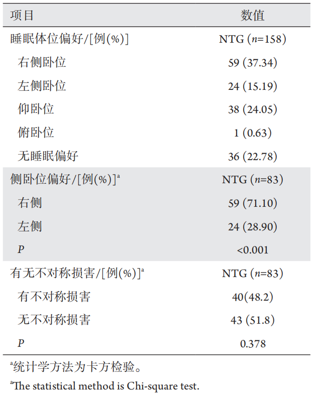
158例符合入组条件的患者中,122例(77.22%)存在睡眠体位偏好;存在睡眠体位偏好的患者中,8 3例(68.03%)存在侧卧位偏好;存在侧卧位偏好的患者中,59例(71.1%)偏好右侧卧位,24例(28.9%)偏好左侧卧位(P<0.001);40(48.2%)例患者双眼有不对称损害,43例(51.8%)无不对称损害(P=0.378,表1)。

表1 NTG患者睡眠偏好及双眼不对称损害分布情况
Table 1 Distribution of sleep preference and binocular asymmetry damage in NTG patients

20230526103554_8426.png

2.2 存在侧卧位偏好的患者中,卧位高侧眼与卧位低侧眼的眼部参数比较

在83例存在侧卧位偏好的患者中,男3 5例(42.17%),女48例(57.83%)例,年龄(63.54±10.83)岁。
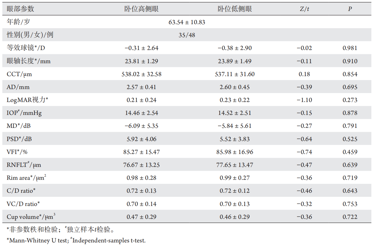
将存在侧卧位偏好的NTG患者分为卧位高侧眼组和卧位低侧眼组,比较其眼部参数,结果示等效球镜、眼轴长度、中央角膜厚度(central corneal thickness,CCT)、LogMAR视力两组间差异无统计学意义(P >0.05)。卧位低侧眼的眼压(intraocular pressure,IOP)高于卧位高侧眼的眼压(14.52±2.51 mmHg vs 14.46±2.54 mmHg),差异无统计学意义( P > 0.05)。卧位低侧眼的神经纤维层厚度(retinal nerve fiber layer thickness,RNFLT)、视野平均偏差(mean deviation,MD)、视野指数(visual field index,VFI)均高于卧位高侧眼(77.65±13.47 μm vs 76.67±13.25 μm;-5.84±5.61 dB vs -6.09±5.35 dB;85.98%±16.9% vs 85.27%±15.47%),差异均无统计学意义(P>0.05,表2)。

表2 卧位高侧眼与卧位低侧眼的眼部参数比较(n=83)
Table 2 Comparison of ocular parameters between the dependent eyes and the non-dependent eyes in recumbent position (n=83)

20230526103626_2020.png

2.3 规律随访患者的卧位高侧眼与卧位低侧眼的视野进展速率比较

在存在侧卧位偏好的患者中,剔除晚期青光眼患者(MD<?12 dB),44例(87眼)随访次数≥5,分析卧位高侧眼与卧位低侧眼的视野进展速率发现,卧位低侧眼的视野进展速率(VFI、MD)均慢于卧位高侧眼[(0.48±1.66)%/年vs (-0.45±3.07)%/年;0.54±0.96 dB/年vs 0.2±1.15 dB/年],但差异均无统计学意义(P>0.05,表3)。

表3 规律随访患者的卧位高侧眼与卧位低侧眼的视野进展速率比较
Table 3 Comparison of the rate of visual field progression between the dependent eyes and the non-dependent eyes in patients who have regularly followed-up

20230526103654_0769.png

2.4 同时存在侧卧位偏好及双眼不对称损害的患者,卧位高侧眼与卧位低侧眼的眼部参数比较

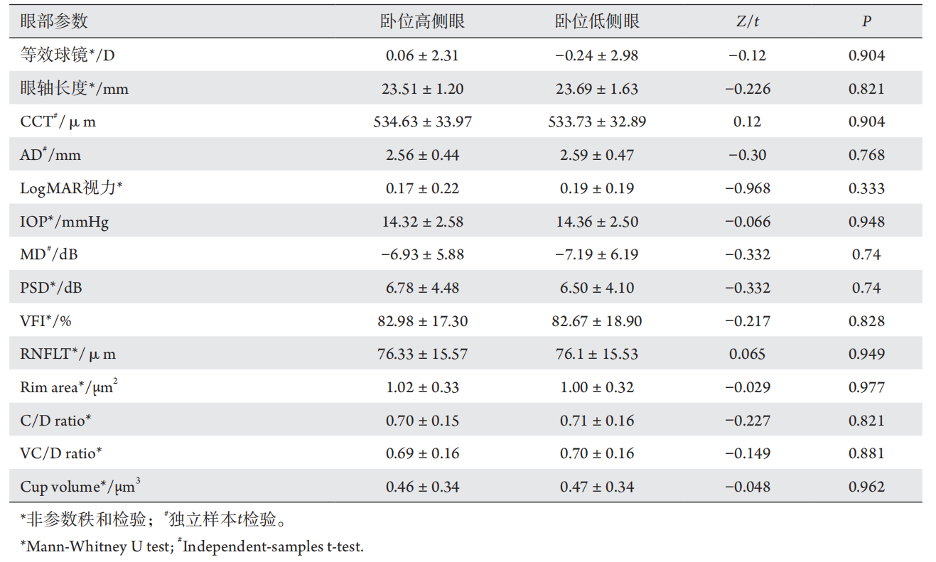
在存在侧卧位偏好的患者中,40例存在双眼不对称损害,比较同时存在侧卧位偏好及双眼不对称损害患者卧位高侧眼与卧位低侧眼的眼部参数,结果示:等效球镜、眼轴长度、CCT、LogMAR视力两组间差异无统计学意义(P >0.05)。卧位低侧眼的IOP高于卧位高侧眼的眼压(14.36±2.50 mmHg vs 14.32±2.58 mmHg)、RNFLT、MD及VFI低于卧位高侧眼(76.1±15.53 μm vs 76.33±15.57 μm;-7.19±6.19 dB vs ?6.93±5.88 dB;82.67%±18.90% vs 82.98%±17.30%),差异均无统计学意义(P>0.05,表4)。

表4 同时存在侧卧位偏好及双眼不对称损害的患者,卧位高侧眼与卧位低侧眼的眼部参数比较(n=40)
Table 4 Comparison of ocular parameters between the dependent eyes and the non-dependent eyes in patients with both lateral preference and asymmetric damage (n=40)

20230526103725_4830.png

2.5 同时存在侧卧位偏好及双眼不对称损害的患者,卧位低侧较好眼与较坏眼占比及卧位高侧眼与卧位低侧眼的视野进展速率的比较

同时存在侧卧位偏好及双眼不对称损害的患者有4 0例,比较卧位低侧较好眼与卧位低侧较坏眼患者的个数及占比,发现卧位低侧较好眼的个数及占比高于卧位低侧较坏眼[23 (57.5%) vs 17 (42.5%)],但差异无统计学意义(χ2=1.81,P = 0.132);40例患者中,剔除晚期青光眼患者(MD<?12 dB),随访次数≥5的有22例(42眼),分析卧位高侧眼与卧位低侧眼的视野进展速率发现:卧位低侧眼的视野进展速率(VFI、MD)均慢于卧位高侧眼[(1.19±1.65)%/年vs (?0.86±3.65)%/年,Z = ? 1.85;0.71 ± 1.13 dB/年vs 0.13 dB/年,Z=?0.81],但差异均无统计学意义(P>0.05)。

3 讨论

本研究对NTG患者睡眠体位与双眼不对称损害的关系进行了横断面及纵向分析,发现存在侧卧位偏好的患者,大多数偏好右侧卧位。但并未发现睡眠体位与其双眼不对称损害及其疾病进展之间存在相关性。本研究对睡眠体位与双眼不对称损害关系的研究做了进一步补充,也表明睡眠体位对青光眼疾病影响的不确定性。
本研究发现存在睡眠体位偏好的患者中,68.03%存在侧卧位偏好;存在侧卧位偏好的患者中,71.1%偏好右侧卧位。Kaplowitz等[11]调查了178名原发性开角型青光眼患者,发现73%的NTG患者和58%的高眼压性青光眼患者存在侧卧位偏好,并且通过睡眠实验室对5 4名受试者的睡姿进行视频监测,观察到大多数受试者偏好右侧卧位,与本研究结果相似。人们选择右侧卧位的原因很多,研究[19-20]发现:对于存在心力衰竭的患者,选择右侧卧位可以降低交感神经兴奋,增加迷走神经兴奋,这种机制可以一定程度上平衡由心力衰竭引起的交感神经兴奋增加,迷走神经兴奋降低。对于存在阻塞性睡眠呼吸暂停综合征的患者,选择侧卧睡姿可明显改善由舌根、喉等结构引起的梗阻症状[2]
本研究并未发现睡眠体位与NTG患者双眼不对称损害存在相关性。对存在侧卧位偏好的患者进行分析,发现卧位高侧眼与卧位低侧眼的眼部参数之间差异无统计学意义;卧位低侧眼的视野进展速率(VFI、MD)均慢于卧位高侧眼,但差异也无统计学意义。对同时存在侧卧位偏好及双眼不对称损害的患者进行分析,发现卧位高侧眼与卧位低侧眼的眼部参数之间差异无统计学意义;卧位低侧较好眼的个数及占比高于卧位低侧较坏眼,但差异无统计学意义;卧位低侧眼的视野进展速率(VFI、MD)均也慢于卧位高侧眼,差异也无统计学意义。不论横断面分析还是纵向研究均未发现侧卧睡姿对青光眼患者双眼不对称损害的影响。
既往研究[8-9]表明:睡眠体位可能影响青光眼疾病的进展,认为睡眠体位与不对称视野损害有关,且与高侧卧位眼相比,低侧卧位眼会有更严重的视野损害。Kim等[9]发现:在同时存在不对称视野损害和侧卧位偏好的青光眼患者中,71.9%偏好较差眼低侧卧位。Tang等[10]通过问卷调查了131名POAG患者的睡眠体位偏好,发现85.7%的左侧卧位患者有较严重的左眼视野损害,71.4%的右侧卧位患者有较严重的右眼视野损害,认为青光眼患者的侧卧位睡眠偏好可能与不对称性视野损害有关。其主要理论依据为:体位改变时眼压会发生相应变化,卧位时眼压明显高于坐位时眼压[3-4];与正常人相比,青光眼患者的眼压对体位的改变更为敏感[6,21]。研究者[22-23]认为:仰卧位可导致巩膜静脉压和眼动脉压升高,进而引起眼压发生相应升高。此外,卧位时由于脉络膜血容量增加,导致葡萄膜巩膜途径的房水流出减少,从而引起眼压升高[24]。然而,Kaplowitz等[11]通过睡眠监测仪对存在不对称视野损害的青光眼患者的睡姿进行客观监测发现,偏好右侧卧位的患者左眼视野损害较重(VFI较低),与前面的研究结果相矛盾。研究[25-26]表明:体位从直立位或坐位变为卧位时,可造成较大幅度的眼压升高。但侧卧睡姿引起的眼压升高幅度相对较小。Malihi等[7]发现:右侧卧位时,较低侧眼眼压与较高侧眼眼压分别为18.8±2.9 mmHg和17.7±3.1 mmHg(P=0.016);而左侧卧位时,卧位低侧眼眼压与卧位高侧眼眼压间的差异无统计学意义。Lee等[27]也发现:身体处于完全侧卧状态时(头位与脊柱平行),POAG患者低侧卧位眼眼压仅高于高侧卧位眼1.3 mmHg。有学者[27-28]对不同头位睡姿(高于30°、与脊柱平行、低于30°)进行研究,发现侧卧位时,较低的头位会导致更高的眼压。Yoo[29]也认为:用头位来判定相关方向可能跟单纯用侧卧睡姿来判断睡眠体位与眼压及不对称视野损害的关系更加准确。当体位由坐位变为卧位时,引起眼压升高的同时眼部灌注压也会相应升高[6]。升高的眼压是青光眼的危险因素,而较高的眼部灌注压可能对青光眼有益[30]。因此,体位改变对青光眼患者造成的影响,暂不适合做定性结论。人们晚上睡觉时难免翻身,可能使眼压升高的持续时间不够,从而削弱侧卧睡姿对不对称视野损害的影响。总之,睡眠体位是人们的生理习惯,人们会倾向于舒适放松的睡眠姿势,不可能按照试验设计的标准睡姿进行。因此,本研究认为睡眠体位与NTG双眼不对称损害及其进展的关系并不确定。本研究的局限性,睡眠体位数据来自问卷,虽然Kaplowitz等[11]对患者睡姿进行客观监测,并同时给予问卷调查发现,问卷与睡眠监测的一致性为77%。但仍具有一定主观性,缺乏客观的睡眠监测数据。
综上所述,存在睡眠体位偏好的NTG患者中,大部分存在侧卧位偏好,存在侧卧位偏好的患者中,大部分偏好右侧卧位。但本研究并未发现睡眠体位与青光眼患者双眼不对称损害及其疾病进展存在相关性。
1、Basner M, Fomberstein KM, Razavi FM, et al. American time use survey: sleep time and its relationship to waking activities[ J]. Sleep, 2007, 30(9): 1085-1095.Basner M, Fomberstein KM, Razavi FM, et al. American time use survey: sleep time and its relationship to waking activities[ J]. Sleep, 2007, 30(9): 1085-1095.
2、Lee CH, Kim DK, Kim SY, et al. Changes in site of obstruction in obstructive sleep apnea patients according to sleep position: A DISE study[ J]. Laryngoscope, 2015, 125(1): 248-254.Lee CH, Kim DK, Kim SY, et al. Changes in site of obstruction in obstructive sleep apnea patients according to sleep position: A DISE study[ J]. Laryngoscope, 2015, 125(1): 248-254.
3、Jain MR, Marmion VJ. Rapid pneumatic and Mackey-Marg applanation tonometry to evaluate the postural effect on intraocular pressure[ J]. Br J Ophthalmol, 1976, 60(10): 687-693.Jain MR, Marmion VJ. Rapid pneumatic and Mackey-Marg applanation tonometry to evaluate the postural effect on intraocular pressure[ J]. Br J Ophthalmol, 1976, 60(10): 687-693.
4、Kiuchi T, Motoyama Y, Oshika T. Relationship of progression of visual field damage to postural changes in intraocular pressure in patients with normal-tension glaucoma[ J]. Ophthalmology, 2006, 113(12): 2150-2155.Kiuchi T, Motoyama Y, Oshika T. Relationship of progression of visual field damage to postural changes in intraocular pressure in patients with normal-tension glaucoma[ J]. Ophthalmology, 2006, 113(12): 2150-2155.
5、Lee JY, Yoo C, Jung JH, et al. The effect of lateral decubitus position on intraocular pressure in healthy young subjects[ J]. Acta Ophthalmol, 2012, 90(1): e68-72.Lee JY, Yoo C, Jung JH, et al. The effect of lateral decubitus position on intraocular pressure in healthy young subjects[ J]. Acta Ophthalmol, 2012, 90(1): e68-72.
6、Lee TE, Yoo C, Kim YY. Effects of different sleeping postures on intraocular pressure and ocular perfusion pressure in healthy young subjects[ J]. Ophthalmology, 2013, 120(8): 1565-1570.Lee TE, Yoo C, Kim YY. Effects of different sleeping postures on intraocular pressure and ocular perfusion pressure in healthy young subjects[ J]. Ophthalmology, 2013, 120(8): 1565-1570.
7、Malihi M, Sit AJ. Effect of head and body position on intraocular pressure[ J]. Ophthalmology, 2012, 119(5): 987-991.Malihi M, Sit AJ. Effect of head and body position on intraocular pressure[ J]. Ophthalmology, 2012, 119(5): 987-991.
8、Kim KN, Jeoung JW, Park KH, et al. Effect of lateral decubitus position on intraocular pressure in glaucoma patients with asymmetric visual field loss[ J]. Ophthalmology, 2013, 120(4): 731-735.Kim KN, Jeoung JW, Park KH, et al. Effect of lateral decubitus position on intraocular pressure in glaucoma patients with asymmetric visual field loss[ J]. Ophthalmology, 2013, 120(4): 731-735.
9、Kim KN, Jeoung JW, Park KH, et al. Relationship between preferred sleeping position and asymmetric visual field loss in open-angle glaucoma patients[ J]. Am J Ophthalmol, 2014, 157(3): 739-745.Kim KN, Jeoung JW, Park KH, et al. Relationship between preferred sleeping position and asymmetric visual field loss in open-angle glaucoma patients[ J]. Am J Ophthalmol, 2014, 157(3): 739-745.
10、Tang J, Li N, Deng YP, et al. Effect of body position on the pathogenesis of asymmetric primary open angle glaucoma[ J]. Int J Ophthalmol, 2018, 11(1): 94-100.Tang J, Li N, Deng YP, et al. Effect of body position on the pathogenesis of asymmetric primary open angle glaucoma[ J]. Int J Ophthalmol, 2018, 11(1): 94-100.
11、Kaplowitz K, Blizzard S, Blizzard DJ, et al. Time spent in lateral sleep position and asymmetry in glaucoma[ J]. Invest Ophthalmol Vis Sci, 2015, 56(6): 3869-3874.Kaplowitz K, Blizzard S, Blizzard DJ, et al. Time spent in lateral sleep position and asymmetry in glaucoma[ J]. Invest Ophthalmol Vis Sci, 2015, 56(6): 3869-3874.
12、Liang YB, Jiang JH, Ou W, et al. Effect of community screening on the demographic makeup and clinical severity of glaucoma patients receiving care in urban China[ J]. Am J Ophthalmol, 2018, 195: 1-7.Liang YB, Jiang JH, Ou W, et al. Effect of community screening on the demographic makeup and clinical severity of glaucoma patients receiving care in urban China[ J]. Am J Ophthalmol, 2018, 195: 1-7.
13、Pan XF, Xu K, Wang X, et al. Evening exercise is associated with lower odds of visual field progression in Chinese patients with primary open angle glaucoma[ J]. Eye Vis (Lond), 2020, 7: 12.Pan XF, Xu K, Wang X, et al. Evening exercise is associated with lower odds of visual field progression in Chinese patients with primary open angle glaucoma[ J]. Eye Vis (Lond), 2020, 7: 12.
14、Lin SG, Cheng HH, Zhang SD, et al. Parapapillar y choroidal microvasculature dropout is associated with the decrease in retinal nerve fiber layer thickness: a prospective study[ J]. Invest Ophthalmol Vis Sci, 2019, 60: 838-842.Lin SG, Cheng HH, Zhang SD, et al. Parapapillar y choroidal microvasculature dropout is associated with the decrease in retinal nerve fiber layer thickness: a prospective study[ J]. Invest Ophthalmol Vis Sci, 2019, 60: 838-842.
15、周堃, 尚晓, 王晓燕, 等. 温州地区原发性开角型青光眼患者视野 缺损进展的危险因素分析[J]. 中华眼科杂志, 2019, 50(10): 777-784.
ZHOU K, SHANG X, WANG XY, et al. Risk factors for visual field loss progression in patients with primary open-angle glaucoma in Wenzhou area[ J]. Chinese Journal of Ophthalmology, 2019, 50(10): 777-784.
周堃, 尚晓, 王晓燕, 等. 温州地区原发性开角型青光眼患者视野 缺损进展的危险因素分析[J]. 中华眼科杂志, 2019, 50(10): 777-784.
ZHOU K, SHANG X, WANG XY, et al. Risk factors for visual field loss progression in patients with primary open-angle glaucoma in Wenzhou area[ J]. Chinese Journal of Ophthalmology, 2019, 50(10): 777-784.
16、De Leon JM, Cheung CY, Wong TY, et al. Retinal vascular caliber between eyes with asymmetric glaucoma[ J]. Graefes Arch Clin Exp Ophthalmol, 2015, 253(4): 583-589.De Leon JM, Cheung CY, Wong TY, et al. Retinal vascular caliber between eyes with asymmetric glaucoma[ J]. Graefes Arch Clin Exp Ophthalmol, 2015, 253(4): 583-589.
17、Bengtsson B, Lindgren A, Heijl A, et al. Perimetric probability maps to separate change caused by glaucoma from that caused by cataract[ J]. Acta Ophthalmol Scand, 1997, 75(2): 184-188.Bengtsson B, Lindgren A, Heijl A, et al. Perimetric probability maps to separate change caused by glaucoma from that caused by cataract[ J]. Acta Ophthalmol Scand, 1997, 75(2): 184-188.
18、Katz J. A comparison of the pattern- and total deviation-based Glaucoma Change Probability programs[ J]. Invest Ophthalmol Vis Sci, 2000, 41(5): 1012-1026.Katz J. A comparison of the pattern- and total deviation-based Glaucoma Change Probability programs[ J]. Invest Ophthalmol Vis Sci, 2000, 41(5): 1012-1026.
19、Fujita M, Miyamoto S, Sekiguchi H, et al. Effects of posture on sympathetic nervous modulation in patients with chronic heart failure[ J]. Lancet, 2000, 356(9244): 1822-1823.Fujita M, Miyamoto S, Sekiguchi H, et al. Effects of posture on sympathetic nervous modulation in patients with chronic heart failure[ J]. Lancet, 2000, 356(9244): 1822-1823.
20、Miyamoto S, Fujita M, Sekiguchi H, et al. Effects of posture on cardiac autonomic nervous activity in patients with congestive heart failure[ J]. J Am Coll Cardiol, 2001, 37(7): 1788-1793.Miyamoto S, Fujita M, Sekiguchi H, et al. Effects of posture on cardiac autonomic nervous activity in patients with congestive heart failure[ J]. J Am Coll Cardiol, 2001, 37(7): 1788-1793.
21、Liu JHK, Sit AJ, Weinreb RN. Variation of 24-hour intraocular pressure in healthy individuals: right eye versus left eye[ J]. Ophthalmology, 2005, 112(10): 1670-1675.Liu JHK, Sit AJ, Weinreb RN. Variation of 24-hour intraocular pressure in healthy individuals: right eye versus left eye[ J]. Ophthalmology, 2005, 112(10): 1670-1675.
22、Sultan M, Blondeau P. Episcleral venous pressure in younger and older subjects in the sitting and supine positions[ J]. J Glaucoma, 2003, 12(4): 370-373.Sultan M, Blondeau P. Episcleral venous pressure in younger and older subjects in the sitting and supine positions[ J]. J Glaucoma, 2003, 12(4): 370-373.
23、Krieglstein GK, Waller WK, Leydhecker W. The vascular basis of the positional influence on the intraocular pressure[ J]. Albrecht Von Graefes Arch Klin Exp Ophthalmol, 1978, 206(2): 99-106.Krieglstein GK, Waller WK, Leydhecker W. The vascular basis of the positional influence on the intraocular pressure[ J]. Albrecht Von Graefes Arch Klin Exp Ophthalmol, 1978, 206(2): 99-106.
24、Longo A, Geiser MH, Riva CE. Posture changes and subfoveal choroidal blood flow[ J]. Invest Ophthalmol Vis Sci, 2004, 45(2): 546.Longo A, Geiser MH, Riva CE. Posture changes and subfoveal choroidal blood flow[ J]. Invest Ophthalmol Vis Sci, 2004, 45(2): 546.
25、Hara T, Hara T, Tsuru T. Increase of peak intraocular pressure during sleep in reproduced diurnal changes by posture[ J]. Arch Ophthalmol, 2006, 124(2): 165-168.Hara T, Hara T, Tsuru T. Increase of peak intraocular pressure during sleep in reproduced diurnal changes by posture[ J]. Arch Ophthalmol, 2006, 124(2): 165-168.
26、Kiuchi T, Motoyama Y, Oshika T. Postural response of intraocular pressure and visual field damage in patients with untreated normaltension glaucoma[ J]. J Glaucoma, 2010, 19(3): 191-193.Kiuchi T, Motoyama Y, Oshika T. Postural response of intraocular pressure and visual field damage in patients with untreated normaltension glaucoma[ J]. J Glaucoma, 2010, 19(3): 191-193.
27、Lee TE, Yoo C, Lin SC, et al. Effect of different head positions in lateral decubitus posture on Intraocular pressure in treated patients with openangle glaucoma[ J]. Am J Ophthalmol, 2015, 160(5): 929-936.e4.Lee TE, Yoo C, Lin SC, et al. Effect of different head positions in lateral decubitus posture on Intraocular pressure in treated patients with openangle glaucoma[ J]. Am J Ophthalmol, 2015, 160(5): 929-936.e4.
28、Seo H, Yoo C, Lee TE, et al. Head position and intraocular pressure in the lateral decubitus position[ J]. Optom Vis Sci, 2015, 92(1): 95-101.Seo H, Yoo C, Lee TE, et al. Head position and intraocular pressure in the lateral decubitus position[ J]. Optom Vis Sci, 2015, 92(1): 95-101.
29、Yoo C. Lateral sleep position and asymmetry in glaucoma[ J]. Invest Ophthalmol Vis Sci, 2016, 57(6): 2543-2544.Yoo C. Lateral sleep position and asymmetry in glaucoma[ J]. Invest Ophthalmol Vis Sci, 2016, 57(6): 2543-2544.
30、Ramli N, Nurull BS, Hairi NN, et al. Low nocturnal ocular perfusion pressure as a risk factor for normal tension glaucoma[ J]. Prev Med, 2013, 57: S47-S49.Ramli N, Nurull BS, Hairi NN, et al. Low nocturnal ocular perfusion pressure as a risk factor for normal tension glaucoma[ J]. Prev Med, 2013, 57: S47-S49.
1、浙江省卫生高层次创新人才计划 (2016025);温州医科大学附属眼视光医院院内创新课题 (YNCX201308)。
This work was supported by Zhejiang Provincial Health Innovation Talents Project (2016025) and the Innovation Research Project of the Eye Hospital of Wenzhou Medical University (YNCX201308), China.()
上一篇
下一篇
其他期刊
  • 眼科学报

    主管:中华人民共和国教育部
    主办:中山大学
    承办:中山大学中山眼科中心
    主编:林浩添
    主管:中华人民共和国教育部
    主办:中山大学
    浏览
  • Eye Science

    主管:中华人民共和国教育部
    主办:中山大学
    承办:中山大学中山眼科中心
    主编:林浩添
    主管:中华人民共和国教育部
    主办:中山大学
    浏览
推荐阅读
出版者信息
目录