您的位置: 首页 > 2021年9月 第36卷 第9期 > 文字全文
2023年7月 第38卷 第7期11
目录

血管生成拟态与翼状胬肉初发型及复发型的相关性

Correlation of vasculogenic mimicry in the primary and recurrent pterygium

来源期刊: 眼科学报 | 2021年9月 第36卷 第9期 727-737 发布时间: 收稿时间:2023/6/21 11:50:14 阅读量:3361
作者:
关键词:
翼状胬肉血管生成拟态三维培养
pterygium vasculogenic mimicry three-dimensional culture
DOI:
10.3978/j.issn.1000-4432.2021.09.08
收稿时间:
 
修订日期:
 
接收日期:
 
目的:探讨血管生成拟态(vasculogenic mimicry,VM)与翼状胬肉初发型及复发型的相关性。方法:采用血小板-内皮细胞黏附分子/过碘酸雪夫(platelet endothelial cell adhesion molecule-1/periodic acid-schiff,CD31/PAS)免疫组织化学双重染色法检测139例翼状胬肉组织(初发型105例;复发型34例)和10例正常结膜中VM的表达,分析VM与初发型及复发型翼状胬肉的相关性及其与患者性别、年龄等因素的关系。原代培养人翼状胬肉成纤维细胞(human pterygium fibroblasts,HPFs),免疫细胞化学染色法鉴定,利用三维培养及PAS染色观察初发型和复发型HPFs构成VM管腔个数的差异。结果:10例正常结膜均未见VM结构,初发型翼状胬肉VM阳性率43.81%,复发型翼状胬肉VM阳性率82.35%,差异具有统计学意义(P<0.001)。相关性分析显示VM与复发型翼状胬肉呈显著正相关(r=0.332)。不同性别、年龄及病程的翼状胬肉患者VM的表达差异均无统计学意义(均P>0.05)。原代培养的HPFs Vimentin表达阳性,符合成纤维细胞特性。细胞三维培养及PAS染色结果提示HPFs具有构建体外VM模型的能力,且复发型HPFs构成的VM管腔数明显高于初发型,差异具有统计学意义(P<0.01)。结论:翼状胬肉组织中存在VM结构,可作为其血供途径之一。VM与翼状胬肉的复发具有密切关系。
Objective: The purpose of this study was to investigate the correlation of vasculogenic mimicry in the primary and recurrent pterygium. Methods: Platelet endothelial cell adhesion molecule-1/periodic acid-schiff (CD31/PAS)immunohistochemical double staining method was adopted to detect the expression of VM in 139 cases of pterygium (105 cases of primary pterygium and 34 cases of recurrent pterygium)and 10 cases of normal conjunctival tissues. The correlation between VM and primary pterygium, recurrent pterygium and the factors such as gender and age of patients were analyzed. Human pterygium fibroblasts (HPFs) were primary cultured and identified by immunocytochemical staining. The differences in the number of VM channels between primary HPFs and recurrent HPFs were observed by three-dimensional culture and PAS staining. Results: There was no VM structure in 10 normal conjunctiva and the positive rate of VM was 43.81% in primary pterygium and 82.35% in recurrent pterygium with a significantly difference (P<0.001). Correlation analysis showed a significant positive correlation between VM and recurrent pterygium (r=0.332). There was no significant difference in the expression of VM in pterygium patients with different sex, age and course (all P>0.05). Vimentin was positive in the primary cultured cells, which was consistent with the characteristics of fibroblasts. The results of three-dimensional culture and PAS staining indicated that HPFs had the ability to construct VM model in vitro, and the number of VM channels constituted by recurrent HPFs was significantly higher than that by primary HPFs, the difference was statistically significant (P<0.01). Conclusion: VM exists in pterygium tissues, and it can be used as one of the blood supply routes, which is closely related to the recurrence of pterygium.
翼状胬肉是常见的眼表疾病之一,临床上表现为睑裂区球结膜与角膜上的一种赘生组织[1]。翼状胬肉初期通常无不适或仅有轻微的干涩感、异物感等症状,当胬肉发展侵入角膜则会导致角膜散光[2]、视力下降,若增生组织越过瞳孔遮盖视轴则会严重影响患者的生活质量。翼状胬肉局部浸润发展、术后易复发的行为学特点,以及微卫星不稳定性、杂合性丢失、p53基因突变及端粒酶表达上调等发病机制均与肿瘤相类似,故部分学者认为其是一种肿瘤样病变[3]。VM是Maniotis等[4]于1999年在葡萄膜黑色素瘤中发现的一种肿瘤微循环模式,它是由高度侵袭性的肿瘤细胞通过自身变形及细胞外基质重塑形成,管腔无内皮细胞衬覆,且可与宿主血管相连通使肿瘤获得血液供应[5]。VM的管壁呈PAS染色阳性,而血管内皮细胞标志物CD31则不表达,符合其不依赖血管内皮细胞的特性,因此CD31/PAS双重染色常作为组织V M的检测方法。有研究发现,V M也存在于翼状胬肉组织中,参与胬肉的血液供应[6],且VM与进展期翼状胬肉呈显著正相关[7]。但VM与翼状胬肉复发的关系未见报道,本研究从组织水平及细胞水平对比初发型和复发型翼状胬肉血管生成拟态的情况,以探讨VM与翼状胬肉复发的相关性。

1 对象与方法

1.1 对象

收集2019年6月至2020年1月于南通大学附属医院眼科行翼状胬肉切除手术患者的初发型翼状胬肉标本105例,男35例,女70例,年龄(60.46±9.21)岁;以及2019年1月至2020年1 2月于本院行同一手术的复发型翼状胬肉标本34例,男12例,女22例,年龄(63.47±9.66)岁;另选10例于本院捐献眼球的正常结膜组织作为对照,男5例,女5例,年龄(68.20±8.52)岁。所有患者胬肉位于鼻侧,且排除其他眼表疾病及眼部外伤史、手术史和长期用药史,手术均由同一医生完成,所取标本均为完整的翼状胬肉组织。所有对照组患者无眼表疾病、眼部外伤史、手术史和长期用药史。本研究遵循赫尔辛基宣言,通过南通大学附属医院伦理委员会审查批准,患者签署知情同意书。

1.2 主要试剂与仪器

柠檬酸钠抗原修复液、PAS染色试剂盒购自北京索莱宝科技有限公司;GTVisionTMIII抗鼠/抗兔通用型免疫组织化学检测试剂盒购自上海基因科技有限公司;CD31抗体、Vimentin抗体、Cytokeratin抗体购自英国Abcam公司;DAB显色试剂盒购自北京中杉金桥生物技术有限公司;胎牛血清、青霉素-链霉素混合液、0.25%胰蛋白酶-EDTA、DMEM/F12培养基及DMSO购自美国Gibco公司;Matrigel胶购自美国BectonDickinson公司;DM3000光学显微镜及显微摄像系统购自德国Leica公司。

1.3 CD31/PAS 免疫组织化学双重染色

将术中取下的正常结膜及翼状胬肉组织用4%多聚甲醛固定,常规脱水,石蜡包埋,以组织长轴连续纵切,制成厚度约为4 μm的切片,脱蜡至水,65 ℃烘箱中干燥40 min,PBST(含0.05%Triton-100的PBS溶液)漂洗3次,每次5 min,柠檬酸钠缓冲液抗原修复15~20 min,PBST漂洗5 min×3次,3%H2O2消除内源性过氧化物酶活性20 min(避光),PBST漂洗3次,每次5 min,10%山羊血清封闭40 min,不洗,加入CD31(1:50)一抗,4 ℃孵育过夜。复温,PBST漂洗3次,每次5 min,滴加抗鼠/抗兔通用型二抗37 ℃孵育30 min,PBST漂洗3次,每次5 min,DAB显色,蒸馏水浸洗,高碘酸氧化5 min,蒸馏水浸洗,Schiff氏液避光染色10 min,蒸馏水浸洗,苏木精染核5 min,稀盐酸乙醇分化2~5 s,自来水漂洗,蒸馏水返蓝,梯度乙醇脱水,二甲苯透明,自然干燥后中性树胶封片,光学显微镜下观察。

1.4 人翼状胬肉成纤维细胞原代培养及鉴定

将手术切除的新鲜翼状胬肉组织用含1%青霉素-链霉素的PBS缓冲液漂洗3次,洗净血液,去除上皮,剪碎,加入0.08%胰蛋白酶-EDTA,室温消化8 min后终止,1 500 r/min离心5 min弃上清,利用牙科探针将组织小块均匀接种于培养瓶内,加入少许完全培养基,将培养瓶底朝上置于37 ℃,5%CO2培养箱中,24 h后待组织小块贴壁,加入适量完全培养基,缓慢翻转培养瓶,静置培养,每3 d换液一次。
取传至第二代的细胞接种爬片,4%多聚甲醛固定1 h,PBS漂洗3次,每次5 min,0.2%Triton-100通透20 min,PBS漂洗3次,每次5 min,消除内源性过氧化物酶活性、封闭同1.3,加入Vimentin(1:500)、Cytokeratin(1:50)及PBS(阴性对照),4 ℃孵育过夜。复温,PBS漂洗3次,每次5 min,二抗37 ℃孵育30 min,PBS漂洗3次,每次5 min,DAB显色,染核,分化,返蓝,脱水,透明,封片,观察同1.3。

1.5 细胞三维培养及 PAS 染色

取出Matrigel胶,4 ℃解冻过夜。将Matrigel胶以50 μL的体积缓慢加至96孔板,不可产生气泡。为避免Matrigel胶受热凝固,所有操作必须在冰上进行,且所用的枪头及9 6孔板均需预冷。将铺好胶的96孔板置于37 ℃孵箱内30 min使其固化。将初发型HPFs及复发型HPFs分别以1×10⁵个/孔的数量接种于Matrigel胶上,置细胞培养箱内培养5 h后,倒置显微镜下观察细胞的管状结构排列及成管的完整程度,随机取3个不同视野(×100)计数成管的数量,计算平均值。观察完成后,将上述培养5 h后的细胞行PAS染色。染色步骤为高碘酸氧化5 min,蒸馏水稍洗,Schiff氏液避光染色10 min,稀盐酸酒精分化2~5 s,水洗2次。

1.6 统计学处理

采用SPSS 23.0软件进行统计学分析,计量资料以均数±标准差(x±s)表示。计量资料采用独立样本t检验,计数资料采用Pearson Chi-Square检验,双变量相关性分析采用Spearman’s相关性分析,以P<0.05为差异具有统计学意义。

2 结果

2.1 VM 在翼状胬肉中的表达

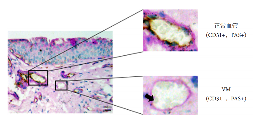
CD31/PAS免疫组织化学双重染色结果见图1,可见血管内皮细胞CD31染色阳性呈棕黄色,管壁PAS染色阳性呈紫红色,为具有内皮细胞的正常血管;CD31染色阴性,PAS染色阳性的管道状结构,是VM的特有形态。本研究统计得出,10例正常结膜均未见V M结构,初发型翼状胬肉V M阳性率43.81%(46/105),复发型翼状胬肉VM阳性率82.35%(28/34),差异具有统计学意义(P<0.05,表1)。Spearman’s相关性分析显示VM与复发型翼状胬肉(r=0.332,P<0.001)呈显著正相关。
20230626113153_8335.png

图1 翼状胬肉中VM的表达(×400)
Figure 1 Expression of VM in pterygium (×400)
CD31阳性且PAS阳性的管腔为具有血管内皮细胞的血管;CD31阴性PAS阳性且管腔中可见红细胞(箭头)的管道为VM结构。
CD31+/PAS+ channels were defined as endothelial vessels. CD31–/PAS+ and red blood cells (arrow) within the cavities were confirmed as VM vessels.

表1 翼状胬肉中VM的表达
Table 1 Expression of VM in pterygium

20230626113249_0685.png

2.2 VM 在翼状胬肉中的表达与患者一般资料的关系

根据患者的性别、年龄及病程,分别观察V M在不同指标分组中的表达情况。Pearson Chi-Square检验结果提示,不同性别、年龄及病程的翼状胬肉患者V M的表达差异均无统计学意义(均P>0.05,表2)。

表2 翼状胬肉VM与患者一般资料的关系
Table 2 Correlation between the VM and the general information of pterygium patients

20230626113331_5683.png

2.3 HPFs 原代培养及鉴定

翼状胬肉组织块接种后10 d,初发组可见细胞从组织块向周围发散爬出,数量较少,分布较为零星(图2A)。复发组爬出的细胞较初发组多,且相互连接,细胞形态多为长梭形、三角形,逐渐向四周延伸(图2B)。由于成纤维细胞对胰酶的耐受性较差,故当细胞传至第2代时已基本纯化,细胞呈长梭形,长满后呈旋涡状(图2C)。免疫细胞化学染色结果显示PBS空白对照组及Cytokeratin染色均为阴性(图3A,3B),Vimentin染色阳性,表现为胞质棕黄色(图3C),符合成纤维细胞特性。
20230626113411_0214.png

图2 HPFs原代培养(×100)
Figure 2 Primary culture of HPFs (×100)
(A)原代培养第10天,初发型胬肉组织中细胞爬出情况;(B)原代培养第10天,复发型胬肉组织中细胞爬出情况;(C)传至第二代的细胞。
(A) Cell growth of primary pterygium on day 10 of primary culture; (B) Cell growth of recurrent pterygium on day 10 of primary culture; (C)Cells well grew after 2 times of passages.

20230626113513_0527.png

图3 HPFs鉴定(×200)
Figure 3 Identification of HPFs (×200)
(A)以PBS为一抗,染色阴性;(B)Cytokeratin表达阴性;(C)Vimentin表达阳性,细胞质棕黄色。
(A) Negative expression of PBS; (B) Negative expression of Cytokeratin; (C) Positive expression of Vimentin and the cytoplasm was yellowish-brown.

2.4 不同类型 HPFs 构建 VM 的差异

细胞三维培养结果显示:HPFs能够黏附于Matrigel胶表面,通过细胞自身变形、彼此相连形成大小不等的网格样、环状管腔结构,且PAS染色呈阳性,提示管腔样结构是由HPFs构成,即V M结构(图4A)。图4 B可见复发型HPFs所构成的VM管腔较初发型大,且数量较初发型多。初发型和复发型HPFs形成VM的个数分别为21.00±2.65、30.33±1.53,差异具有统计学意义( t =-5.292,P=0.006;图4C)。
20230626113618_2875.png

图4 HPFs构建体外VM模型(×100)
Figure 4 VM model was constructed by HPFs in vitro (×100)
(A)初发型HPFs构建体外VM,PAS染色阳性;(B)复发型HPFs构建体外VM;(C)初发型及复发型HPFs构建VM管腔个数量化图。**P<0.01。
(A) VM model was constructed by primary HPFs and the PAS staining was positive; (B) VM model was constructed by recurrent HPFs; (C)Quantification of the number of VM channels constructed by primary and recurrent HPFs. **P<0.01.

3 讨论

翼状胬肉是全球范围内较为多发的眼科疾病[8]。据最新统计,中国4 0岁及以上人群翼状胬肉患病率为13.4%[9],处于较高水平,且随着年龄的增长而不断升高,好发于长期紫外线暴露的人群,日光、粉尘等的慢性刺激[10]可能是其主要诱因。翼状胬肉类肿瘤样特性为其表达VM奠定了一定的理论基础。
VM是最新发现的肿瘤微循环模式,其主要特点如下[11-12]:1 )管道由肿瘤细胞及细胞外基质构成;2 )管腔内壁没有血管内皮细胞衬里;3 )肿瘤细胞变形成条索状并模仿正常血管结构围成管腔,可输送血液;4 )肿瘤细胞在管壁成分外表面排列;5 )管道周围无明显的炎症细胞浸润。许多学者发现,VM表达阳性的肿瘤存在更强的侵袭、转移及复发的能力[13-14]。目前,在乳腺癌[15]、卵巢癌[16]、肺癌[17]、前列腺癌[18]、肝癌[19]等许多恶性肿瘤中均检测到VM结构。VM的管壁呈PAS染色阳性,而血管内皮细胞标志物CD31则不表达,符合其不依赖血管内皮细胞的特性,因此CD31/PAS双重染色常作为组织VM的检测方法。
本研究采用CD31/PAS双重染色检测翼状胬肉中的V M结构,结果显示:翼状胬肉中存在V M结构,而正常结膜中未见,且复发型翼状胬肉VM阳性率大于初发型,VM与复发型翼状胬肉呈显著正相关。本研究结果与李永平等[6]的结论共同支持了“VM结构不是肿瘤组织所独有的血供模式”这一观点。推测VM是由于机体局部组织由于某些原因需要大量的营养供应,但自身血管不能满足其需要而形成的一种供给方式。复发型翼状胬肉组织中细胞增殖水平较初发型翼状胬肉显著升高[20],即复发型胬肉进展速度快、时间短,需要较多的血液供应,故VM形成率较高。同时,VM结构的产生,使胬肉获得充分的血液供应,进一步促进了翼状胬肉的进展。一般资料统计结果得出,不同性别、年龄及病程的翼状胬肉患者VM的表达无显著差异。这些结果与肿瘤组织中VM阳性者具有更强的侵袭及复发的能力,VM阳性率与肿瘤患者一般资料无显著相关性相对应。
李永平等[6]指出,翼状胬肉VM主要表现为PAS阳性的细胞外基质包绕在成纤维细胞伸出的突起表面形成的管道状结构,即成纤维细胞是构成翼状胬肉VM的主要成分,因此本研究使用原代培养的HPFs构建体外VM。对于体外培养的肿瘤细胞而言,其可与Matrigel基质胶或鼠尾I型胶原蛋白等模拟的细胞外基质成分一起构建体外细胞培养的三维模型,如果肿瘤细胞可以在Matrigel基质胶或鼠尾I型胶原蛋白中通过自身变形、彼此相互连接而围成环状、网状结构,且可被PAS染成紫红色,则可将其认定为VM结构,这种方法常被用来作为体外VM的检测。本研究发现:在翼状胬肉中,具有侵袭性的HPFs也能够黏附于Matrigel基质胶表面,通过细胞自身变形、彼此相连形成大小不等的网格样、环状管腔结构,且PAS染色呈阳性,提示HPFs具有在体外形成VM结构的能力,进一步验证了李永平等[6]的观点。经过对初发型及复发型HPFs构成VM管腔数量的统计得出,复发型HPFs构成的VM管腔数量明显较初发型多。可能原因是复发型翼状胬肉向角膜及深层浸润发展迅速[21],其成纤维细胞具有更强的侵袭及迁移能力,故可形成较多的VM结构。
综上所述,翼状胬肉组织中存在VM结构,可作为其血供途径之一,且复发型翼状胬肉VM阳性率及复发型HPFs构建VM的数量均高于初发型,即VM与翼状胬肉的复发密切相关。因此,进一步研究针对VM的靶向药物,可能对预防翼状胬肉复发具有重大意义。

开放获取声明

本文适用于知识共享许可协议(Creative Commons),允许第三方用户按照署名(BY)-非商业性使用(NC)-禁止演绎(ND)(CC BY-NC-ND)的方式共享,即允许第三方对本刊发表的文章进行复制、发行、展览、表演、放映、广播或通过信息网络向公众传播,但在这些过程中必须保留作者署名、仅限于非商业性目的、不得进行演绎创作。详情请访问:https://creativecommons.org/licenses/by-nc-nd/4.0/
1、Golu T, Mogoant? L, Streba CT, et al. Pterygium: histological and immunohistochemical aspects[ J]. Rom J Morphol Embryol, 2011, 52(1): 153-158.Golu T, Mogoant? L, Streba CT, et al. Pterygium: histological and immunohistochemical aspects[ J]. Rom J Morphol Embryol, 2011, 52(1): 153-158.
2、Garg P, Sahai A, Shamshad MA, et al. A comparative study preoperative and postoperative changes in corneal astigmatism after pterygium excision by different techniques[ J]. Indian J Ophthalmol, 2019, 67(7): 1036-1039.Garg P, Sahai A, Shamshad MA, et al. A comparative study preoperative and postoperative changes in corneal astigmatism after pterygium excision by different techniques[ J]. Indian J Ophthalmol, 2019, 67(7): 1036-1039.
3、Chui J, Coroneo MT, Tat LT, et al. Ophthalmic pterygium: a stem cell disorder with premalignant features[ J]. Am J Pathol, 2011, 178(2): 817-827.Chui J, Coroneo MT, Tat LT, et al. Ophthalmic pterygium: a stem cell disorder with premalignant features[ J]. Am J Pathol, 2011, 178(2): 817-827.
4、Maniotis AJ, Folberg R, Hess A, et al. Vascular channel formation by human melanoma cells in vivo and in vitro: vasculogenic mimicry[ J]. Am J Pathol, 1999, 155(3): 739-752. Maniotis AJ, Folberg R, Hess A, et al. Vascular channel formation by human melanoma cells in vivo and in vitro: vasculogenic mimicry[ J]. Am J Pathol, 1999, 155(3): 739-752.
5、Fernández-Cortés M, Delgado-Bellido D, Oliver FJ, et al. Vasculogenic mimicry: become an endothelial cell "But Not So Much"[ J]. Front Oncol, 2019, 9: 803.Fernández-Cortés M, Delgado-Bellido D, Oliver FJ, et al. Vasculogenic mimicry: become an endothelial cell "But Not So Much"[ J]. Front Oncol, 2019, 9: 803.
6、李永平, 朱哲, 张文忻. 翼状胬肉组织中血管拟态的初步研 究[ J]. 中华眼科杂志, 2007, 43(10): 872-875.
LI YP, ZHU Zhe, ZHANG Wenxin. Preliminary study of vascular mimicry in pterygium[ J]. Chinese Journal of Ophthalmology, 2007, 43(10): 872-875.
李永平, 朱哲, 张文忻. 翼状胬肉组织中血管拟态的初步研 究[ J]. 中华眼科杂志, 2007, 43(10): 872-875.
LI YP, ZHU Zhe, ZHANG Wenxin. Preliminary study of vascular mimicry in pterygium[ J]. Chinese Journal of Ophthalmology, 2007, 43(10): 872-875.
7、陈俊杰, 蓝育青, 吴共发, 等. 血管生成拟态与翼状胬肉进行期及 静止期的相关性研究[ J]. 国际眼科杂志, 2015, 15(3): 414-417.
CHEN JJ, LAN YQ, WU GF, et al. Correlation of vasculogenic mimicry in the aggressive and quiescent period of pterygium[ J]. International Eye Science, 2015, 15(3): 414-417.
陈俊杰, 蓝育青, 吴共发, 等. 血管生成拟态与翼状胬肉进行期及 静止期的相关性研究[ J]. 国际眼科杂志, 2015, 15(3): 414-417.
CHEN JJ, LAN YQ, WU GF, et al. Correlation of vasculogenic mimicry in the aggressive and quiescent period of pterygium[ J]. International Eye Science, 2015, 15(3): 414-417.
8、Rezvan F, Khabazkhoob M, Hooshmand E, et al. Prevalence and risk factors of pterygium: a systematic review and meta-analysis[ J]. Surv Ophthalmol, 2018, 63(5): 719-735.Rezvan F, Khabazkhoob M, Hooshmand E, et al. Prevalence and risk factors of pterygium: a systematic review and meta-analysis[ J]. Surv Ophthalmol, 2018, 63(5): 719-735.
9、杨梅, 管宇, 康丽华, 等. 中国40岁及以上人群翼状胬肉患病率 Meta分析[ J]. 中华实验眼科杂志, 2019, 37(3): 190-196.
YANG M, GUAN Y, KANG LH, et al. Meta-analysis of prevalence of pterygium among people aged over 40 in China[ J]. Chinese Journal of Experimental Ophthalmology, 2019, 37(3): 190-196.
杨梅, 管宇, 康丽华, 等. 中国40岁及以上人群翼状胬肉患病率 Meta分析[ J]. 中华实验眼科杂志, 2019, 37(3): 190-196.
YANG M, GUAN Y, KANG LH, et al. Meta-analysis of prevalence of pterygium among people aged over 40 in China[ J]. Chinese Journal of Experimental Ophthalmology, 2019, 37(3): 190-196.
10、Zhou WP, Zhu YF, Zhang B, et al. The role of ultraviolet radiation in the pathogenesis of pterygia (Review)[ J]. Mol Med Rep, 2016, 14(1): 3-15.Zhou WP, Zhu YF, Zhang B, et al. The role of ultraviolet radiation in the pathogenesis of pterygia (Review)[ J]. Mol Med Rep, 2016, 14(1): 3-15.
11、Luo Q, Wang J, Zhao W, et al. Vasculogenic mimicry in carcinogenesis and clinical applications[ J]. J Hematol Oncol, 2020, 13(1): 19.Luo Q, Wang J, Zhao W, et al. Vasculogenic mimicry in carcinogenesis and clinical applications[ J]. J Hematol Oncol, 2020, 13(1): 19.
12、Treps L, Faure S, Clere N, et al. Vasculogenic mimicry, a complex and devious process favoring tumorigenesis - Interest in making it a therapeutic target[ J]. Pharmacol Ther, 2021, 223: 107805.Treps L, Faure S, Clere N, et al. Vasculogenic mimicry, a complex and devious process favoring tumorigenesis - Interest in making it a therapeutic target[ J]. Pharmacol Ther, 2021, 223: 107805.
13、Zhang J, Qiao L, Liang N, et al. Vasculogenic mimicry and tumor metastasis[ J]. J BUON, 2016, 21(3): 533-541.Zhang J, Qiao L, Liang N, et al. Vasculogenic mimicry and tumor metastasis[ J]. J BUON, 2016, 21(3): 533-541.
14、Qiao L, Liang N, Zhang J, et al. Advanced research on vasculogenic mimicry in cancer[ J]. J Cell Mol Med, 2015, 19(2): 315-326.Qiao L, Liang N, Zhang J, et al. Advanced research on vasculogenic mimicry in cancer[ J]. J Cell Mol Med, 2015, 19(2): 315-326.
15、Andonegui-Elguera MA, Alfaro-Mora Y, Cáceres-Gutiérrez R, et al. An overview of vasculogenic mimicry in breast cancer[ J]. Front Oncol, 2020, 10: 220.Andonegui-Elguera MA, Alfaro-Mora Y, Cáceres-Gutiérrez R, et al. An overview of vasculogenic mimicry in breast cancer[ J]. Front Oncol, 2020, 10: 220.
16、Ayala-Domínguez L, Olmedo-Nieva L, Mu?oz-Bello JO, et al. Mechanisms of vasculogenic mimicry in ovarian cancer[ J]. Front Oncol, 2019, 9: 998.Ayala-Domínguez L, Olmedo-Nieva L, Mu?oz-Bello JO, et al. Mechanisms of vasculogenic mimicry in ovarian cancer[ J]. Front Oncol, 2019, 9: 998.
17、Williamson SC, Metcalf RL, Trapani F, et al. Vasculogenic mimicry in small cell lung cancer[ J]. Nat Commun, 2016, 7: 13322.Williamson SC, Metcalf RL, Trapani F, et al. Vasculogenic mimicry in small cell lung cancer[ J]. Nat Commun, 2016, 7: 13322.
18、Wang H, Lin H, Pan J, et al. Vasculogenic mimicry in prostate cancer: the roles of EphA2 and PI3K[ J]. J Cancer, 2016, 7(9): 1114-1124.Wang H, Lin H, Pan J, et al. Vasculogenic mimicry in prostate cancer: the roles of EphA2 and PI3K[ J]. J Cancer, 2016, 7(9): 1114-1124.
19、Chen Q, Lin W, Yin Z, et al. Melittin inhibits hypoxia-induced vasculogenic mimicry formation and epithelial-mesenchymal transition through suppression of HIF-1α/Akt pathway in liver cancer[ J]. Evid Based Complement Alternat Med 2019;2019:9602935.Chen Q, Lin W, Yin Z, et al. Melittin inhibits hypoxia-induced vasculogenic mimicry formation and epithelial-mesenchymal transition through suppression of HIF-1α/Akt pathway in liver cancer[ J]. Evid Based Complement Alternat Med 2019;2019:9602935.
20、沈韵之, 许咪, 孙松. 原发性与复发性翼状胬肉的临床指标及实 验室指标[ J]. 国际眼科杂志, 2020, 20(4): 639-642.
SHEN YZ, XU M, SUN S. Differences in clinical characteristics and laboratory parameters of primary and recurrent pterygium[ J]. International Eye Science, 2020,20(4):639-642.
沈韵之, 许咪, 孙松. 原发性与复发性翼状胬肉的临床指标及实 验室指标[ J]. 国际眼科杂志, 2020, 20(4): 639-642.
SHEN YZ, XU M, SUN S. Differences in clinical characteristics and laboratory parameters of primary and recurrent pterygium[ J]. International Eye Science, 2020,20(4):639-642.
21、Nuhoglu F, Turna F, Uyar M, et al. Is there a relation between histopathologic characteristics of pterygium and recurrence rates?[ J]. Eur J Ophthalmol, 2013, 23(3): 303-308.Nuhoglu F, Turna F, Uyar M, et al. Is there a relation between histopathologic characteristics of pterygium and recurrence rates?[ J]. Eur J Ophthalmol, 2013, 23(3): 303-308.
1、南通市科技项目 (MS12020033)。
This work was supported by the Nantong Science and Technology Commission Project, China (MS12020033).()
上一篇
下一篇
其他期刊
  • 眼科学报

    主管:中华人民共和国教育部
    主办:中山大学
    承办:中山大学中山眼科中心
    主编:林浩添
    主管:中华人民共和国教育部
    主办:中山大学
    浏览
  • Eye Science

    主管:中华人民共和国教育部
    主办:中山大学
    承办:中山大学中山眼科中心
    主编:林浩添
    主管:中华人民共和国教育部
    主办:中山大学
    浏览
推荐阅读
出版者信息
目录