您的位置: 首页 > 2022年1月 第37卷 第1期 > 文字全文
2023年7月 第38卷 第7期11
目录

飞秒激光小切口基质透镜取出术和飞秒制瓣联合准分子激光原位角膜磨镶术术后有效光学区及角膜像差的比较

Comparison of effective optical zone and high order aberrations after small incision lenticule extraction and femtosecond laser-assisted in situ keratomileusis

来源期刊: 眼科学报 | 2022年1月 第37卷 第1期 36-43 发布时间: 收稿时间:2022/9/26 15:53:20 阅读量:4147
作者:
关键词:
飞秒激光小切口基质透镜取出术飞秒制瓣联合准分子激光原位角膜磨镶术有效光学区像差
small incision lenticule extraction femtosecond laser-assisted in situ keratomileusis effective optical zone aberration
DOI:
10.3978/j.issn.1000-4432.2021.12.11
收稿时间:
 
修订日期:
 
接收日期:
 
目的:比较不同预设光学区组的飞秒激光小切口基质透镜取出术(small incision lenticule extraction,SMILE)和飞秒激光制瓣联合准分子激光原位角膜磨镶术(femtosecond laser-assisted in situ keratomileuses,FS-LASIK)术后有效光学区(effective optical zone,EOZ)大小与角膜高阶像差(high order aberrations,HOAs)变化以及有效光学区大小对角膜高阶像差的影响。方法:收集2019年2月 至2020年5月来佛山市第二人民医院行激光手术的患者80例,均取右眼入组,SMILE组43例,FSLASIK组37例,按预设光学区大小分为6.5 mm和6.0 mm组,分别于术前和术后1个月应用Pentacam三维眼前节分析系统测量有效光学区及总角膜高阶像差、彗差、球差,分析不同预设光学区下SMILE组和FS-LASIK组有效光学大小及与角膜高阶像差的关系。结果:在相同预设光学区下,术后1个月SMILE组的有效光学区均大于FS-LASIK组,差异均有统计学意义(均P<0.05),且SMILE组的角膜总高阶像差、球差、彗差均较FS-LASIK组低,差异均有统计学意义(均P<0.05);对于相同手术方式,预设光学区6.0 mm较6.5 mm组,术后1个月角膜总高阶像差、球差、彗差均升高,差异均有统计学意义(均P<0.05)。结论:SMILE组和FS-LASIK组术后1个月的有效光学区均小于预设光学区,SMILE组大于FS-LASIK组;术后1个月有效光学区越大,角膜高阶像差越小;在相同预设光学区下,SMILE组术后1个月角膜高阶像差小于FS-LASIK组。
Objective: To compare the effective optical zone (EOZ) and the changes in corneal high order aberrations (HOAs) after small incision lenticule extraction (SMILE) with those after femtosecond laser-assisted in situ keratomileuses (FS-LASIK). Methods: This study included 80 subjects who underwent laser refractive surgery at the Second People’s Hospital of Foshan between February 2019 and May 2020. Only data from the right eye of each subject were analyzed. A total of 43 eyes underwent SMILE while 37 eyes received FS-LASIK. The eyes were further stratified into subgroups based on different programmed optical zones: the 6.5 mm group and the 6.0 mm group. EOZ, coma, and spherical aberration were measured with Pentacam 3D anterior segment analysis system preoperatively and one month postoperatively. In addition, the relationship between EOZ and corneal HOAs was analyzed and compared between different optical-zone groups after SMILE and FS-LASIK. Results: For the same programmed optical zone, the SMILE group achieved a significantly greater EOZ than the FS-LASIK group who was measured 1-month postoperatively did (P<0.05). Meanwhile, corneal HOAs, spherical aberration, and coma in the SMILE group are significantly lower than those in the FS-LASIK group (P<0.05). For the same procedure (SMILE or FS-LASIK), the 6.0 mm group demonstrated significantly higher corneal total HOAs, spherical aberration, and coma than the 6.5 mm group did 1-month after the surgery (P<0.05). Conclusion: In both the SMILE and the FS-LASIK groups, 1-month postoperative EOZ was smaller than the programmed optical zone. EOZ in the SMILE group was larger than that in the FS-LASIK group. The larger the 1-month postoperative EOZ was, the lower corneal HOAs were. For the same programmed optical zone, 1-month postoperative corneal HOAs in the SMILE group is lower than that in the FS-LASIK group.
    小切口基质透镜取出术(small incision lenticule extraction,SMILE)在角膜屈光手术中所占的比例越来越高[1],相对于飞秒制瓣联合准分子激光原位角膜磨镶术(femtosecond laser-assisted in situ keratomileuses,FS-LASIK),SMILE手术切口仅2~4 mm,无瓣,更好的保留了角膜前部的形态结构。SMILE和FS-LASIK手术均是角膜板层手术,通过切削角膜组织改变角膜形态,从而达到矫正屈光不正的目的[2-3]。屈光手术的目的不仅要提升患者的视力,更要考虑到患者的视觉质量。
    角膜的高阶像差在一定程度上可以反映术后的视觉质量,有研究表明角膜屈光术后角膜高阶像差会增加,影响患者术后视觉质量,发生眩光、光晕、夜间视力下降等现象[4-5],术后高阶像差增加的原因之一可能与术后有效光学区减少有[6],有效光学区(effective optical zone,EOZ)被认为是产生像差最少,视觉质量最好,角膜屈光全部矫正的区域[7]
    因手术的有效光学区及角膜的高阶像差对患者术后的视觉质量影响关系密切,手术切口和切削方式对于有效光学区和角膜高阶像差有无影响,以及他们之间的关系如何,本研究想对比SMILE和FS-LASIK两种手术方式在不同预设光学区下所产生的有效光学区大小及与角膜高阶像差的关系。

1 对象与方法

1.1 对象

    选取2019年2月 至2020年5月来佛山市第二人民医院行激光手术的8 0例患者,SMILE组4 3例,FS-LASIK组3 7例。年龄18~50岁。等效球镜度数为?0.50 DS至?12.00 DS。屈光度数稳定,2年内增长小于?0.50 D,排除眼部器质性病变,圆锥角膜病史,全身免疫系统病史,1周内未佩戴隐形眼镜,1个月内未佩戴硬性角膜接触镜(rigid gas permeable contact lens,RGP),3个月内未佩戴角膜塑形镜(orthokeratology,OK镜)。根据手术方式分为SMILE和FS-LASIK组,根据手术预设光学区大小分为6.5 mm和6.0 mm组。本研究符合医学伦理学要求,所有患者签署手术同意书。

1.2 术前检查及准备

    所有患者术前进行裸眼视力检查,裂隙灯检查,小瞳下客观验光,小瞳下主觉验光,散瞳下客观验光,散瞳下主觉验光,眼压,角膜厚度,眼底,角膜地形图,眼轴长度等检查。符合条件手术患者可选择SMILE或FS-LASIK手术。手术患者术前滴3 d抗生素眼水及人工泪液眼水,每天4次。

1.3 手术过程

1.3.1 SMILE 手术过程

    使用德国蔡司Visumax激光系统,能量135 nJ,扫描频率500 Hz,线点间距4.5 μm,角膜帽直径7.0~7.6 mm,厚度110~120 μm,治疗光学区直径6.0 mm或6.5 mm,透镜基底厚度10~20 μm,切口位于130°,边切角度90°,散光过渡区0.1 mm。摆正患者头位,对患者进行消毒铺巾,操作操纵杆将患者推至治疗镜下,上开睑器,嘱患者注视绿色灯,术者上升手术床,至角膜与负压吸引环接触产生水印,中心定位,当水印超过80%时,启动
负压吸引并发射激光,激光扫描结束后,将患者移至观察镜下,进行透镜分离,取出透镜,完成手术。

1.3.2 FS-LASIK 手术过程

    第一步制作角膜瓣,使用德国蔡司Visumax激光系统,能量140 nJ,扫描频率500 Hz,线点间距4.5 μm,角膜瓣直径8.1 mm,厚度110~120 μm,蒂在90°方向,蒂宽度3.53 mm,边切角度60°。摆正患者头位,对患者进行消毒铺巾,操作操纵杆将患者推至治疗镜下,上开睑器,嘱患者注视绿色灯,术者上升手术床,至角膜与负压吸引环接触产生水印,中心定位,当水印超过80%时启动负压吸引发射激光,激光扫描结束后将治疗床移至德国蔡司MEL90准分子激光系统下,分离器分离角膜瓣,进行激光消融,频率250~500 Hz,治疗光学区直径6.0 mm或6.5 mm,激光消融结束后进行冲洗,复位角膜瓣,结束手术。

1.4 术后随访及处理

    术后随访1 d、1周 及1个月进行裸眼视力检查,裂隙灯检查,小瞳下客观及主观验光,眼压检查,术后1个月时进行Pentacam三维眼前节系统角膜地形图及角膜高阶像差检查,与术前进行比较。
    术后用药:0.5%左氧氟沙星滴眼液4次/d,1周;氯替泼诺混悬滴眼液6次/d,3 d,后改为4次/d,1周;0.3%的玻璃酸钠滴眼液4次/d,1~3个月。
    Pentacam检查:检查在暗室中进行,患者在自然瞳孔状态下,将下颌放置在下颌托上,前额紧贴前额带,睁开双眼注视蓝色视标。检查者使用操纵杆按电脑屏幕提示进行对焦,旋转的Scheimpflug相机在2 s内拍摄25张图像,只接受成像质量显示OK的检测结果。
    EOZ计算:以手术前后角膜地形图的切向曲率差异图显示的切削轮廓为术后的EOZ。在差异图上移动光标定位,分别在0°到330°子午线上,每相差3 0 °测量一次,沿子午线自中心向周边移动,当角膜曲率差值为0时,停止移动,记录该点坐标值( x,y),同一子午线另一端同样记录角膜曲率差值为0时坐标( x’,y ’ ) ,共记录1 2个值,同一子午线上半径求和,
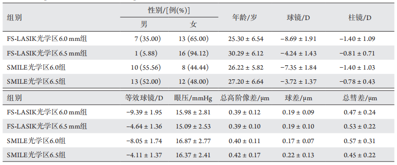
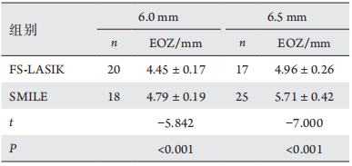
EOZ直径为6条主子午线EOZ直径的平均值,记为EOZ。
    像差记录:Pentacam系统像差分析模式,记录角膜直径6 mm内的角膜总高阶像差(tHOA),球差(Z40),水平彗差(Z31),垂直彗差(Z3?1),彗差记为

1.5 统计学处理

    使用IBM SPSS 24.0软件分析数据。采用均数±标准差(x±s)描述服从正态分布的数值变量、中位数(全距)描述不服从正态分布的数值变量,采用频数 (%) 描述分类变量。数据正态性采用Kolmogorov-Smirnov法验证。术前术后比较采用配对t检验,术后1个月角膜总高阶像差、球差、彗差的比较采用多重线性回归对混杂因素进行校正。P<0.05为差异有统计学意义。

2 结果

     手术前患者的基本资料比较见表1。本研究的80例患者手术顺利,无术中并发症发生,术后无感染及弥漫性角膜炎发生。预设光学区6.5 mm组,术后1个月时SMILE组EOZ为(5.71±0.42) mm,FS-LASIK组的EOZ为(4.96±0.26) mm,差异有统计学意义(P<0.05,表2)。
    预设光学区6.0 mm组,术后1个月时SMILE组的EOZ为(4.79±0.19) mm,FS-LASIK组的有效光学区为(4.45±0.17) mm,差异有统计学意义(P<0.05,表2)。

表1 数据的基本情况描述(n=80)
Table 1 Preoperative characteristics of the patients (n=80)

20221214155814_9860.png

表2 不同组别术后1个月的EOZ
Table 2 EOZ at 1 month in each group

20221214155854_9440.png
    SMILE和FS-LASIK组术后1个月与术前相比三种像差均有增加,差异有统计学意义(P<0.05,表3)。术后1个月FS-LASIK组三种像差较SMILE组高,且差异有统计学意义(P<0.05,表4)。
    与术前相比,SMILE和FS-LASIK组术后1个月三种像差均有增加,差异有统计学意义(P<0.05,表3),且FS-LASIK组三种像差较SMILE组高,差异有统计学意义(P<0.05,表4)。
    术后1个月时,FS-LASIK组预设光学区6.0 mm组三种像差均高于6.5 mm组,差异有统计学意义(P<0.05,表5)。术后1个月时,SMILE组预设光学区6.0 mm组三种像差均大于6.5 mm组,差异有统计学意义(P<0.05,表5)。
    术后1个月裸眼视力≥1.0者77眼(96%),最佳矫正视力≥1.0者79眼(99%),术后73眼(91.25%)等效球镜在±0.50D以内,69眼(86.3%)散光屈光度在0.50D以内(图1)。

表3 不同组别术前和术后1个月的角膜总高阶像差、球差、彗差
Table 3 tHOA, SA, and coma before and one month after operation

20221214160440_4257.png
20221214161117_1161.png

表4 不同预设光学区下两种术式的术后1个月角膜总高阶像差、球差及彗差
Table 4 tHOA, SA, and coma at one month postoperatively for the two procedures with difffferent programmed optical zones

20221214161319_5630.png

表5 两种术式在不同预设光学区组的术后1个月角膜总高阶像差、球差及彗差
Table 5 tHOA, SA, and coma at 1 month postoperatively with difffferent programmed optical zones after FS-LASIK or SMILE

20221214162206_9680.png
20221214162146_6168.png
图1 术后1个月患者视力、球镜、柱镜及等效球镜情况
Figure 1 VA, sphero-cylindrical refraction and SE at 1 month postoperatively

3 讨论

    屈光手术的目的都是提高患者的视力及视觉质量,术后的视觉质量与患者瞳孔大小,屈光度数高低,手术的光学区大小都有密切关系[8]。其中光学区是可以控制的因素,并且光学区的大小会直接影响术后的角膜像差,影响患者术后的视觉质量[9]
    本研究分析了在不同预设光学区下,SMILE和FS-LASIK的实际有效光学区的大小及有无差别,以及不同光学区组SMILE和FS-LASIK的总角膜高阶像差、球差及彗差之间有无差异。
    EOZ 的测量有多种方法 [3],本研究使用的是角膜地形图切线差异图法。对于预设光学区为6.5 mm组别,SMILE组术后1个月EOZ平均值较FS-LASIK组 大0.75 mm,对于预设光学区为6.0 mm组别,SMILE组术后1个月EOZ平均值较FS-LASIK组大0.34 mm。Hou等[10]的研究表明:术后1个月时,对于预设光学区为6.5 mm的组别,SMILE和FS-LASIK的EOZ均较预设光学区缩小,SMILE组为(5.33±0.30) mm,FS-LASIK组为(4.98±0.39) mm,与
本研究结果相似。Qian等[11]研究了6.5 mm和6.2 mm预设光学区下,高度和低度近视组别的光学区差异,以与瞳孔中心屈光力像差1.5D以内的区域定义为功能光学区,结果表明不论高度还是低度近视,术后功能光学区均缩小,且度数越高,功能光学区减少的越多。Fu等[12]以角膜顶点为中心,与直径4 mm环的平均屈光力相差0.5D的区域记为功能光学区,对比了不同屈光度下,术后功能光学区的变化,结果表明高度、中度和低度近视组功能光学区均缩小,且度数越高,功能光学区缩小越多。后两种计算有效光学区的方法与本研究有所不同,但得到的结
果都是相似的。考虑SMILE术后EOZ大于FS-LASIK的原因可能有:1)SMILE是透镜取出的方式,能量是连续的,比较稳定,无过渡区,能量衰减较准分子激光少,尤其是周边部,而FS-LASIK是激光消融的方式,有过渡区,激光能量从中央到周边逐渐衰减[13-14];2)不同手术方式诱发的愈合反应不同,SMILE无角膜瓣,角膜前部基质层损伤小,对角膜生物力学影响小,修复反应更轻[15]
    角膜屈光手术改变了角膜的形态,会引起角膜像差的改变,有研究表明:EOZ越小,角膜像差会更大[16],SMILE较FS-LASIK引入更少的球差和彗差[17]。本研究显示:对于SMILE和FS-LASIK,预设光学区越小,术后1月角膜总高阶像差、球差、彗差越高,差异有统计学意义。赵艳等[18]使用Sirius三维眼前节分析系统分析预设光学区直径为6.0 mm和6.5 mm下FS-LASIK术后像差,与本研究结果相同,三组像差均有增加。Seo等[19]分析不同光学区组的LASEK术后球差、彗差、三叶草差,结果发现光学区越大引入的像差越少。
    本研究还发现在相同预设光学区下,术后1月FS-LASIK组的角膜总高阶像差、球差、彗差均较SMILE组高。陈弯等[20]研究发现SMILE组和FS-LASIK组,球差结果与本研究结果相似,但彗差及三叶草差无差别,可能与准分子激光系统不同有关。Wu等[21]的结果也与本研究类似,SMILE组术后的角膜总高阶像差、球差、水平彗差较FS-LASIK组明显减少,且该研究发现SMILE组术后的垂直彗差明显增加,FS-LASIK组的水平彗差明显增加,考虑为SMILE手术切口的位置和FS-LASIK角膜瓣蒂的位置有关。引起像差改变不同的原因可能有:1)SMILE手术更好的保留了角膜的非球面性,减少了角膜像差的引入[17];2)手术方式不同,SMILE手术切口只有2~4 mm,无瓣,更大程度地保持了角膜前部结构,对角膜生物力学影响小,FS-LASIK手术因为角膜瓣的原因,可能会引入更多的角膜像差[22-23]
    总结来说,本研究发现:1)在相同预设光学区下,SMILE组的术后EOZ大于半飞秒;2)在相同预设光学区下,SMILE组的术后角膜高阶像差增加少于FS-LASIK组;3)EOZ越大,术后的角膜高阶像差增加越少。这提示在对患者进行手术设计时特别是对一些高度近视的患者,如何选择合适的手术方式,手术参数,让患者达到最佳的视力和视觉质量。
    本研究仍有一些不足:1 )没有考虑到不同屈光度数,切削厚度对有效光学区和角膜像差的影响;2 )观察时间不够长,没有考虑手术早期患者泪液等情况对角膜像差的影响。在接下来的研究中希望可以搜集更多的样本,观察更长的时间,按不同屈光度分组,研究不同的屈光度数及术后不同时间段角膜像差及有效光学区有无差异。

开放获取声明

    本文适用于知识共享许可协议 (Creative Commons),允许第三方用户按照署名(BY)-非商业性使用(NC)-禁止演绎(ND)(CC BY-NC-ND)的方式共享,即允许第三方对本刊发表的文章进行复制、发行、展览、表演、放映、广播或通过信息网络向公众传播,但在这些过程中必须保留作者署名、仅限于非商业性目的、不得进行演绎创作。详情请访问:https://creativecommons.org/licenses/by-nc-nd/4.0/
1、Reinstein DZ, Archer TJ, Gobbe M. Small incision lenticule extraction(SMILE) history, fundamentals of a new refractive surgery technique and clinical outcomes[ J]. Eye Vis (Lond), 2014, 1: 3.Reinstein DZ, Archer TJ, Gobbe M. Small incision lenticule extraction(SMILE) history, fundamentals of a new refractive surgery technique and clinical outcomes[ J]. Eye Vis (Lond), 2014, 1: 3.
2、Boxer Wachler BS, Huynh VN, El-Shiaty AF, et al. Evaluation of corneal functional optical zone after laser in situ keratomileusis[ J]. J Cataract Refract Surg, 2002, 28(6): 948-953.Boxer Wachler BS, Huynh VN, El-Shiaty AF, et al. Evaluation of corneal functional optical zone after laser in situ keratomileusis[ J]. J Cataract Refract Surg, 2002, 28(6): 948-953.
3、Tabernero J, Klyce SD, Sarver EJ, et al. Functional optical zone of the cornea[ J]. Invest Ophthalmol Vis Sci, 2007, 48(3): 1053-1060.Tabernero J, Klyce SD, Sarver EJ, et al. Functional optical zone of the cornea[ J]. Invest Ophthalmol Vis Sci, 2007, 48(3): 1053-1060.
4、Oliveira CM, Ferreira A, Franco S. Wavefront analysis and Zernike polynomial decomposition for evaluation of corneal optical quality[ J].J Cataract Refract Surg, 2012, 38(2): 343-356.Oliveira CM, Ferreira A, Franco S. Wavefront analysis and Zernike polynomial decomposition for evaluation of corneal optical quality[ J].J Cataract Refract Surg, 2012, 38(2): 343-356.
5、Kamiya K, Umeda K, Igarashi A, et al. Factors influencing the changes in coma-like aberrations after myopic laser in situ keratomileusis[ J].Curr Eye Res, 2011, 36(10): 905-909.Kamiya K, Umeda K, Igarashi A, et al. Factors influencing the changes in coma-like aberrations after myopic laser in situ keratomileusis[ J].Curr Eye Res, 2011, 36(10): 905-909.
6、Jin HY, Wan T, Wu F, et al. Comparison of visual results and higher-order aberrations after small incision lenticule extraction (SMILE): high myopia vs. mild to moderate myopia[J]. BMC Ophthalmol, 2017, 17(1): 118.Jin HY, Wan T, Wu F, et al. Comparison of visual results and higher-order aberrations after small incision lenticule extraction (SMILE): high myopia vs. mild to moderate myopia[J]. BMC Ophthalmol, 2017, 17(1): 118.
7、Racine L, Wang L, Koch DD. Size of corneal topographic effective optical zone: comparison of standard and customized myopic laser in situ keratomileusis[ J]. Am J Ophthalmol, 2006, 142(2): 227-232.Racine L, Wang L, Koch DD. Size of corneal topographic effective optical zone: comparison of standard and customized myopic laser in situ keratomileusis[ J]. Am J Ophthalmol, 2006, 142(2): 227-232.
8、Chan TCY, Wan KH, Kang DSY, et al. Effect of corneal curvature on optical zone decentration and its impact on astigmatism and higher-order aberrations in SMILE and LASIK[ J]. Graefes Arch Clin Exp Ophthalmol, 2019, 257(1): 233-240.Chan TCY, Wan KH, Kang DSY, et al. Effect of corneal curvature on optical zone decentration and its impact on astigmatism and higher-order aberrations in SMILE and LASIK[ J]. Graefes Arch Clin Exp Ophthalmol, 2019, 257(1): 233-240.
9、Camellin M, Arba Mosquera S. Aspheric optical zones: the effective optical zone with the SCHWIND AMARIS[ J]. J Refract Surg, 2011,27(2): 135-146.Camellin M, Arba Mosquera S. Aspheric optical zones: the effective optical zone with the SCHWIND AMARIS[ J]. J Refract Surg, 2011,27(2): 135-146.
10、Hou J, Wang Y, Lei Y, et al. Comparison of effective optical zone after small-incision lenticule extraction and femtosecond laser-assisted laser in situ keratomileusis for myopia[ J]. J Cataract Refract Surg, 2018,44(10): 1179-1185.Hou J, Wang Y, Lei Y, et al. Comparison of effective optical zone after small-incision lenticule extraction and femtosecond laser-assisted laser in situ keratomileusis for myopia[ J]. J Cataract Refract Surg, 2018,44(10): 1179-1185.
11、Qian Y, Huang J, Zhou X, et al. Corneal power distribution and functional optical zone following small incision lenticule extraction for myopia[ J]. J Refract Surg, 2015, 31(8): 532-538.Qian Y, Huang J, Zhou X, et al. Corneal power distribution and functional optical zone following small incision lenticule extraction for myopia[ J]. J Refract Surg, 2015, 31(8): 532-538.
12、Fu D, Wang L, Zhou X, et al. Functional optical zone after small-incision lenticule extraction as stratified by attempted correction and optical zone[ J]. Cornea, 2018, 37(9): 1110-1117.Fu D, Wang L, Zhou X, et al. Functional optical zone after small-incision lenticule extraction as stratified by attempted correction and optical zone[ J]. Cornea, 2018, 37(9): 1110-1117.
13、Dong Z, Zhou X, Wu J, et al. Small incision lenticule extraction(SMILE) and femtosecond laser LASIK: comparison of corneal wound healing and inflammation[ J]. Br J Ophthalmol, 2014, 98(2): 263-269.Dong Z, Zhou X, Wu J, et al. Small incision lenticule extraction(SMILE) and femtosecond laser LASIK: comparison of corneal wound healing and inflammation[ J]. Br J Ophthalmol, 2014, 98(2): 263-269.
14、Mohamed-Noriega K, Riau AK, Lwin NC, et al. Early corneal nerve damage and recovery following small incision lenticule extraction(SMILE) and laser in situ keratomileusis (LASIK)[ J]. Invest Ophthalmol Vis Sci, 2014, 55(3): 1823-1834.Mohamed-Noriega K, Riau AK, Lwin NC, et al. Early corneal nerve damage and recovery following small incision lenticule extraction(SMILE) and laser in situ keratomileusis (LASIK)[ J]. Invest Ophthalmol Vis Sci, 2014, 55(3): 1823-1834.
15、Yu M, Chen M, Liu W, et al. Comparative study of wave-front aberration and corneal Asphericity after SMILE and LASEK for myopia: a short and long term study[ J]. BMC Ophthalmol, 2019, 19(1): 80.Yu M, Chen M, Liu W, et al. Comparative study of wave-front aberration and corneal Asphericity after SMILE and LASEK for myopia: a short and long term study[ J]. BMC Ophthalmol, 2019, 19(1): 80.
16、Ang M, Tan D, Mehta JS. Small incision lenticule extraction (SMILE) versus laser in-situ keratomileusis (LASIK): study protocol for a randomized, non-inferiority trial[ J]. Trials, 2012, 13: 75.Ang M, Tan D, Mehta JS. Small incision lenticule extraction (SMILE) versus laser in-situ keratomileusis (LASIK): study protocol for a randomized, non-inferiority trial[ J]. Trials, 2012, 13: 75.
17、Gyldenkerne A, Ivarsen A, Hjortdal J?. Comparison of corneal shape changes and aberrations induced by FS-LASIK and SMILE for myopia[ J]. J Refract Surg, 2015, 31(4): 223-229.Gyldenkerne A, Ivarsen A, Hjortdal J?. Comparison of corneal shape changes and aberrations induced by FS-LASIK and SMILE for myopia[ J]. J Refract Surg, 2015, 31(4): 223-229.
18、赵艳, 孙西宇, 王晓睿, 等. FS-LASIK两种光学区直径术后视觉质量的比较[ J]. 国际眼科杂, 2019, 19(8): 1373-1376.
ZHAO Yan, SUN Xining, WANG Xiaorui, et al. Comparison of two optical zones in visual quality after femtosecond laser-assisted Lasik[ J].International Eye Science, 2019, 19(8): 1373-1376.
赵艳, 孙西宇, 王晓睿, 等. FS-LASIK两种光学区直径术后视觉质量的比较[ J]. 国际眼科杂, 2019, 19(8): 1373-1376.
ZHAO Yan, SUN Xining, WANG Xiaorui, et al. Comparison of two optical zones in visual quality after femtosecond laser-assisted Lasik[ J].International Eye Science, 2019, 19(8): 1373-1376.
19、Seo KY, Lee JB, Kang JJ, et al. Comparison of higher-order aberrations after LASEK with a 6.0 mm ablation zone and a 6.5 mm ablation zone with blend zone[ J]. J Cataract Refract Surg, 2004, 30(3): 653-657.Seo KY, Lee JB, Kang JJ, et al. Comparison of higher-order aberrations after LASEK with a 6.0 mm ablation zone and a 6.5 mm ablation zone with blend zone[ J]. J Cataract Refract Surg, 2004, 30(3): 653-657.
20、陈弯, 李莉, 刘胜旭, 等. SMILE与FS-LASIK术后光学区特性比较[ J]. 中华眼视光学与视觉科学杂志, 2020, 22(5): 333-340.
CHEN Wan, LI Li, LIU Shengxu, et al. Comparison of the actual optical zones after SMILE and FS-LASIK for myopia[ J]. Chinese Journal of Optometry Ophthalmology and Visual Science, 2020, 22(5):333-340.
陈弯, 李莉, 刘胜旭, 等. SMILE与FS-LASIK术后光学区特性比较[ J]. 中华眼视光学与视觉科学杂志, 2020, 22(5): 333-340.
CHEN Wan, LI Li, LIU Shengxu, et al. Comparison of the actual optical zones after SMILE and FS-LASIK for myopia[ J]. Chinese Journal of Optometry Ophthalmology and Visual Science, 2020, 22(5):333-340.
21、Wu W, Wang Y. Corneal higher-order aberrations of the anterior surface, posterior surface, and total cornea after SMILE, FS-LASIK,and FLEx surgeries[ J]. Eye Contact Lens, 2016, 42(6): 358-365.Wu W, Wang Y. Corneal higher-order aberrations of the anterior surface, posterior surface, and total cornea after SMILE, FS-LASIK,and FLEx surgeries[ J]. Eye Contact Lens, 2016, 42(6): 358-365.
22、Waheed S, Chalita MR, Xu M, et al. Flap-induced and laser-induced ocular aberrations in a two-step LASIK procedure[ J]. J Refract Surg,2005, 21(4): 346-352.Waheed S, Chalita MR, Xu M, et al. Flap-induced and laser-induced ocular aberrations in a two-step LASIK procedure[ J]. J Refract Surg,2005, 21(4): 346-352.
23、Wu D, Wang Y, Zhang L, et al. Corneal biomechanical effects: small-incision lenticule extraction versus femtosecond laser-assisted laser in situ keratomileusis[ J]. J Cataract Refract Surg, 2014, 40(6): 954-962.Wu D, Wang Y, Zhang L, et al. Corneal biomechanical effects: small-incision lenticule extraction versus femtosecond laser-assisted laser in situ keratomileusis[ J]. J Cataract Refract Surg, 2014, 40(6): 954-962.
1、刘佳慧,郭秀瑾,高萌蔓.角膜屈光手术前后高阶像差的变化[J].国际眼科纵览,2023,47(3):269-275.LIU Jiahui, GUO Xiujin, GAO (Meng)(ManWan). The change of higher order aberrations before and after corneal refractive surgery[J]. Int Rev Ophthalmol, 2023, 47(3): 269-275.
2、钟洁,刘曦,周跃华等.SMILE术后有效光学区变化及影响因素分析[J].四川医学,2023,44(7):722-727.ZHONG Jie, LIU Xi, ZHOU Yuehua, et al. Analysis of the changes and influencing factors of effective optical zone after SMILE[J]. Sichuan Med J, 2023, 44(7): 722-727.
1、佛山市卫生和计生局医学科研课题 (20190118)。This work was supported by the Foshan Health and Family Planning Bureau Scientific Research Project, China (20190118).()
上一篇
下一篇
其他期刊
  • 眼科学报

    主管:中华人民共和国教育部
    主办:中山大学
    承办:中山大学中山眼科中心
    主编:林浩添
    主管:中华人民共和国教育部
    主办:中山大学
    浏览
  • Eye Science

    主管:中华人民共和国教育部
    主办:中山大学
    承办:中山大学中山眼科中心
    主编:林浩添
    主管:中华人民共和国教育部
    主办:中山大学
    浏览
推荐阅读
出版者信息
目录