您的位置: 首页 > 2016年6月 第31卷 第2期 > 文字全文
2023年7月 第38卷 第7期11
目录

Anti-vascular endothelial growth factor treatment for choroidal neovascularization secondary to angioid streaks in pseudoxanthoma elasticum: a case report and systemic review

Anti-vascular endothelial growth factor treatment for choroidal neovascularization secondary to angioid streaks in pseudoxanthoma elasticum: a case report and systemic review

来源期刊: 眼科学报 | 2016年6月 第31卷 第2期 111-118 发布时间: 收稿时间:2024/11/28 17:21:48 阅读量:79
作者:
关键词:
Bevacizumab choroidal neovascularization (CNV) pseudoxanthoma elasticum (PXE)
DOI:
doi: 10.3978/j.issn.1000-4432.2016.06.03
收稿时间:
2016-02-01 
修订日期:
 
接收日期:
2016-05-28 
The present study reports a case of a patient with choroidal neovascularization (CNV) associated with pseudoxanthoma elasticum (PXE). We observed the functional and anatomical improvement of the patient treated with intravitreal vascular endothelial growth factor (VEGF) inhibitor bevacizumab. The study also systematically searched the database for similar cases to provide a literature review. Data concerning the clinical features, treatment strategies and outcomes were extracted and analyzed. Retrospective interventional case report and systematic literature review. A 56-year-old healthy Chinese woman with CNV secondary to PXE was reported. Examinations included best corrected visual acuity (BCVA), biomicroscopy, optical coherence tomography (OCT), fluorescein and indocyanine green angiography and digital fundus photography. The patient managed with intravitreal anti-vascular endothelial growth factor (anti-VEGF) injections (bevacizumab 1.25 mg/0.05 mL). The Cochrane Library, PubMed, OVID, and UpToDate databases were searched using the term pseudoxanthoma elasticum or Gr?nblad-Strandberg syndrome with the limits English. Articles that predated the databases were gathered from current references. Fundus examination revealed angioid streaks bilaterally and CNV in left eye (LE). After the patient underwent three intravitreal injections of bevacizumab, the LE showed absorption of the subretinal fluid and shrinkage of the CNV. Visual acuity (VA) was improved in her treated LE. Bevacizumab treatment was well tolerated with no adverse events reported. Approximately ten articles about 45 patients (49 eyes) describing CNV secondary to angioid streaks in PXE treated with anti-VEGF were found in the literature search. In the present case, bevacizumab of an initial three injection loading dose, achieved maintenance of visual function in the treatment of CNV associated with angioid streaks in PXE. Literature articles concluded that the intravitreal application of anti-VEGF is highly efficient for improving and stabilizing the lesion as well as the eyesight. So we believe that anti-VEGF therapy can be a great choice of treatment for CNV secondary to angioid streaks related PXE.

Introduction

Pseudoxanthoma elasticum (PXE) is an inherited disorder that is associated with accumulation of mineralized and fragmented elastic fibers in the skin, vascular walls and Bruch’s membrane in the eye (1). It has been linked to mutations in the adenosine triphosphate-binding cassette subtype C number 6 gene, which encodes for an adenosine triphosphate-binding cassette transporter. Although the exact function of this protein remains unknown, studies suggest its dysfunction may result in aberrant elastin or elastic fiber assembly in affected tissues (2-4).

Ocular manifestations include angioid streaks, peau d’orange, optic disc drusen, geographic atrophy and comet lesions (5). A common and serious complication of angioid streaks is the development of macular choroidal neovascularization (CNV), which can result in significant and irreversible impairment of vision (6). This exudative disease of the central retina leads to loss of visual acuity (VA), and results of treatments in the past have been disappointing. CNV in PXE-associated retinopathy is believed to be mediated by the action of vascular endothelial growth factor (VEGF), thus resembling the neovascular form of age-related macular degeneration and proliferative diabetic retinopathy (7). The introduction of intravitreally administered agents that inhibit VEGF has improved outcomes in patients with CNV. Also from the present evidence it may be concluded that intravitreal vascular endothelial growth factor (anti-VEGF) therapy with ranibizumab or bevacizumab is beneficial for the treatment of CNV secondary to angioid streaks associated with PXE (8). Especially in the early stages of the disease, VA can be maintained or even improved over a prolonged period of time, even with a low number of injections.

Since PXE is a rare disease, the controlled clinical trial about anti-VEGF therapy for CNV with PXE is difficult. The present study reports the presentation and management of a case of CNV in a patient with PXE and provides a systematic review of the previously reported cases to study the effectiveness of anti-VEGF therapy for CNV secondary to angioid streaks in PXE. 

Case presentation

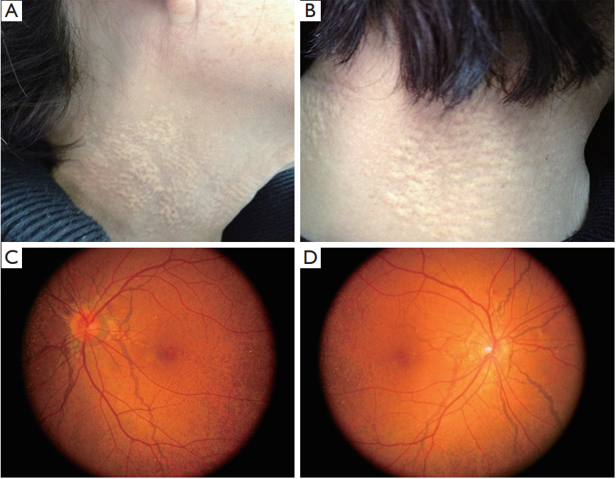
A 53-year-old Chinese woman with PXE and angioid streaks was referred in May 2012 because of noticing visual loss in his left eye (LE) of 2 month’s duration, and the occurrence of metamorphopsia and central scotoma. Her right eye (RE) was unaffected. The patient reported the onset of yellowish asymptomatic micropapules in the cervical region 10 years ago (Figure 1A,B), which later progressed to the axillae and cubital fossae. In her personal records, a history of arterial hypertension or diabetes mellitus was not noticed. The patient denied similar cases in her family but her daughter, who reported the onset of yellowish asymptomatic micropapules in 2 years ago, and the presence of bilateral angioid streaks confirmed by ophthalmoscopy and fundus photography (Figure 1C,D). They two were diagnosed with angioid streaks associated with PXE, which was confirmed by skin biopsy.

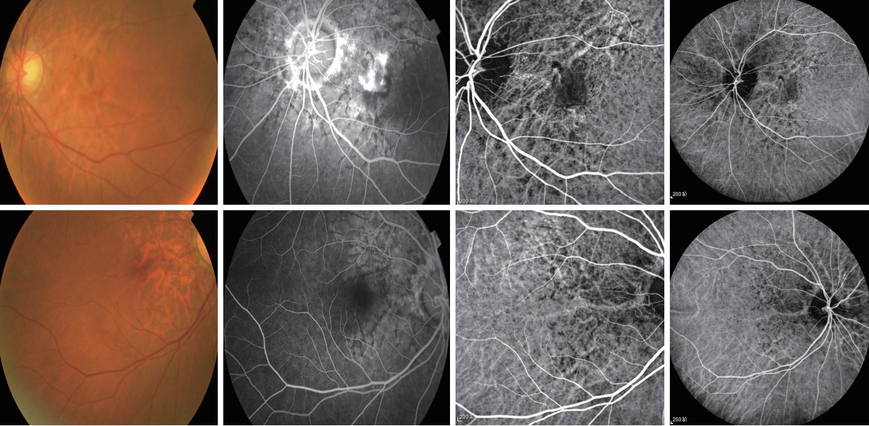
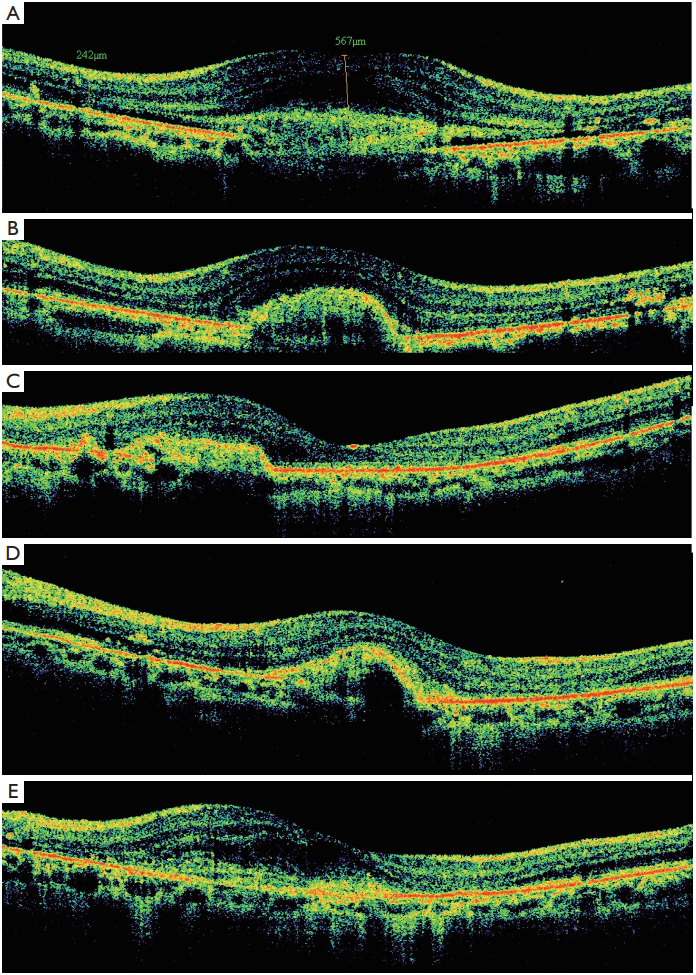
Eye examinations including fundus photography, fluorescein angiography, indocyanine green angiography, and optical coherence tomography (OCT) revealed the presence of CNV in her LE, while no CNV were seen in her RE (Figure 2). Fluorescein angiography revealed a hyper- and hypofluorescent grid due to alteration of the retina pigment epithelium and a hyper-fluorescent lesion with late diffusion in the LE compatible with CNV. OCT (Figure 3) evidenced a loss of structure of the RPEchoriocapillary hyper-reflective strip, and a dense cupshaped lesion in the LE (CNV), surrounded by a small neuroepithelium detachment. At presentation VA was 20/200 in the LE and 6/6 in the RE.

The off-label use of the drug and its potential risks and benefits were discussed with the patient. The study adhered to the tenets of the Declaration of Helsinki, and written informed consent was obtained from the patient. The study was approved by the institutional review board/ethics committee of the Second Affiliated Hospital of Medical College of Xi’an Jiaotong University, China. Treatment of her LE with intravitreal injection of bevacizumab (1.25 mg/50 µL, Avastin, Genentech/Roche, USA) once a month was initiated in May 2012. After 3 months, the patient’s VA was 20/100 stably in her LE and there was no evidence of macular hemorrhage or leaking of the neovascular complex. After 2 years, there still was no evidence of neovascular complex leakage or hemorrhage, and best corrected visual acuity (BCVA) still was 20/100. Central retinal thickness decreased significantly from 567 µm at baseline to 314 µm at the last followup as measured by OCT (Figure 3). His VA was stable in his bevacizumab-treated LE 2-year later. Intraocular pressure was normal (16 mmHg) in both eyes. Bevacizumab treatment was well tolerated with no adverse events reported.

Figure 1 Skin changes of the patient and the retinography of the patient’s daughter. Irregular skin surface of inelastic consistency, presenting plaques constituted of symmetric small yellowish papules located in the cervical region of the patient (A,B); the retinography of the patient’s daughter (C,D) showed that angioid streaks visualized in retinography. 


Figure 2 Eye examinations of the patients. Angioid streaks visualized in retinography bilaterally and highlighted in the fluorescein angiography. Fluorescein angiography of the right eye at presentation shows no obvious leakage. The color fundus image of LE shows the macular involvement by the CNV. The CNV of LE is visible by fluorescein angiography and indocyanine green angiography.

Figure 3 Spectral-domain OCT at presentation and post treatment. Baseline OCT (A) showed diffuse retinal thickening with subfoveal CNV. The patient reported subjective improvement of vision after one injection; OCT (B after one injection and C after three injections) after treatment showed CNV thinning and diminished retinal thickness; OCT of 1 year (D) and 2 years (E) after presentation revealed stable CNV, but retinal structural disorder. 


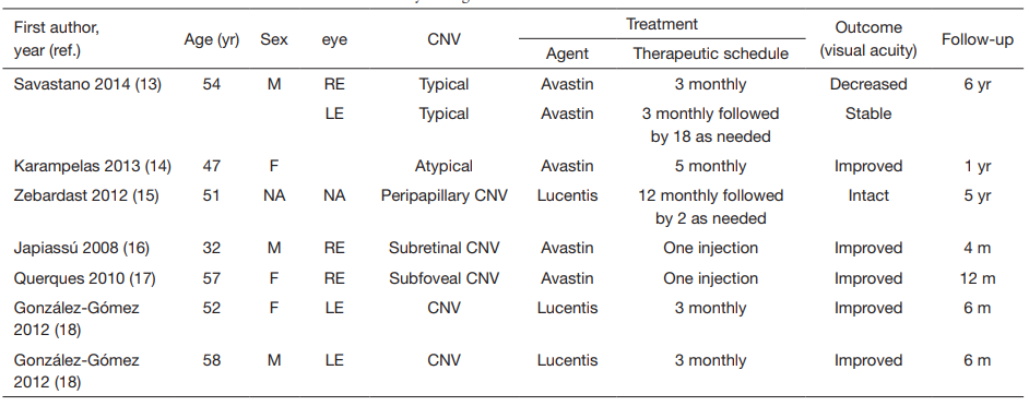
Table 1 Characteristics and clinical data of CNV secondary to angioid streaks in PXE treated with anti-VEGF

Systematic literature review

A systematic review of the cases reported in the literature was conducted to examine the safety and effectiveness of intravitreal anti-VEGF treatment for CNV associated with PXE. We searched Cochrane Library, PubMed, OVID, and UpToDate databases (from inception to July 2015) using combinations of search terms including pseudoxanthoma elasticum or Grönblad-Strandberg syndrome and VEGF or CNV. Electronic searches were supplemented by perusal of the references of the retrieved papers. All potentially eligible studies and their references were carefully scanned to identify other eligible studies.

The systematic review included studies that fulfilled all of the following criteria: (i) a focus on anti-VEGF treatment of CNV associated with angioid streaks in PXE; (ii) a diagnosis verified by eye examinations including fundus photography, fluorescein angiography, and OCT; and (iii) a previously unreported patient. We included studies irrespective of study design and regardless type of the literature. However, we excluded studies without primary data, i.e., commentaries, or letters to the editor. We included only papers in English.

All items were extracted by one investigator (Aiyi Zhou) and were confirmed by two other investigators (Yanlong Quan and Chenjing Zhou). Discrepancies were resolved after consensus. For each study, we extracted the name of first author, publication year, country, and type of research center or department where the study was conducted. We also recorded sample size, age of participants, gender, type of CNV, anti-VEGF drugs, times and frequency of intravitreal treatment, outcomes about VA and fundus examinations including OCT and fluorescein angiography, follow-up period. In addition, we recorded intravitreal treatment complications or specific outcome were reported.

Ten papers (four for case series, and six for case reports) were eligible and finally included. There was no randomized controlled clinical trial about CNV secondary to PXE. Four papers (9-12) about three case series including 41 eyes of 38 patients investigated the efficacy of intravitreal bevacizumab injections for treating CNV secondary to PXE ,wherein two papers (11,12) were the same case series of short-term and long-term observation respectively. Six papers of case reports reported 7 patients (8 eyes) of CNV associated with angioid streaks in PXE treated with bevacizumab or ranibizumab. 

Tables 1,2 summarizes the patients characteristics, the therapies implemented and the outcomes recorded. As detailed in Tables 1,2, almost all published cases reported positive response to treatment with anti-VEGF in CNV secondary to PXE. The majority of published cases applied bevacizumab. Three references involving ranibizumab for the treatment of this disease were found.

Finger RP started an uncontrolled investigator-initiated clinical trial to test the efficacy and safety of intravitreal ranibizumab for CNV due to PXE in 2007 at Department of Ophthalmology, University of Bonn, Bonn, Germany (PXE-CNV 06; http://www.clinicaltrials.gov). And they (Retina, 2011) (10) compared the group of patients with early disease stage and limited fundus changes in PXE with that of those with advanced disease stage and more pronounced fundus alterations, and concluded that patients with early disease have better treatment outcomes with a marked improvement in VA. Two papers (11,12) about a same series of 9 patients (9 eyes) with PXE and CNV related to angioid streaks managed with intravitreal bevacizumab were from Retina Research Center of the Manhattan Eye, Ear, and Throat Hospital, New York University School of Medicine. They reported short-term (6 months) and long-term (28.6 months) efficacy of intravitreal anti-VEGF in this disease respectively. A total of eight of nine eyes developed recurrence of CNV during the study period. The mean recurrence rate in eyes on the PRN regimen was 2 (range, 0–4) during the follow-up period. The PRN regimen group and the maintenance regimen group did not have significant differences in VA outcomes.

Table 2 Case series of CNV secondary to angioid streaks in PXE treated with anti-VEGF

Discussion

Previous methods of treatment for CNV in patients with PXE such as thermal photocoagulation and verteporfin PDT had poor outcomes (19-22). This presumably resulted from the treatment itself creating tissue injury and high recurrence rates after these procedures. This case report demonstrates that intravitreal bevacizumab is effective and safe treatment options for CNV secondary to PXE. The presented case report supports and confirms so far published evidence for the effectiveness of bevacizumab in CNVs secondary to PXE.

In patients with PXE who have an increased cardiovascular risk profile, possible complications of intravitreal treatment with bevacizumab need to be considered. Patients need to be aware of the possibility of systemic side effects such as myocardial infarction or a deterioration of their cardiovascular; although at present no definite proof has been documented of this higher risk in patients with or without PXE treated with intravitreal anti-VEGF. No drug- or injection-related complications were noted in these literatures. No patients with PXE experienced increased intraocular pressure, inflammation, endophthalmitis, retinal breaks, retinal detachment, or ocular or systemic occlusive events (including myocardial infarction and cerebral ischemia).

The risk of recurrent exudation of CNV associated with angioid streaks in PXE and likely need for repeated injections is obvious (12). Most eyes required repeated injections to achieve CNV quiescence and visual stabilization. Despite the higher recurrence rate in the eyes that received intravitreal anti-VEGF as needed for recurrence, both maintenance and PRN treatment regimens seemed capable of stabilizing or improving final VA outcomes in most patients. However, the ideal timetable and criteria for injections have yet to be determined. Although there seem to be arguments to treat selected patients with a maintenance treatment of intravitreal injections once a month, an as-needed regimen is the most used strategy. Further prospective, randomized controlled clinical trials with defined treatment protocols would be necessary to elucidate the ideal timing and criteria for intravitreal antiVEGF administration for managing CNV from PXErelated angioid streaks. Specifically, studies with close regular follow-up comparing the efficacy of a maintenance regimen versus an as needed regimen are required.

A limitation of these case series is the retrospective study design. Ideally, effectiveness of bevacizumab in CNVs because of PXE should be assessed in a randomized controlled clinical trial. However, because withholding treatment for CNVs because of PXE is very problematic, clinical trials are most likely to be implemented uncontrolled. PXE is a rare disease, and therefore, largescale trials appear unrealistic. Continued experience with intravitreal bevacizumab or ranibizumab in this population will help establish long-term efficacy and better define optimal dosing strategies.

Overall, based on the evidence available, intravitreal treatment with anti-VEGF seems to be the best choice at present to treat patients with CNV secondary to angioid streaks of PXE. Further studies and larger case series are needed to clarify the role of anti-VEGF therapy in managing CNV secondary to PXE.
1、Finger RP, Charbel Issa P, Ladewig MS, et al. Pseudoxanthoma elasticum: genetics, clinical manifestations and therapeutic approaches. Surv Ophthalmol 2009;54:272-85
2、Chassaing N, Martin L, Calvas P, et al. Pseudoxanthoma elasticum: a clinical, pathophysiological and genetic update including 11 novel ABCC6 mutations. J Med Genet 2005;42:881-92
3、Le Saux O, Urban Z, Tschuch C, et al. Mutations in a gene encoding an ABC transporter cause pseudoxanthoma elasticum. Nat Genet 2000;25:223-7.
4、Struk%20B%2C%20Cai%20L%2C%20Z%C3%A4ch%20S%2C%20et%20al.%20Mutations%20of%20the%20gene%20encoding%20the%20transmembrane%20transporter%20protein%20ABC-C6%20cause%20pseudoxanthoma%20elasticum.%20J%20Mol%20Med%20(Berl)%20%0A2000%3B78%3A282-6.
5、Schoenberger SD, Agarwal A. Geographic chorioretinal atrophy in pseudoxanthoma elasticum. Am J Ophthalmol 2013;156:715-23.
6、Georgalas I, Papaconstantinou D, Koutsandrea C, et al. Angioid streaks, clinical course, complications, and current therapeutic management. Ther Clin Risk Manag 2009;5:81-9.
7、Zarbock R, Hendig D, Szliska C, et al. Vascular endothelial growth factor gene polymorphisms as prognostic markers for ocular manifestations in pseudoxanthoma elasticum. Hum Mol Genet 2009;18:3344-51
8、Verbraak FD. Antivascular endothelial growth factor treatment in pseudoxanthoma elasticum patients. Dev Ophthalmol 2010;46:96-106.
9、Finger RP, Charbel Issa P, Ladewig M, et al. Intravitreal bevacizumab for choroidal neovascularisation associated with pseudoxanthoma elasticum. Br J Ophthalmol 2008;92:483-7.
10、Finger RP, Charbel Issa P, Schmitz-Valckenberg S, et al. Long-term effectiveness of intravitreal bevacizumab for choroidal neovascularization secondary to angioid streaks in pseudoxanthoma elasticum. Retina 2011;31:1268-78.
11、Bhatnagar P, Freund KB, Spaide RF, et al. Intravitreal bevacizumab for the management of choroidal neovascularization in pseudoxanthoma elasticum. Retina 2007;27:897-902.
12、Myung JS, Bhatnagar P, Spaide RF, et al. Longtermoutcomes of intravitreal antivascular endothelial growth factor therapy for the management of choroidal neovascularization in pseudoxanthoma elasticum. Retina 2010;30:748-55.
13、Savastano MC, Minnella AM, Zinzanella G, et al. Successful long-term management of choroidal neovascularization secondary to angioid streaks in a patient with pseudoxanthoma elasticum: a case report. J Med Case Rep 2014;8:458.
14、Karampelas M, Soumplis V, Karagiannis D, et al. An atypical case of choroidal neovascularization associated with pseudoxanthoma elasticum treated with intravitreal bevacizumab: a case report. BMC Res Notes 2013;6:530.
15、Zebardast N, Adelman RA. Intravitreal ranibizumab for treatment of choroidal neovascularization secondary to angioid streaks in pseudoxanthoma elasticum: five-year follow-up. Semin Ophthalmol 2012;27:61-4.
16、Japiass%C3%BA%20RM%2C%20Japiass%C3%BA%20MA%2C%20Pecego%20MG.%20Intravitreal%20bevacizumab%20in%20choroidal%20neovascularization%20secondary%20to%20Gr%C3%B6nblad-Strandberg%20syndrome%3A%20case%20report.%20Arq%20Bras%20%0AOftalmol%202008%3B71%3A427-9.
17、Querques G, Bux AV, Prascina F, et al. Intravitreal avastin for choroidal neovascularization associated with stargardtlikeretinal abnormalities in pseudoxanthoma elasticum. Middle East Afr J Ophthalmol 2010;17:387-9.
18、González-Gómez A, Morillo MJ, González-Escobar AB, et al. Choroidal neovascularization secondary to pseudoxanthoma elasticum treated with ranibizumab: a report of 2 cases. Arch Soc Esp Oftalmol 2012;87:153-6.
19、Lee JM, Nam WH, Kim HK. Photodynamic therapy with verteporfin for choroidal neovascularization in patients with angioid streaks. Korean J Ophthalmol 2007;21:142-5.
20、Jurklies B, Bornfeld N, Schilling H. Photodynamic therapy using verteporfin for choroidal neovascularization associated with angioid streaks--long-term effects. Ophthalmic Res 2006;38:209-17.
21、Arias L, Pujol O, Rubio M, et al. Long-term results of photodynamic therapy for the treatment of choroidal neovascularization secondary to angioid streaks. Graefes Arch Clin Exp Ophthalmol 2006;244:753-7.
22、Pece A, Avanza P, Galli L, et al. Laser photocoagulation of choroidal neovascularization in angioid streaks. Retina 1997;17:12-6.
上一篇
下一篇
其他期刊
  • 眼科学报

    主管:中华人民共和国教育部
    主办:中山大学
    承办:中山大学中山眼科中心
    主编:林浩添
    主管:中华人民共和国教育部
    主办:中山大学
    浏览
  • Eye Science

    主管:中华人民共和国教育部
    主办:中山大学
    承办:中山大学中山眼科中心
    主编:林浩添
    主管:中华人民共和国教育部
    主办:中山大学
    浏览
推荐阅读
出版者信息
目录