您的位置: 首页 > 2016年9月 第31卷 第3期 > 文字全文
2023年7月 第38卷 第7期11
目录

Delayed presentation of intracameral graphite foreign body in a child: school trauma

Delayed presentation of intracameral graphite foreign body in a child: school trauma

来源期刊: 眼科学报 | 2016年9月 第31卷 第3期 189-191 发布时间: 收稿时间:2024/11/30 21:22:12 阅读量:346
作者:
关键词:
Graphite foreign body intraocular foreign body in children school trauma
DOI:
doi: 10.3978/j.issn.1000-4432.2016.09.03
收稿时间:
2016-08-02 
修订日期:
 
接收日期:
2016-08-31 
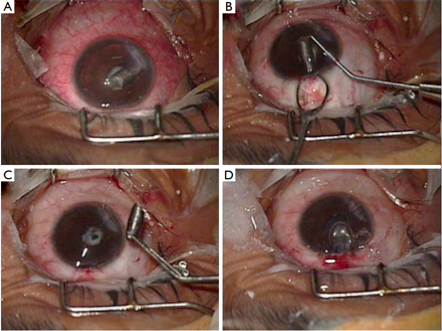
We report a case of large graphite foreign body (FB) in the anterior chamber of eye of a 4-yearold child, incurred during unsupervised play. Despite delayed presentation, the eye had few signs of resolved inff ammation which allowed safe extraction of the FB bimanually through limbus. School play, especially in young children, should be under supervision and free of sharp objects. Graphite is inert while inside the eye, and even large pieces can be well tolerated for long time in absence of infection.  

Case presentation

We report a case of a 4-year-old girl child who was presented to our ocular emergency department with blurred vision in left eye (LE). There was history of trauma in school with a graphite based pencil 21 days back. Trauma had occurred while playing in class room and delayed presentation was due to late referral and economic reasons. Child did not allow complete examination or accurate assessment of visual acuity. However, supersonic inspection of the LE posterior segment was found to be normal. Hence keeping a suspicion of open globe injury, parents were counseled for examination under anesthesia and possible surgery for primary repair in operation room. A written informed consent was obtained from the parents for the surgery. During examination under anesthesia, right eye was within normal limits while LE was found to have a full thickness corneal laceration in the inferonasal quadrant along with a large piece of pencil lead in anterior chamber and cataract (Figure 1A). The pencil lead was more than half the size of the corneal diameter. There were no gross signs of inff ammation.

The wound was secured with a single 10–0 mono filament nylon suture and the anterior chamber formed with visco-elastic. A superior limbal incision was made and the long axis of foreign body (FB) was aligned perpendicular to it. The FB was delivered bimanually using a hook and a non-irrigating wire Vectis (Figure 1B). The laceration and superior incision were then repaired and anterior chamber formed with air and saline (Figure 1C,D). Multiple long sutures were needed to repair the wound. The anterior chamber was well formed, intra ocular pressure normal on digital palpation with visual acuity of counting ff ngers close to face on ff rst postoperative day. She subsequently underwent lens aspiration with intraocular lens implant 6 weeks later, and her visual acuity improved to at least 6/18. 

Figure 1 Intraoperative removal of foreign body. (A) Full thickness corneal laceration along with intra cameral graphite foreign body (FB). Note sharp end of FB is towards the wound; (B) wound repaired with single suture and FB being removed bimanually with sinskey hook and wire Vectis; (C) FB successfully removed without complications; (D) wound and incision site repaired with nylon sutures and anterior chamber formed with air bubble.

Discussion

School trauma during unsupervised play is common and can be blinding (1). Such children are especially prone to trauma with sharp objects like pencils which are commonly available in the school premises. Unsupervised play with sharp objects should therefore be avoided. Penetrating injuries may present with lack of pain or redness or pertinent history, especially in less than 5 years age group and should not stop us from suspicion of a FB. One ff fth of patients with ocular FB may present without any symptoms (2). Although superficial FB on the conjunctiva or cornea may be easily seen and at times managed in the emergency room itself, supratarsal and intraocular FB may require specialized slit lamp examination and expert surgical removal. Appropriate referral should be made as early as possible for the same as delayed management increases the possibility of vision threatening complications like endophthalmitis (3)

Graphite being inert does not incite inflammatory reactions (4), unless infective, explaining a relatively stable anterior chamber. The only evidence of previous inflammation in our case was a thin membrane over the pupil, which was easily removed. The eye overall appeared to be stable at presentation, notwithstanding the size of the FB or prolonged exposure to graphite. Wound apposition however was difficult, presumably due to delayed repair. Orienting the long axis of FB perpendicular to the removal site (Figure 1B) followed by bimanual extraction is easy and off ers an uncomplicated removal of large anterior chamber foreign bodies. 

Conclusions

School trauma is a cause of preventable vision loss in young children and school environment needs to be appropriately controlled. Intraocular graphite FB is well tolerated, though delayed wound repair may be difficult.
1、Ayanniyi AA, Mahmoud OA, Olatunji FO, et al. Pattern of ocular trauma among primary school pupils in Ilorin, Nigeria. Afr J Med Med Sci 2009;38:193-6.
2、Upshaw JE, Brenkert TE, Losek JD. Ocular foreign bodies in children. Pediatr Emerg Care 2008;24:409-14; quiz 415-7.
3、Li X, Zarbin MA, Bhagat N. Pediatric open globe injury: A review of the literature. J Emerg Trauma Shock 2015;8:216-23.
4、Han ER, Wee WR, Lee JH, et al. A case of retained graphite anterior chamber foreign body masquerading as stromal keratitis. Korean J Ophthalmol 2011;25:128-31.
上一篇
下一篇
其他期刊
  • 眼科学报

    主管:中华人民共和国教育部
    主办:中山大学
    承办:中山大学中山眼科中心
    主编:林浩添
    主管:中华人民共和国教育部
    主办:中山大学
    浏览
  • Eye Science

    主管:中华人民共和国教育部
    主办:中山大学
    承办:中山大学中山眼科中心
    主编:林浩添
    主管:中华人民共和国教育部
    主办:中山大学
    浏览
推荐阅读
出版者信息
目录

点击右上角菜单,浏览器打开下载

我知道了