您的位置: 首页 > 2025年3月 第40卷 第3期 > 文字全文
2023年7月 第38卷 第7期11
目录

非感染性葡萄膜炎继发脉络膜新生血管的多模式影像特征

Multimodal imaging features of choroidal neovascularization secondary to non-infectious uveitis

来源期刊: 眼科学报 | 2025年3月 第40卷 第3期 241-251 发布时间:2025-3-28 收稿时间:2025/3/25 8:56:33 阅读量:84
作者:
关键词:
脉络膜新生血管眼底多模式影像光学相干断层扫描血管成像
choroidal neovascularization multimodal imaging optical coherence tomography angiography
DOI:
10.12419/24111902
收稿时间:
2024-10-04 
修订日期:
2024-12-23 
接收日期:
2025-01-10 
目的:评估炎非感染性葡萄膜炎继发炎性脉络膜新生血管(inflammatory choroidal neovascularization, iCNV)的临床特征及眼底多模式影像表现。方法:采用回顾性观察性研究,采用眼底荧光素血管造影(fundus fluorescein angiography, FFA)、吲哚菁绿血管造影(Indocyanine green angiography, ICGA)、谱域相干光断层扫描(spectral domain optical coherence tomography, SD-OCT)联合光学相干断层扫描血管成像(optical coherence tomography angiography, OCTA)等多种眼底影像学方法,对纳入患者的眼底进行检查,分析非感染性iCNV的面积、分型、位置及形态等影像学特征与临床特征的关系。结果:研究共纳入39例患者,对48只患眼中的51处iCNV病灶进行了评估。纳入患者年龄为(35.28±13.62)岁。其中3例患眼出现多灶性CNV。SD-OCT显示92.16%(47/51)的iCNV为2型CNV,17.65%(9/51)的iCNV出现海绵征,13.72%(7/51)的iCNV伴有局灶脉络膜凹陷。ICGA造影期间,74.50%的iCNV病灶(38/51)伴有弱荧光病灶,25.49%的病例(13/51)显示脉络膜高通透性表现。OCTA enface图像显示iCNV形态多样,包括焦点状(15例,29.41%)、盘状/海扇状(16例,31.37%)、枯树状(9例,17.65%)、星状(9例,17.65%)及弥漫网状(2例,3.92%)。其中,枯树状及星状iCNV提示iCNV为非活动性(P<0.01)。结论:非感染性iCNV与炎性病灶关系密切,在SD-OCT,ICGA上皆具特征性的影像表现。OCTA能直观地观察到iCNV的形态。这些多模式影像特征为临床医生提供了对于非感染性iCNV重要的鉴别诊断依据,有助于制定有效的诊疗方案。
Objective: To evaluate the clinical characteristics and multimodal imaging features of non-infectious inflammatory choroidal neovascularization (iCNV). Methods: In this study retrospective, observational study, multimodal imaging examinations, including fluorescein angiography (FFA), indocyanine green angiography (ICGA), spectral-domain optical coherence tomography (SD-OCT), and optical coherence tomography angiography (OCTA), were used to observe the morphology of non-infectious iCNV in patients diagnosed with uveitis. The area of iCNV, CNV types, CNV morphology and other imaging characteristics were further analyzed. Results: A total of 39 patients were included, with 48 affected eyes and 51 iCNV were identified. The average age of the included patients was 35.28±13.62 years. Among the affected eyes, 3 presented with multifocal CNV, and 92.16% of iCNV were classified as type 2 CNV. iCNV exhibited diverse morphologies, including focal-like pattern (15 cases, 29.41%),sea-fan pattern(16 cases, 31.37%), dead-tree pattern CNV(9 cases, 17.65%), stellar pattern (9 cases, 17.65%), and diffuse reticular (2 cases, 3.92%). Notably, tree-like and stellar pattern iCNV showed a significant correlation with non-active CNV (P < 0.01). Conclusions: Non-infectious iCNV is closely related to inflammatory lesions, exhibiting characteristic imaging features on SD-OCT and ICGA. OCTA allows for direct observation of the morphology of iCNV. These multimodal imaging characteristics provide important diagnostic criteria for clinicians, aiding in the formulation of effective treatment plans.

文章亮点

1. 关键发现

 • 本文总结了非感染性葡萄膜炎继发脉络膜新生血管 (inflammatory choroidal neovascularization, iCNV) 的多模式影像特征,揭示了 光学相干断层扫描血管成像 (optical coherence tomography angiography, OCTA) 上 iCNV 形态的多样性,包括焦点状、盘状 / 海扇状、枯树状、星状及弥漫网状。其中,枯树状和星状形态与非活动性 iCNV 显著相关。

2. 已知与发现

 • iCNV 是葡萄膜炎的严重并发症,可显著损害视力;抗 VEGF 治疗是 iCNV 的有效疗法,但早期诊断对预后至关重要。
 • OCTA作为一种非侵入性的眼底检查技术,通过探测眼底血流信号变化构建可视化的血管网,已被证明在诊断 iCNV 中具有高灵敏度和特异度。

3. 意义与改变

 • iCNV特异性的多模式影像特征为临床医生提供了对于其鉴别诊断的重要依据,同时基于OCTA的形态-活动性关联框架,利于推动炎 iCNV 精准诊疗体系的完善。

       炎性脉络膜新生血管(inflammatory choroidal neovascularization, iCNV)是指与葡萄膜炎相关的继发性脉络膜新生血管(uveitis - related choroidal neovascularization)。iCNV 是葡萄膜炎严重损伤患者视力的临床并发症[1-4]。目前,关于继发于感染性葡萄膜炎的iCNV发生率的数据很少,主要来自病例报告,而在非感染性后葡萄膜炎(non-infectious uveitis)患者中,iCNV 的发病率约为2%[2,5-6];iCNV的发生率主要决于葡萄膜炎的类型,在点状内脉络膜病变(punctate inner choroidopathy, PIC)、多灶性脉络膜炎(multifocal choroiditis,MFC)、匍行性脉络膜炎(serpiginous choroiditis,SC)及小柳-原田综合征(vogtkoyanagi-harada disease,VKH)等类型中,iCNV 的发生率高达9%~75%[4,7-8]。目前,玻璃体腔注射抗血管内皮生长因子(anti-vascular endothelial growth factor, antiVEGF)已被证实在iCNV中有较好的疗效[2],因此,临床工作中对葡萄膜炎患者进行监测,尽早诊治iCNV是非常重要的。
       既往研究表明,炎症是脉络膜新生血管(choroidal neovascularization, CNV)的第三大常见原因,发病率仅次于年龄相关性黄斑变性(age-related macular degeneration, AMD)和病理性近视(pathological myopia, PM)[2,4,9]。然而,由于炎症因素的参与,iCNV的诊断仍具挑战性[4],如何将iCNV与炎症病灶相鉴别,以及iCNV与病理性近视继发性CNV(pathological myopia choroidal neovascularization, PM-CNV)的鉴别一直是临床工作中的难点。传统的荧光素血管造影(fluorescein angiography, FFA)和吲哚菁绿血管造影术(indocyanine green angiography, ICGA)结合相干光断层扫描(spectraldomain optical coherence tomography, SD-OCT)对监测葡萄膜炎的疾病进展和治疗反应至关重要[1]。而近年来光学相干断层扫描血管成像(optical coherence tomography angiography, OCTA)作为一种非侵入性、可量化的眼底检查技术,通过探测眼底血流信号变化构建可视化的血管网,已被证明在诊断iCNV中具有高灵敏度和特异度[9-11],但关于iCNV的眼底多模式影像表现尤其是OCTA上的形态改变,目前鲜有相关研究报道。
       既往大多数与iCNV相关的研究数据来自不同类型葡萄膜炎的病例报告或研究。然而,总体iCNV的临床特征及多模式影像表现尚未得到充分记录及报道。本研究旨在总结iCNV的临床特征及多模式影像表现,尤其是OCTA上的形态特征,以期为iCNV的早期诊断提供更多依据。

1 资料与方法

1.1 研究对象

       采用回顾性分析,收集2017年1月—2024年1月转诊至中国广州中山眼科中心的所有诊断为“葡萄膜炎继发CNV”“炎性脉络膜血管”“炎性CNV”的患者资料。该研究方案已获得中山大学中山眼科中心机构审查委员会的批准(批件号:2018KYPJO98)。本研究遵守了赫尔辛基宣言中关于人类参与者的相关规定。纳入标准:患者年龄<80岁,无耳聋、痴呆、精神疾病,能在检查及随访过程中明确回应、知情并同意其信息用于临床研究的患者,屈光间质清晰并具备完善的眼底影像检查并诊断明确为iCNV。排除标准:患有AMD、病理性近视CNV、中心性浆液性脉络膜视网膜病变、糖尿病视网膜病变等其他视网膜疾病者。
       葡萄膜炎的诊断及分类基于葡萄膜炎命名标准化(standardization of uveitis nomenclature, SUN)工作组 [7,12]。感染及非感染性葡萄膜炎的诊断基于详细的病史分析,结合全面的眼部查体、眼科辅助检查、实验室检查及胸部X线检查结果,经临床和影像学检查确诊为CNV,CNV眼底诊断依据包括检眼镜下见深层视网膜灰白病灶伴视网膜浆液性脱离、出血,对应病灶在FFA造影期间表现为早期强荧光伴或不伴造影期间荧光素渗漏,在ICGA上表现为晚期强荧光病灶染色,在SD-OCT上为视网膜色素上皮(retinal pigment epithelium, RPE)连续性中断、伴其上中高反射的病灶。OCTA可清晰呈现CNV,表现为外层视网膜enface图像上的异常血管网信号。

1.2 临床资料及眼底多模式影像资料分析

       所有临床及影像学资料均在患者同一次就诊中获得。每例患者在此次就诊中均接受了系统且全面的眼科检查。眼科临床评估项目主要涵盖裂隙灯显微检查、散瞳后眼底检查及最佳矫正视力(bestcorrected visual acuity, BCVA)检查。在视觉功能评估环节,最佳矫正视力(BCCV)的测定严格遵循国际标准视力表系统,采用最小分辨角对数(logarithm of the minimum angle of resolution, logMAR)作为标准化记录单位。同时,患者还接受了眼底多模式影像检查,包括眼底照相(使用Zeiss FF450 plus眼底照相机,德国Carl Zeiss, Jena,或Topcon 50 VT眼底照相机,Topcon Corp, Tokyo,日本)、SD-OCT、FFA、ICGA(Spectralis HRA + OCT,海德堡工程)及OCTA(AngioVue, RTVue XR Avanti,Optovue,Fremont,美国加利福尼亚州)。所有患者的OCTA图像均由同一位经验丰富的临床技术员在患者充分散大瞳孔后进行采集。检查过程中,采用“HD-Angio Retina 6 mm×6 mm”进行黄斑区扫描,以覆盖更多病灶。使用机器内置软件进行视网膜分层,如发现分层错误,将进行手动调整。
       基于光学相干断层扫描血管成像(OCTA)的视网膜分层分析,系统预设将视网膜结构划分为四个解剖层次进行观察:浅层视网膜结构定义为内界膜(internal limiting membrane, ILM)至内丛状层(inner plexiform layer, IPL)基底面下方10微米区间;深层视网膜结构涵盖IPL基底面下10微米至外丛状层(outer plexiform layer, OPL)上10微米范围;外层视网膜(无血管层)结构由OPL顶面上10微米延伸至布鲁赫膜(Bruch's membrane)基底面下10微米;而脉络膜毛细血管层的检测区间则设定为布鲁赫膜基底面下10-30微米深度。针对iCNV的影像学评估,需重点观察外层视网膜结构层面呈现的异常血管网络,该区域是检测病理性脉络膜新生血管改变的关键解剖部位。
       根据患者的临床资料及多模式眼底影像资料,评估iCNV的特征如下:1) iCNV的位置,根据B-scan中CNV和黄斑中心凹、视盘的位置,将CNV的位置分为中心凹下(中心凹无血管区下方)、中心凹旁(距中心凹无血管区 1~199 μm内)、中心凹外(中心凹无血管区外 200 μm)、视盘旁(距离盘边缘1个视盘直径)[9] ;2) iCNV活动性,FFA是评估 CNV 活动性的传统金标准,评估标准为在FFA造影期间,CNV处是否伴有荧光素渗漏;3) iCNV的形态,依据OCTA 外层视网膜层面的enface图像,将iCNV分为焦点状、盘状/海扇状、枯树状、星状及弥漫网状,CNV的形态学评估标准见图1;4) iCNV的面积,使用OptVue内置定量软件在外层视网膜enface层面勾勒iCNV的边缘,并由软件测算iCNV的面积;5) CNV类型,按MNV分类标准,根据B-scan中CNV累及视网膜的层次将CNV分为1型、2型及混合型[13–16];6) iCNV处中心凹下脉络膜厚度,使用Spectralis OCT内置软件测量中心凹下脉络膜厚度;7) 通过SD-OCT/B-scan OCT分析iCNV是否伴有视网膜下液(视网膜神经上皮脱离伴视网膜下液性暗腔)、视网膜层间液体(视网膜层间出现低反射性区域)、脉络膜高穿透性(hypertransmission in choroid)、脉络膜海绵征(sponge sign,炎症活动期iCNV下脉络膜厚度显著增加,炎症消退期该区域脉络膜厚度显著减少,这种脉络膜厚度随炎症活动性显著变化称为“脉络膜海绵征”)[2-3]、局灶脉络膜凹陷(脉络膜组织局灶性缺失伴其上视网膜外层以及RPE/脉络膜毛细血管复合体光带局限向脉络膜层凹陷)[17-20]、干草叉征(pitchfork sign,指在B-scan OCT上活动性CNV病灶中自外层视网膜的高反射叉柱状突起)[4,21-24];8)FFA特征,iCNV处是否有荧光素渗漏、病灶染色等;9) ICGA特征,造影晚期iCNV是否染色、是否伴有脉络膜血管高通透、点状弱荧光等。

1.3 统计学分析

       统计学分析使用SPSS 16.0软件。正态分布的定量资料以x±s进行统计描述;非正态分布的定量资料以中位数(极差)描述;计数资料以n(%)描述。与患者相关的临床数据依据数据类型合理进行独立样本检验,Kruskal-Wallis检验,χ2检验或Fisher确切概率法。P<0.05被认为差异有统计学意义。

2 结果

2.1 纳入患者的基本资料及临床特征

       本研究共纳入39名患者的48例患眼。纳入患者的平均年龄为(35.28±13.62)岁(范围为13~68岁),主要为女性(30例,76.92%)。本研究纳入病例经全面临床检查后诊断为非感染性葡萄膜炎,纳入iCNV常见的病因学诊断包括PIC(31例,64.58%),MFC(5例,10.42%),VKH(4例,8.33%),多发性一过性白点综合征(multiple evanescent white dot syndrome,MEWDS)(3例,6.25%)。同时本研究亦发现iCNV在一些较罕见的葡萄膜炎中发生,纳入了弥漫性视网膜下纤维化综合征(diffuse subretinal fibrosis syndrome,DSFS)2例,匐行性脉络膜炎(serpiginous choroiditis,SC)2例及持续性鳞状黄斑病变(persistent placoid maculopathy,PPM)1例。其中出现眼部症状到本次就诊平均时间为(14.21±11.24)个月。纳入患眼中有34眼(70.83%)中iCNV具有活动性,余下14眼(29.17%)为非活动性。21眼(43.75%)眼底炎性病灶具有活动性,余下27眼眼底炎性病变为非活动性。iCNV的活动和眼底炎性病变活动性相关(χ2=4.18,P<0.05)。纳入患眼的平均屈光度为(-4.46±4.78)D(范围为-23~0 D),基线视力为logMAR(0.45±0.39)。纳入患者的基本信息见表1。

表 1 39 例 (48 眼 ) 炎性脉络膜新生血管患者的基本信息
Table 1 Clinical characteristics of 39 included patients (48 eyes) with inflammatory choroidal neovascularization

项目


结果

患者数量,人次(患眼数目,只)


3948

诊断时的年龄/岁


35.28±13

性别,人次(%)


女性


30,76.92%

男性


9, 23.08%

屈光度,D(范围)


-4.46±4.78(-23D~0D)

双侧iCNV,人次(%)


9(23.08%

多灶iCNV,眼(%)


36.25%

诊断iCNV时的视力,BCVA (logMAR)


0.45±0.39

iCNV活动性


活动性CNV,眼数(%)


3470.83%

陈旧性CNV,眼数(%)


1429.17%

出现症状到就诊平均时间,月(平均值±标准差)


14±11.24

是否曾行抗VEGF治疗


12(25%


36(75%

是否为活动性炎症


21(43.75%


27(56.25%

病因,眼数(%)


n,%

PIC


3164.58%

MFC


5 10.42%

VKH


4 8.33%

MEWDS


3 6.25%

DSFS


2 4.17%

SC


2 6.25%

PPM


1,2.08%

注:iCNV,炎性脉络膜新生血管;BCVA,最佳矫正视力,PIC,点状内层脉络膜病变;MFC,多灶脉络膜炎;VKH,小柳-原田综合征;MEWDS,多发性一过性白点综合征;DSFS,弥漫性视网膜下纤维化综合征;SC,匍行性脉络膜炎;PPM,持续性鳞状黄斑病变。
Notes: iCNV,inflammatory choroidal neovascularization;BCVA: best corrected visual acuity;PIC,punctate inner choroidopathy;MFC, multifocal choroiditis;VKH: Vogt-Koyanagi-Harada syndrome; MEWDS: multiple evanescent white dot syndrome; DSFS: diffuse subretinal fibrosis syndrome;SC: serpiginous choroiditis; PPM: persistent plaque-like macular lesion.

2.2 iCNV多模式影像特征分析

       对纳入的48例患眼进行SD-OCT、OCTA、FFA及ICGA的多模式影像评估,其眼底多模式影像特征见表2。其中3例患眼呈多灶CNV,其病因为PIC(2/48,4.17%)和VKH(1/48,2.08%)。在51处iCNV病灶中,多数iCNV为2型CNV(47/51,92.16%),即CNV穿过RPE层于视网膜神经上皮层下浸润、生长,仅4处iCNV病灶为1型CNV;解剖位置方面,36处(70.59%)iCNV病灶位于视网膜中心凹下,11处(21.57%)iCNV位于中心凹旁,3处(5.88%)iCNV位于中心凹外而仅有1处(1.96%)iCNV位于视盘旁。同时,多数CNV表现为视网膜层间水肿28例(54.90%),仅有11例iCNV患眼(21.57%)伴有明显视网膜下液。部分iCNV(9/51,17.65%)在B-scan OCT中观察到海绵征,即iCNV下脉络膜厚度随炎症消退、CNV活动性消退而变薄。有7处(13.73%)iCNV伴有局灶脉络膜凹陷,9处iCNV(17.65%)伴有干草叉征,28处iCNV(54.90%)伴有脉络膜高穿透性。iCNV病灶在FFA上表现为荧光素渗漏(32/51,62.74%)或病灶染色(19/51,37.26%),ICGA晚期病灶染色(51/51,100%),多数iCNV病灶(38/51,74.50%)旁伴有弱荧光病灶,25.49%iCNV(13/51,25.49%)在ICGA造影期间伴有脉络膜高通透表现(图3)。

表 2 - 炎性脉络膜新生血管的多模式影像表现
Table 2 - Multimodal imaging features of inflammatory choroidal neovascularization

病因

iCNV病灶数

SD-OCT(B-scan )特征

OCTA enface图像CNV形态

FFA特征

ICGA特征


海绵征

脉络膜高穿透性

干草叉征

局灶脉络膜凹陷

视网膜下液

视网膜层间液体

焦点状

盘状/海扇状

枯树状

星状

弥漫网状

荧光素渗漏

病灶染色

病灶染色

脉络膜高通透

炎性弱荧光病灶

PIC


33

5

15.15%

22

(66.67%)

5

(15.15%)

5

(15.15%)

2

(6.06%)

15

(45.45%)

12

(36.36%)

7

(21.21%)

9

(27.27%)

5

(15.15%)

0

18

(54.44%)

15

(45.56%)

33


7

30

MFC

6

0

3(50%)

1

(16.67%)

1

(16.67%)

2

(33.33%)

3

(50.00%)

0

2

(33.33%

0

4

(66.67%)

0

4(66.67%)

2(33.33%)

6

(100%)

2

(33.33%)

5

(83.33%)

VKH

4

4

(100%)

1(25%)

1

(25%)

0

4

(100%)

4

(100%)

0

4(80.00%)

0

0

0

4

(100%)

0

4

(100%)

4

(100%)

0

MEWDS

3

0

1(33.33%)

1

(33.33%)

1

(33.33%)

1

(33.33%)

1

(33.33%)

3

(100%)

0

0

0

0

2(66.67%)

1

(33.33%)

3

(100%)

0

3

(100%)

其他(DSFS,SC,PPM)

5

0

1(20%)

1

(20%)

0

2

(50.00%)

5

(100%)

1

(20%)

2

(40%)

0


0


2

(40%)

4

(80%)

1

(20%)

5

(100%)

0

0

注:iCNV,炎性脉络膜新生血管;BCVA,最佳矫正视力,PIC,点状内层脉络膜病变;MFC,多灶性脉络膜炎;VKH,小柳-原田综合征;MEWDS,多发性一过性白点综合征;DSFS,弥漫性视网膜下纤维化综合征;SC,匍行性脉络膜炎;PPM,持续性鳞状黄斑病变。
Notes: iCNV,inflammatory choroidal neovascularization;BCVA: best corrected visual acuity; PIC, punctate inner choroidopathy; MFC, multifocal choroiditis; VKH: Vogt-Koyanagi-Harada syndrome; MEWDS: multiple evanescent white dot syndrome; DSFS: diffuse subretinal fibrosis syndrome; SC: serpiginous choroiditis; PPM: persistent plaque-like macular lesion.

       48眼51处iCNV病灶在OCTA enface图像上的表现。如图所示,iCNV呈现形态多样(图1),包括焦点状15例(29.41%)、盘状/海扇状16例(31.37%)、枯树状9例(17.65%)、星状9例(17.65%)及弥漫网状2例(3.92%)。各形态iCNV的临床特征见表3。通过OCTA内置软件测量iCNV面积为(1.43±2.71)mm2(范围为0.02~13.90 mm2)。焦点状CNV面积(0.08±0.05)mm2远小于其他类型CNV,而星状及弥漫网状iCNV面积较大,差异具有统计学意义(P<0.01)。枯树状及星状iCNV多为非活动性CNV,与其他形态CNV比较差异有统计学意义(P<0.01)。

图1 OCTA中不同形态的iCNV示意图
Figure 1 Illustration of different forms of iCNV in OCTA


(A, B)焦点状CNV(focal-like pattern CNV)呈现为局部的、圆形或椭圆形的、直径不超过1/3PD大小的CNV病灶,其内有通短小纤细的血管结构,脉络膜高透射征(绿色箭头指示);(C, D)盘状CNV/海扇状CNV(sea-fan CNV)呈现为偏心的主要血管干及细小的毛细血管网分支,通常面积比焦点状CNV更大,iCNV干草叉征(黄色箭头指示);(E, F) 枯树状CNV(dead-tree pattern CNV)中仅残存粗大的成熟血管而细小的分支血管较少,枯树状CNV的整体形态呈长条形;(G, H)星状CNV(stellar pattern CNV)中血管网在多个炎性病灶间缠结,最终形成多角星形的血管网结构;(I, J)弥漫网状(diffuse reticular CNV)为大片视网膜下的纤维血管网络,由细小交错的血管网分支构成,通常面积较大。
(A, B) Focal-like pattern CNV appears as localized, round or oval lesions (≤1/3 disc diameter) with short, delicate vascular structures, accompanied by choroidal hypertransmission (green arrows). (C, D) Sea-fan CNV features an eccentric main vascular trunk with fine branching capillary networks, typically larger than focal lesions, and demonstrates the "trident sign" (yellow arrow). (E, F) Dead-tree pattern CNV retains only thickened mature vessels with sparse distal branches, presenting an elongated morphology. (G, H) Stellar pattern CNV exhibits tangled vascular networks between multiple inflammatory foci, forming polygonal star-shaped structures. (I, J) Diffuse reticular CNV manifests as extensive subretinal fibrovascular networks composed of densely interwoven fine vessels, often covering large areas.

表 3 OCTA 上不同形态 iCNV 的临床特征
Table 3  Clinical features of different morphologies of iCNV on OCTA

     项目

OCTA中 iCNV形态

统计分析

P


焦点状

盘状/海扇状

星状

枯木状

弥漫网状


CNV活动性

活动/非活动,n

11/4

14/2

3/6

2/7

2/0

χ2=21.80

<0.01

CNV面积,mm2,

mean±SD

0.08±0.05

1.04±0.82

2.18±2.73

0.67±0.45

12.56±1.90

F=44.17

<0.01

屈光度数,D,median(range)

0

(-23.00~0.00)

0

(-9.00~0.00)

-6.00

-12.25~-3.00

-6.50

(-11.50~0.00)

0

(0.00~0.00)

H=7.19


0.126


BCVA,

logMAR

mean±SD

0.43±0.55

0.49±0.26

0.47±0.30

0.25±0.22


1.06±0.09

F=2.07

0.100

3 讨论

       iCNV是葡萄膜炎患者中导致严重视力损伤的重要并发症。已有研究表明,iCNV的形成机制可为局部炎症对RPE-Bruch膜-脉络膜毛细血管复合体(choroidal capillaries, CC)复合体的直接损伤,或炎症细胞及其分泌的细胞因子介导的外层视网膜的损伤[21]。同时,炎症细胞释放的促血管内皮生长因子等可进一步促进新生血管的生长,使iCNV通过受损的外层视网膜组织向视网膜下浸润并生长[22]。此外部分研究指出,炎症浸润可能导致脉络膜毛细血管的血流供应减少,从而形成视网膜与脉络膜之间的缺氧梯度,进一步促进CNV的形成[23]。因此,全葡萄膜炎和后葡萄膜炎患者中iCNV的发生率较高[6]。与既往研究结果类似,本研究中iCNV的病因主要包括PIC(31眼,64.58%),MFC(5眼,10.42%),VKH(4眼,8.33%),为累及外层视网膜及脉络膜的非感染性炎症[1, 9]。同时,本研究纳入iCNV患者主要为中青年[年龄为(35.28±13.62岁)]、女性(占76.92%),和非感染性葡萄膜炎患者流行病学分布类似[24],样本具有一定代表性。

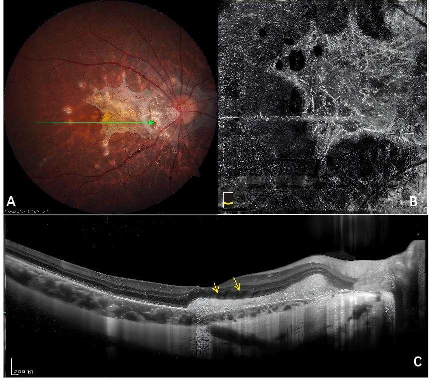
图2 一例PIC患眼中的星状炎性脉络膜新生血管
Figure 2 A case of iCNV secondary to PIC with stellar pattern

20250327095323_2672.png
(A)眼底彩照可见右眼后极部视网膜散在点状炎性病灶。黄斑区和视盘周围见大片视网膜下纤维血管网,“桥接”各炎性病灶,呈星形;(B)对应OCTA enface图像可清晰呈现星形脉络膜新生血管网。C.B-scan OCT图像上见黄斑区中脉络膜血新生血管病灶伴黄斑区低反射液性暗腔(黄色箭头指示)。
(A) The fundus color photograph shows scattered punctate inflammatory lesions in the posterior pole of the right eye. A large subretinal fibrous vascular network is observed in the macular region and around the optic disc, 'bridging' the various inflammatory lesions, forming a stellar pattern; (B) The corresponding OCTA en face image clearly presents the stellar pattern CNV network. (C) The B-scan OCT image shows choroidal neovascular lesions in the macular region accompanied by a hypo-reflective fluid dark area (indicated by the yellow arrow).
       本研究通过多模式影像学分析发现,大多数iCNV与炎性病灶密切相关。同时iCNV活动性也和眼底炎症活动性相关(χ2=4.18,P<0.05)。这些多模式影像学表现进一步支持了iCNV可能源自炎性病灶对RPE-Bruch膜-CC复合体造成损伤的观点。本研究结果显示,iCNV主要位于视网膜中心凹下或中心凹旁(47/51,92.15%)。其原因可能是由于黄斑区的高代谢需求及丰富密集的脉络膜血管网络,导致免疫复合物及炎症因子更容易在此沉积、发生局部炎症反应及RPE-Bruch-CC复合体的破坏[1]。在13.72%的iCNV处研究者观察到了局灶脉络膜凹陷的OCT征象,其形成与炎症浸润造成局灶内层脉络膜缺失及纤维血管膜形成的牵拉密切相关[15]。“干草叉征”是由Hoang等研究者于2013年首次提出的鉴别iCNV的特征性OCT指标[28],指在B-scan OCT上的活动性iCNV病灶可见“干草叉”样的多个自外层视网膜的高反射突起,本研究在9处iCNV病灶处观察到此征象,且均为活动性CNV。此外,本研究还在9例iCNV(17.64%)处发现了脉络膜海绵征。“脉络膜海绵征”是由Giuffre等[3]提出的iCNV具有诊断意义的OCT指标,为iCNV下的下脉络膜厚度如同海绵般在炎症活动期变厚、炎症消退期变薄的表现。Giuffre等[3]指出,形成iCNV处形成脉络膜海绵征的原因可能是炎症过程中释放的前列腺素等炎性介质导致脉络膜血管扩张、脉络膜增厚,而在iCNV下方这种脉络膜厚度改变更为明显,可能是由于在此区域炎症反应更严重。本研究在ICGA上的发现亦佐证该观点。本研究显示,部分iCNV病灶(13/51,25.49%)处存在脉络膜血管通透性增强的影像表现(图3)。这种ICGA表现可能为炎症因子导致的脉络膜血管扩张、通透性增强,故在ICGA造影中期呈斑片状强荧光的表现。同时,在ICGA上,多数iCNV病灶(38/51,74.50%)旁伴有弱荧光病灶。笔者推测弱荧光病灶可能是炎性病变,或者是包裹在CNV外缘的反应性RPE肥厚或增生。

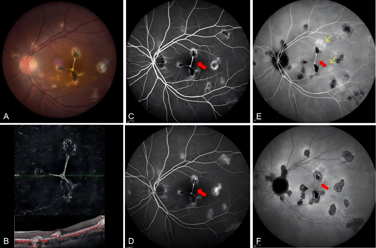
图3 一例MFC患眼中的星状脉络膜新生血管
Figure 3 A case of iCNV secondary to MFC with stellar pattern

20250327095439_9853.png
(A,B)在眼底彩照(A)及OCTA enface图像上(B)可清晰看到源自3处斑状炎性病灶的纤维血管网交织呈星状iCNV,对应B-scan上可见血管信号;(C,D)FFA造影期间红色箭头所指的iCNV呈稍强荧光,造影期间未见明显荧光素渗漏,提示星状iCNV为非活动性CNV;(E)ICGA造影早期可见炎性病灶处局灶脉络膜血管扩张、通透性增强呈斑状强荧光(黄色箭头);(F)ICGA造影晚期可见iCNV染色呈稍强荧光,周围炎性病灶呈全程弱荧光。
(A, B) The fundus color photograph (A) and the OCTA en face image (B) clearly show a stellar pattern iCNV formed by a fibrous vascular network originating from three punctate inflammatory lesions, with vascular signals visible on the corresponding B-scan; (C, D) During the FFA imaging, the iCNV indicated by the red arrow shows slightly hyperfluorescence, and no significant leakage is observed during the FFA phase, suggesting that the stellar pattern iCNV is non-active CNV; (E) Early ICGA imaging reveals focal choroidal vascular dilation and increased permeability at the site of the inflammatory lesions, appearing as patchy hyperfluorescence (yellow arrow); (F) Late phase of ICGA shows the iCNV staining with slightly hyperfluorescence, while the surrounding inflammatory lesions exhibit hypofluorescence throughout.
       本研究还通过OCTA手段评估了iCNV的血管形态,发现iCNV的血管形态极具特征。首先,除在DSFS患眼发现的大片弥漫网状CNV外,多数iCNV血管网较小。本研究表明,平均面积仅为0.08 mm2的焦点状CNV占iCNV病灶比例的29.41%。Wang等[25]的研究亦通过比较iCNV和AMD中的CNV面积,指出继发于炎症的CNV由于面积较AMD中的CNV更小。因此焦点状的、小的CNV提示CNV可能源于炎性病灶。其次,本研究还在iCNV中发现了既往未被描述过的“星状CNV(stellar pattern CNV)”。在AMD和PM-CNV相关的研究中,研究者们将OCTA上CNV的形态描述为“美杜莎(Medusa-shaped)”“海扇状(seafan pattern)”“花环状(lace-wheel pattern)”等,这些形态常常指具有许多微小毛细血管和血管环/袢的CNV;研究者们还将仅具有粗大血管而无细小分支的CNV描述为“枯树状(dead tree pattern)”“不规则形(irregular shape)” [13,26-27];或简单总结OCTA上CNV为“缠结或交织状(interlacing and tangled)”[28–30]。然而,由于iCNV特殊的病理生理机制和解剖学特征,其具有在OCTA上独特的形态表现。Olsen等[23]最早根据iCNV的病理结果提出了葡萄膜炎继发性iCNV形成的五期过程:Ⅰ期为脉络膜炎症或RPE处的炎症反应造成局灶的损伤;Ⅱ期为新生血管芽自损伤的Bruch膜向视网膜下生长;Ⅲ期为独立的新生血管灶相互“桥接(bridging vessels)”,连结为较大的“脉络膜新生血管复合体(vessels complex)”。同时在逐渐成熟的脉络膜新生血管网中,其边缘发生反应性视网膜色素上皮增生,而血管网内逐渐被纤维细胞浸润。Ⅳ期为纤维收缩期,即反应性增生的视网膜色素上皮细胞已包围新血管复合体,成纤维细胞渗透新生血管复合体并开始收缩。这个过程导致“星状”或“哑铃形”视网膜下纤维血管的形成;Ⅴ期为瘢痕形成期,临床上表现为伴有色素沉着的视网膜下纤维瘢痕,伴有其上视网膜光感受器细胞的破坏。而本研究中发现了9例(17.65%)星状iCNV病灶(图2、3),且多为非活动性,说明星状iCNV可能是炎性CNV特征性的形态,并且发生于iCNV血管化较为成熟的过程中。此外,本研究还发现各形态iCNV与患眼屈光度、视力情况无关,而与CNV活动性相关。星状、枯树状CNV主要为非活动性CNV。Coscas等研究曾指出,细小、缠结的毛细血管是OCTA观测CNV活动性的重要指标[13],而在经抗VEGF治疗或新生血管逐渐成熟的过程中,细小的毛细血管逐渐消退,剩下粗大的血管主干。因此无论是在iCNV中还是已有相关报道的mCNV(myopia choriodal neovascularization)及AMD相关CNV中,由粗大血管构成的枯树状CNV均可提示CNV趋于成熟、非活动[13,29]。在本研究中,以enface OCTA中CNV形态评估iCNV活动性的诊断敏感度为94.12%和特异性为77.78%。
       综上所述,本研究总结了48例葡萄膜炎患眼中的51处iCNV病灶的眼底多模式影像特征。其中SD-OCT上iCNV的脉络膜海绵征和局灶脉络膜凹陷等特征均提示iCNV与炎性病灶密切相关。同时本研究还发现ICGA造影期间,74.50%的iCNV病灶(38/51)伴有弱荧光病灶,25.49%的病例(13/51)显示脉络膜高通透性表现。这些表现均佐证了炎症在iCNV形成过程中的病理生理作用。同时本研究还揭示了OCTA enface图像上多样的iCNV形态,发现了iCNV特征性的星状CNV,同时发现了枯树状及星状iCNV与CNV活动性之间的相关性。本研究的局限性在于,尽管对较大样本量的iCNV进行了评估,本研究仅收集到了非感染性葡萄膜炎继发炎性CNV的病例,对iCNV的形态学评估缺乏感染性炎症的参照。同时本研究缺乏长期随访数据,对于iCNV形成的病理生理过程及其长期预后缺乏随访数据。因此,未来需要进行前瞻性研究,以进一步验证本研究对iCNV特殊形态形成原因的推断。

声明

在论文撰写中无使用生成式人工智能。论文撰写中的所有内容均由作者独立完成,并对出版物的真实性和准确性承担全部责任。

利益冲突

所有作者均声明不存在利益冲突。

开放获取声明

本文适用于知识共享许可协议 (Creative Commons),允许第三方用户按照署名(BY)-非商业性使用(NC)-禁止演绎(ND)(CC BY-NC-ND)的方式共享,即允许第三方对本刊发表的文章进行复制、发行、展览、表演、放映、广播或通过信息网络向公众传播,但在这些过程中必须保留作者署名、仅限于非商业性目的、不得进行演绎创作。
 

1、Agarwal A, Invernizzi A, Singh RB, et al. An update on inflammatory choroidal neovascularization: epidemiology, multimodal imaging, and management. J Ophthalmic Inflamm Infect, 2018, 8(1): 13. DOI: 10.1186/s12348-018-0155-6.Agarwal A, Invernizzi A, Singh RB, et al. An update on inflammatory choroidal neovascularization: epidemiology, multimodal imaging, and management. J Ophthalmic Inflamm Infect, 2018, 8(1): 13. DOI: 10.1186/s12348-018-0155-6.
2、Invernizzi A, Pichi F, Symes R, et al. Twenty-four-month outcomes of inflammatory choroidal neovascularisation treated with intravitreal anti-vascular endothelial growth factors: a comparison between two treatment regimens. Br J Ophthalmol, 2020, 104(8): 1052-1056. DOI: 10.1136/bjophthalmol-2019-315257.Invernizzi A, Pichi F, Symes R, et al. Twenty-four-month outcomes of inflammatory choroidal neovascularisation treated with intravitreal anti-vascular endothelial growth factors: a comparison between two treatment regimens. Br J Ophthalmol, 2020, 104(8): 1052-1056. DOI: 10.1136/bjophthalmol-2019-315257.
3、Giuffrè C, Marchese A, Fogliato G, et al. The “sponge sign”: a novel feature of inflammatory choroidal neovascularization. Eur J Ophthalmol, 2021, 31(3): 1240-1247. DOI: 10.1177/1120672120917621.Giuffrè C, Marchese A, Fogliato G, et al. The “sponge sign”: a novel feature of inflammatory choroidal neovascularization. Eur J Ophthalmol, 2021, 31(3): 1240-1247. DOI: 10.1177/1120672120917621.
4、Karska-Basta%20I%2C%20Pociej-Marciak%20W%2C%20%C5%BBuber-%C5%81askawiec%20K%2C%20et%20al.%20Diagnostic%20%0Achallenges%20in%20inflammatory%20choroidal%20neovascularization.%20Medicina%20%0A(Kaunas)%2C%202024%2C%2060(3)%3A%20465.%20DOI%3A%2010.3390%2Fmedicina60030465.Karska-Basta%20I%2C%20Pociej-Marciak%20W%2C%20%C5%BBuber-%C5%81askawiec%20K%2C%20et%20al.%20Diagnostic%20%0Achallenges%20in%20inflammatory%20choroidal%20neovascularization.%20Medicina%20%0A(Kaunas)%2C%202024%2C%2060(3)%3A%20465.%20DOI%3A%2010.3390%2Fmedicina60030465.
5、Woronkowicz M, Niederer R, Lightman S, et al. Intravitreal antivascular endothelial growth factor treatment for inflammatory choroidal neovascularization in noninfectious uveitis. Am J Ophthalmol, 2022, 236: 281-287. DOI: 10.1016/j.ajo.2021.07.010.Woronkowicz M, Niederer R, Lightman S, et al. Intravitreal antivascular endothelial growth factor treatment for inflammatory choroidal neovascularization in noninfectious uveitis. Am J Ophthalmol, 2022, 236: 281-287. DOI: 10.1016/j.ajo.2021.07.010.
6、Baxter SL , Pistilli M , Pujari SS, et al . Risk of choroidal neovascularization among the uveitides. Am J Ophthalmol, 2013, 156(3): 468-477.e2. DOI: 10.1016/j.ajo.2013.04.040.Baxter SL , Pistilli M , Pujari SS, et al . Risk of choroidal neovascularization among the uveitides. Am J Ophthalmol, 2013, 156(3): 468-477.e2. DOI: 10.1016/j.ajo.2013.04.040.
7、Moorthy RS, Chong LP, Smith RE, et al. Subretinal neovascular membranes in vogt-koyanagi-harada syndrome. Am J Ophthalmol, 1993, 116(2): 164-170. DOI: 10.1016/s0002-9394(14)71280-2.Moorthy RS, Chong LP, Smith RE, et al. Subretinal neovascular membranes in vogt-koyanagi-harada syndrome. Am J Ophthalmol, 1993, 116(2): 164-170. DOI: 10.1016/s0002-9394(14)71280-2.
8、Zhang X, Wen F, Zuo C, et al. Clinical features of punctate inner choroidopathy in Chinese patients. Retina, 2011, 31(8): 1680-1691. DOI: 10.1097/IAE.0b013e31820a67ad.Zhang X, Wen F, Zuo C, et al. Clinical features of punctate inner choroidopathy in Chinese patients. Retina, 2011, 31(8): 1680-1691. DOI: 10.1097/IAE.0b013e31820a67ad.
9、Wu K, Zhang X, Su Y, et al. Clinical characteristics of inflammatory choroidal neovascularization in a Chinese population. Ocul Immunol Inflamm, 2016, 24(3): 261-267. DOI: 10.3109/09273948.2015.1015741.Wu K, Zhang X, Su Y, et al. Clinical characteristics of inflammatory choroidal neovascularization in a Chinese population. Ocul Immunol Inflamm, 2016, 24(3): 261-267. DOI: 10.3109/09273948.2015.1015741.
10、Miyata M, Ooto S, Hata M, et al. Detection of myopic choroidal neovascularization using optical coherence tomography angiography. Am J Ophthalmol, 2016, 165: 108-114. DOI: 10.1016/j.ajo.2016.03.009.Miyata M, Ooto S, Hata M, et al. Detection of myopic choroidal neovascularization using optical coherence tomography angiography. Am J Ophthalmol, 2016, 165: 108-114. DOI: 10.1016/j.ajo.2016.03.009.
11、Gan Y, Zhang X, Su Y, et al. OCTA versus dye angiography for the diagnosis and evaluation of neovascularisation in punctate inner choroidopathy. Br J Ophthalmol, 2022, 106(4): 547-552. DOI: 10.1136/bjophthalmol-2020-318191.Gan Y, Zhang X, Su Y, et al. OCTA versus dye angiography for the diagnosis and evaluation of neovascularisation in punctate inner choroidopathy. Br J Ophthalmol, 2022, 106(4): 547-552. DOI: 10.1136/bjophthalmol-2020-318191.
12、Jabs DA, McCluskey P, Palestine AG, et al. The standardisation of uveitis nomenclature (SUN) project. Clinical Exper Ophthalmology, 2022, 50(9): 991-1000. DOI: 10.1111/ceo.14175.Jabs DA, McCluskey P, Palestine AG, et al. The standardisation of uveitis nomenclature (SUN) project. Clinical Exper Ophthalmology, 2022, 50(9): 991-1000. DOI: 10.1111/ceo.14175.
13、Coscas GJ, Lupidi M, Coscas F, et al. Optical coherence tomography angiography versus traditional multimodal imaging in assessing the activity of exudative age-related macular degeneration: a New Diagnostic Challenge. Retina, 2015, 35(11): 2219-2228. DOI: 10.1097/IAE.0000000000000766.Coscas GJ, Lupidi M, Coscas F, et al. Optical coherence tomography angiography versus traditional multimodal imaging in assessing the activity of exudative age-related macular degeneration: a New Diagnostic Challenge. Retina, 2015, 35(11): 2219-2228. DOI: 10.1097/IAE.0000000000000766.
14、Zhang Y, Gan Y, Zeng Y, et al. Incidence and multimodal imaging characteristics of macular neovascularisation subtypes in Chinese neovascular age-related macular degeneration patients. Br J Ophthalmol, 2024, 108(3): 391-397. DOI: 10.1136/bjo-2022-322392.Zhang Y, Gan Y, Zeng Y, et al. Incidence and multimodal imaging characteristics of macular neovascularisation subtypes in Chinese neovascular age-related macular degeneration patients. Br J Ophthalmol, 2024, 108(3): 391-397. DOI: 10.1136/bjo-2022-322392.
15、Liu Y, Wen F, Huang S, et al. Subtype lesions of neovascular age-related macular degeneration in Chinese patients. Graefes Arch Clin Exp Ophthalmol, 2007, 245(10): 1441-1445. DOI: 10.1007/s00417-007- 0575-8.Liu Y, Wen F, Huang S, et al. Subtype lesions of neovascular age-related macular degeneration in Chinese patients. Graefes Arch Clin Exp Ophthalmol, 2007, 245(10): 1441-1445. DOI: 10.1007/s00417-007- 0575-8.
16、Spaide RF, Jaffe GJ, Sarraf D, et al. Consensus nomenclature for reporting neovascular age-related macular degeneration data: consensus on neovascular age-related macular degeneration nomenclature study group. Ophthalmology, 2020, 127(5): 616-636. DOI: 10.1016/ j.ophtha.2019.11.004.Spaide RF, Jaffe GJ, Sarraf D, et al. Consensus nomenclature for reporting neovascular age-related macular degeneration data: consensus on neovascular age-related macular degeneration nomenclature study group. Ophthalmology, 2020, 127(5): 616-636. DOI: 10.1016/ j.ophtha.2019.11.004.
17、Gan Y, Ji Y, Zuo C, et al. Correlation between focal choroidal excavation and underlying retinochoroidal disease: a Pathological Hypothesis From Clinical Observation. Retina, 2022, 42(2): 348-356. DOI: 10.1097/IAE.0000000000003307.Gan Y, Ji Y, Zuo C, et al. Correlation between focal choroidal excavation and underlying retinochoroidal disease: a Pathological Hypothesis From Clinical Observation. Retina, 2022, 42(2): 348-356. DOI: 10.1097/IAE.0000000000003307.
18、Jampol LM, Shankle J, Schroeder R, et al. Diagnostic and therapeutic challenges. Retina, 2006, 26(9): 1072-1076. DOI: 10.1097/01. iae.0000248819.86737.a5.Jampol LM, Shankle J, Schroeder R, et al. Diagnostic and therapeutic challenges. Retina, 2006, 26(9): 1072-1076. DOI: 10.1097/01. iae.0000248819.86737.a5.
19、Xu H, Zeng F, Shi D, et al. Focal choroidal excavation complicated by choroidal neovascularization. Ophthalmology, 2014, 121(1): 246-250. DOI: 10.1016/j.ophtha.2013.08.014.Xu H, Zeng F, Shi D, et al. Focal choroidal excavation complicated by choroidal neovascularization. Ophthalmology, 2014, 121(1): 246-250. DOI: 10.1016/j.ophtha.2013.08.014.
20、Obata R, Takahashi H, Ueta T, et al. Tomographic and angiographic characteristics of eyes with macular focal choroidal excavation. Retina, 2013, 33(6): 1201-1210. DOI: 10.1097/IAE.0b013e31827b6452.Obata R, Takahashi H, Ueta T, et al. Tomographic and angiographic characteristics of eyes with macular focal choroidal excavation. Retina, 2013, 33(6): 1201-1210. DOI: 10.1097/IAE.0b013e31827b6452.
21、de Groot EL, Ten Dam-van Loon, N H, Kouwenberg CV, et al. Exploring imaging characteristics associated with disease activity in idiopathic multifocal choroiditis: a multimodal imaging approach. Am J Ophthalmol, 2023, 252: 45-58. DOI: 10.1016/j.ajo.2023.03.022.de Groot EL, Ten Dam-van Loon, N H, Kouwenberg CV, et al. Exploring imaging characteristics associated with disease activity in idiopathic multifocal choroiditis: a multimodal imaging approach. Am J Ophthalmol, 2023, 252: 45-58. DOI: 10.1016/j.ajo.2023.03.022.
22、Zhang X, Zuo C, Li M, et al. Spectral-domain optical coherence tomographic findings at each stage of punctate inner choroidopathy. Ophthalmology, 2013, 120(12): 2678-2683. DOI: 10.1016/ j.ophtha.2013.05.012.Zhang X, Zuo C, Li M, et al. Spectral-domain optical coherence tomographic findings at each stage of punctate inner choroidopathy. Ophthalmology, 2013, 120(12): 2678-2683. DOI: 10.1016/ j.ophtha.2013.05.012.
23、Olsen TW, Capone A Jr, Sternberg P Jr, et al. Subfoveal choroidal neovascularization in punctate inner choroidopathy. Surgical management and pathologic findings. Ophthalmology, 1996, 103(12): 2061-2069. DOI: 10.1016/s0161-6420(96)30387-4.Olsen TW, Capone A Jr, Sternberg P Jr, et al. Subfoveal choroidal neovascularization in punctate inner choroidopathy. Surgical management and pathologic findings. Ophthalmology, 1996, 103(12): 2061-2069. DOI: 10.1016/s0161-6420(96)30387-4.
24、Liu B, Zhang X, Peng Y, et al. Etiologies and characteristics of choroidal neovascularization in young Chinese patients. Ophthalmologica, 2019, 241(2): 73-80. DOI: 10.1159/000492133.Liu B, Zhang X, Peng Y, et al. Etiologies and characteristics of choroidal neovascularization in young Chinese patients. Ophthalmologica, 2019, 241(2): 73-80. DOI: 10.1159/000492133.
25、Wang JC, McKay KM, Sood AB, et al. Comparison of choroidal neovascularization secondary to white dot syndromes and agerelated macular degeneration by using optical coherence tomography angiography. Clin Ophthalmol, 2018, 13: 95-105. DOI: 10.2147/ OPTH.S185468.Wang JC, McKay KM, Sood AB, et al. Comparison of choroidal neovascularization secondary to white dot syndromes and agerelated macular degeneration by using optical coherence tomography angiography. Clin Ophthalmol, 2018, 13: 95-105. DOI: 10.2147/ OPTH.S185468.
26、Miere A, Butori P, Cohen SY, et al. Vascular remodeling of choroidal neovascularization after anti-vascular endothelial growth factor therapy visualized on optical coherence tomography angiography. Retina, 2019, 39(3): 548-557. DOI: 10.1097/IAE.0000000000001964.Miere A, Butori P, Cohen SY, et al. Vascular remodeling of choroidal neovascularization after anti-vascular endothelial growth factor therapy visualized on optical coherence tomography angiography. Retina, 2019, 39(3): 548-557. DOI: 10.1097/IAE.0000000000001964.
27、Carnevali%20A%2C%20Cicinelli%20MV%2C%20Capuano%20V%2C%20et%20al.%20Optical%20coherence%20%0Atomography%20angiography%3A%20a%20useful%20tool%20for%20diagnosis%20of%20treatment-Na%C3%AFve%20%0Aquiescent%20choroidal%20neovascularization.%20Am%20J%20Ophthalmol%2C%202016%2C%20169%3A%20%0A189-198.%20DOI%3A%2010.1016%2Fj.ajo.2016.06.042.Carnevali%20A%2C%20Cicinelli%20MV%2C%20Capuano%20V%2C%20et%20al.%20Optical%20coherence%20%0Atomography%20angiography%3A%20a%20useful%20tool%20for%20diagnosis%20of%20treatment-Na%C3%AFve%20%0Aquiescent%20choroidal%20neovascularization.%20Am%20J%20Ophthalmol%2C%202016%2C%20169%3A%20%0A189-198.%20DOI%3A%2010.1016%2Fj.ajo.2016.06.042.
28、Bruyère E, Miere A, Cohen SY, et al. Neovascularization secondary to high myopia imaged by optical coherence tomography angiography. Retina, 2017, 37(11): 2095-2101. DOI: 10.1097/IAE.0000000000001456.Bruyère E, Miere A, Cohen SY, et al. Neovascularization secondary to high myopia imaged by optical coherence tomography angiography. Retina, 2017, 37(11): 2095-2101. DOI: 10.1097/IAE.0000000000001456.
29、Li S, Sun L, Zhao X, et al. Assessing the activity of myopic choroidal neovascularization: comparison between optical coherence tomography angiography and dye angiography. Retina, 2020, 40(9): 1757-1764. DOI: 10.1097/IAE.0000000000002650.Li S, Sun L, Zhao X, et al. Assessing the activity of myopic choroidal neovascularization: comparison between optical coherence tomography angiography and dye angiography. Retina, 2020, 40(9): 1757-1764. DOI: 10.1097/IAE.0000000000002650.
30、Bagchi A, Schwartz R, Hykin P, et al. Diagnostic algorithm utilising multimodal imaging including optical coherence tomography angiography for the detection of myopic choroidal neovascularisation. Eye (Lond), 2019, 33(7): 1111-1118. DOI: 10.1038/s41433-019- 0378-2.Bagchi A, Schwartz R, Hykin P, et al. Diagnostic algorithm utilising multimodal imaging including optical coherence tomography angiography for the detection of myopic choroidal neovascularisation. Eye (Lond), 2019, 33(7): 1111-1118. DOI: 10.1038/s41433-019- 0378-2.
上一篇
下一篇
其他期刊
  • 眼科学报

    主管:中华人民共和国教育部
    主办:中山大学
    承办:中山大学中山眼科中心
    主编:林浩添
    主管:中华人民共和国教育部
    主办:中山大学
    浏览
  • Eye Science

    主管:中华人民共和国教育部
    主办:中山大学
    承办:中山大学中山眼科中心
    主编:林浩添
    主管:中华人民共和国教育部
    主办:中山大学
    浏览
推荐阅读
出版者信息
目录