您的位置: 首页 > 2022年12月 第37卷 第12期 > 文字全文
2023年7月 第38卷 第7期11
目录

超声乳化白内障吸除术对高度近视黄斑劈裂进展影响的临床观察

Clinical observation of the effect of phacoemulsification on the progression of myopic foveoschisis in high myopia

来源期刊: 眼科学报 | 2022年12月 第37卷 第12期 926-933 发布时间: 收稿时间:2023/1/4 16:36:14 阅读量:4855
作者:
关键词:
黄斑劈裂高度近视发病机制自然病程白内障超声乳化吸除术
macular retinoschisis high myopia pathogenesis natural course of disease phacoemulsification
DOI:
10.3978/j.issn.1000-4432.2022.11.15
收稿时间:
 
修订日期:
 
接收日期:
 
目的:评估白内障手术是否影响高度近视黄斑劈裂的进展,为高度近视黄斑劈裂合并白内障患者先行白内障手术改善视觉质量可行性提供依据。方法:选取2018年4月至2021年1月在上海市第十人民医院眼科确诊为高度近视黄斑劈裂患者共36例36眼纳入研究。根据是否行白内障手术分为实验组:男10例10眼,女8例8眼;对照组:男9例9眼,女9例9眼。比较两组初诊及1个月、3个月、6个月、12个月后黄斑中心区视网膜厚度(central subfield thickness,CST)、黄斑区视网膜容积(macular volume,MV)、视网膜平均厚度(average macular thickness,AMT)、最佳矫正视力(best corrected visual acuity,BCVA)、黄斑劈裂形态学进展率情况。结果:两组CST、MV、AMT时间和组别交互效应差异无统计学意义(F=0.825,P=0.370;F=1.266,P=0.268;F=0.612,P =0.657),两组间差异无统计学意义(F =2.519,P =0.122;F =3.318,P =0.077;F =1.378,P=0.249)。两组BCVA时间和组别交互效应差异有统计学意义(P<0.001);做简单效应分析实验组术后1、3、6及12个月BCVA较初诊差异均有统计学意义(均P<0.001);两组1、3、6、12个月BCVA较初诊BCVA的增量差异均有统计学意义(均P<0.001)。两组黄斑劈裂形态进展率差异无统计学意义(P=1.0)。结论:白内障超声乳化吸除术在短期内不影响高度近视黄斑劈裂的进展,且可以提高高度近视黄斑劈裂合并白内障患者的视力。
Objective: To assess whether cataract surgery can affect the progression of myopic foveoschisis, so as to provide evidence for the feasibility of cataract surgery to improve visual quality in patients with myopia foveoschisis accompanied by cataract. Methods: A total of 36 patients (36 eyes) diagnosed with high myopia foveoschisis at the Department of Ophthalmology, Shanghai Tenth People’s Hospital from April 2018 to January 2021 were enrolled in the study. The patients were divided into an experimental group (10 males with 10 eyes, and 8 females with 8 eyes) and an observation group (9 males with 9 eyes, and 9 females with 9 eyes) according to whether cataract surgery was performed. The central subfield thickness (CST), macular volume (MV), average macular thickness (AMT), best corrected visual acuity (BCVA), and the progression rate of foveoschis morphology were compared between the two groups at the initial diagnosis and 1 month, 3 months, 6 months, and 12 months after the surgery. Results: In terms of CST, MV, and AMT in the central macular area, no significant difference was found between the two groups (F=2.519, P=0.122; F=3.318, P=0.077; F=1.378, P=0.249), and there was no significant difference in time and group interaction effects between the two groups (F=0.825, P=0.370; F=1.266, P=0.268; F=0.612, P=0.657). In terms of BCVA, there was significant difference in time and group interaction effects between the two groups (P<0.001). The simple effect analysis showed that, compared with initial diagnosis, the BCVA in the experimental group was statistically significant at 1, 3, 6 and 12 months after the surgery (all P<0.001). In addition, there was a significant difference in the increments of BCVA between the two groups at 1, 3, 6, and 12 months after the surgery when compared with the initial diagnosis (all P<0.001). However, there was no significant difference in the morphological progression between the two groups (P=1.0). Conclusion: Phacoemulsification will not affect the progression of myopic foveoschisis in a short period, and can improve the vision of patients with myopic foveoschisis accompanied by cataract.
    高度近视黄斑劈裂(myopic foveoschisis,MF)是高度近视患者眼底常见的并发症之一。该病发病隐匿,多数患者发病早期没有明显的视觉症状,且病程进展缓慢,可保持数年视力及眼底结构的稳定[1-3]。主流术式玻璃体切除术临床效果较好,但并发症黄斑裂孔常导致患者术后中心视力完全丧失[4]。有研究提出对于MF合并白内障患者可仅先行单纯白内障摘除手术使患者获得良好的复明效果,再根据眼底病变进展决定是否行玻璃体切除术(pars plana vitrectomy,PPV)治疗[5-6]特别是对不愿行眼底手术的患者,这是目前临床较为常用的治疗方法。但白内障手术是否会加速黄斑劈裂患者眼底病程的进展目前尚无报道。因此,本文旨在观察白内障手术对MF患者病程进展的影响。

1 对象与方法

1.1 对象

    选取2018年4月至2021年1月在上海市第十人民医院确诊为M F合并白内障患者共3 6例3 6眼。入组标准:1)等效球镜屈光度≥-6.00 D且眼轴长度 ≥ 26.0 mm ; 2 ) 光学相干断层扫描 (optical coherence tomography,OCT)显示视网膜神经上皮层层间分离伴桥样柱状连接;3 )晶状体混浊,白内障分级在C3、 N3、P3[7]及以上。排除标准 :1 )合并脉络膜新生血管、视网膜裂孔等高度近视黄斑区其他并发症患者;2 )糖尿病、高血压等影响视功能的系统疾病患者;3 )因屈光间质混浊,OCT检查无法获取清晰的黄斑区信号图像患者。本研究遵循《赫尔辛基宣言》,通过上海市第十人民医院伦理委员会审查(审批号:ChiCTR2000036875),患者自愿接受相关检查和手术方案,并签署知情同意书。根据是否进行白内障手术分成行单纯白内障手术的实验组,其中男10例10眼,女8例8眼,年龄(73.78±6.34)岁,眼轴长度(28.21±2.26) mm;未手术行门诊随访观察的对照组,其中男9例9眼,女9例9眼,年龄(74.56±5.46)岁,眼轴长度(29.15±1.42) mm。

1.2 参数测量

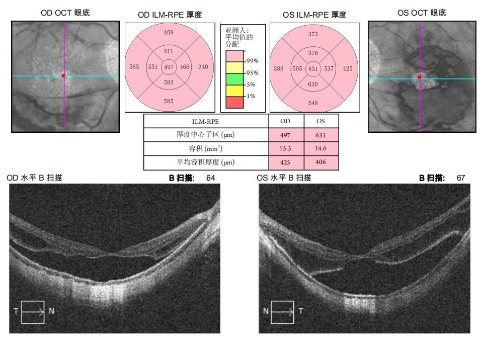
    所有患眼均行医学验光、最佳矫正视力(best corrected visual acuity,BCVA)、间接检眼镜、OCT检查以及眼轴长度测量,BCVA转换为logMAR视力进行统计分析。使用Cirrus HD-OCT 4000的Macular Cube 512*128扫描模式扫描,测量以固视点为圆心1 mm为直径圆周的视网膜平均厚度(central subfield thickness CST)、以固视点为圆心6 mm为直径圆周的视网膜容积(macular volume,MV)、以固视点为圆心6 mm为直径圆周的视网膜平均厚度(average macular thickness,AMT),记录数据(图1)。根据Ruiz-Medrano等[8]将黄斑劈裂形态分为6级:T0为无黄斑劈裂;T1为内层中心凹劈裂;T1为外层中心凹劈裂;T2为内层和外层中心凹劈裂;T3为中心凹视网膜脱离;T4为全层黄斑裂孔;T5为黄斑裂孔性视网膜脱离。

1.2 方法和随访

    实验组患眼行常规白内障摘除+人工晶状体植入手术,对照组门诊随访观察。实验组在初诊,术后1、3、6、12个月随访检查,对照组患者在初诊及初诊后1、3、6、12个月进行随访检查,测量并记录BCVA、CST、MV、AMT及黄斑形态学情况。

1.3 统计学处理

    采用SPSS 25.0统计学软件分析数据。计量资料用均数±标准差(x±s)表示,重复测量资料采用两因素重复测量的方差分析,进一步比较做简单效应分析,两组差异比较采用独立样本t检验(α校正值=0.0125);计数资料用频数描述数据,形态学变化比较采用Fisher确切概率法。P<0.05为差异有统计学意义。
20230104170921_6368.jpg

图1 OCT参数:用HD-OCT仪器自带的黄斑厚度分析系统分析并记录CST、MV、AMT
Figure 1 Parameters of OCT: CST, MV and AMT were analyzed and recorded by macular thickness analysis system of HD-OCT instrument

2 结果

2.1 两组一般资料比较

    两组患者初诊时的性别、年龄、眼轴长度、BCVA(logMAR)、等效球镜屈光度、CST、MV、AMT差异均无统计学意义(P>0.05,表1)。

2.2 两组 CST、MV、AMT 比较

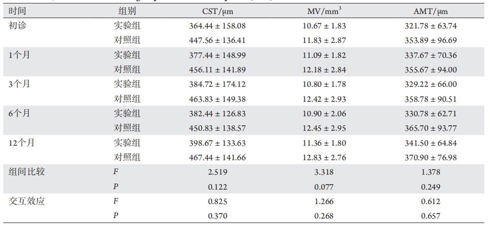
    各组数据服从正态分布且满足球形性假设,采用两因素重复测量方差分析,两组CST、MV、AMT变化程度的时间和组别交互效应差异均无统计学意义(F=0.825,P=0.370,F=1.266,P=0.268;F =0.612,P =0.657,表2),组间差异均无统计学意义(F =2.519,P =0.122,F =3.318,P =0.077;F=1.378,P=0.249,表2)。

2.3 两组手术前后视力变化及比较

    各组数据服从正态分布且满足球形性假设,采用两因素重复测量资料方差分析,时间和组别交互效应均有统计学意义(P<0.001,表3);做简单效应比较实验组术后1、3、6、12个月较初诊时差异均有统计学意义(均P<0.001,表4);两组1、3、6、12个月时BCVA较初诊时差值的差异均有统计学意义(均P<0.001,表5)。

表1 患者一般资料
Table 1 Baseline data of the patients

20230104171133_2407.jpg

表2 两组各时间点CST、MV、AMT(x±s)
Table 2 CST, MV and AMT in both groups at difffferent time points (x±s)

20230104171213_4828.jpg

表3 两组患者各时间点logMAR(x±s)
Table 3 LogMAR in both groups at difffferent time points (x±s)

20230104171249_1491.jpg

表4 实验组各随访时间点logMAR简单效应比较P值
Table 4 P value of logMAR simple effffect comparison in the experiment groups at difffferent time points

20230104171327_7346.jpg

表5 两组各随访时间点较初诊logMAR增值(x±s)
Table 5 Increased logMAR value in both groups at difffferent follow-up points compared with initial diagnosis (x±s)

20230104171403_0459.jpg

2.4 两组黄斑形态学变化比较

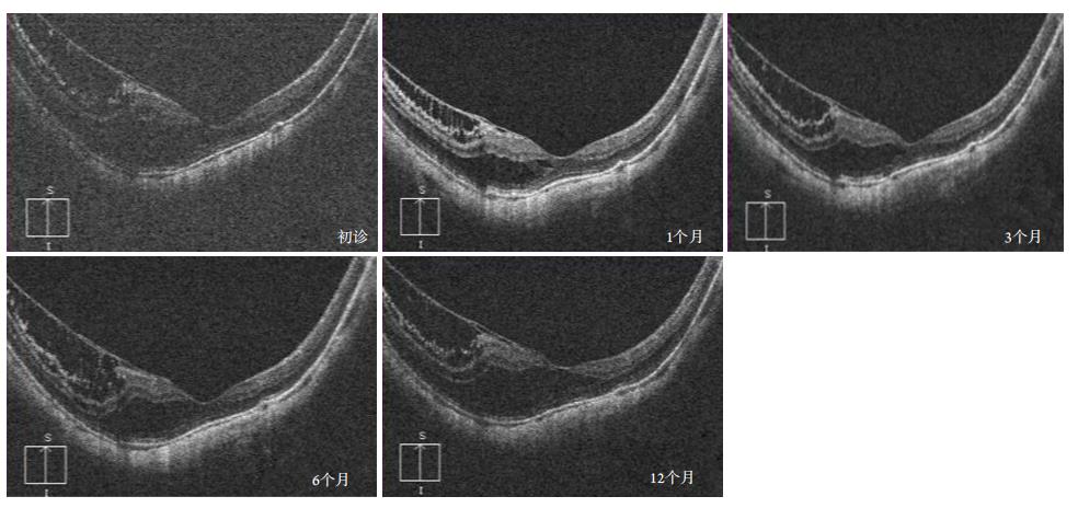
    实验组16例16眼T1,1例1眼T2,1例1眼T3,术后随访12个月黄斑形态均未发生改变(图2)。对照组18例18眼T1,12个月随访中1例1眼发展成局限性中心凹脱离(T3),余患者黄斑形态稳定。所有患者均未观察到黄斑裂孔,孔源性视网膜脱离的形成。两组患者黄斑劈裂病程进展率的差异无统计学意义(P=1.00,表6)。

图2 一名男性患者术前黄斑形态(T2),术后随访黄斑形态未发生改变
Figure 2 Preoperative macula morphology of a male patient (T2), and the postoperative macula morphology did not change during the follow-up at 1, 3, 5 and 12 months

20230104171520_2317.jpg

表6 两组黄斑形态学变化的比较
Table 6 Comparison of macula morphology change in both groups

20230104171558_2984.jpg

3 讨论

    白内障摘除术会增加玻璃体后脱离[9-10]、黄斑水肿和视网膜脱离的风险。白内障超声乳化术后随诊10年,视网膜脱离的年发病率为0.36%~2.9%[11-12]。探讨原因,术后产生眼底并发症可能是术中对玻璃体干扰、增加玻璃体后脱离以及玻璃体视网膜牵引的结果。这些因素是黄斑劈裂发病机制、病程进展的相关因素[13-14]。既往有研究[5,15]采用黄斑中心凹厚度,劈裂最显著处视网膜神经上皮层厚度观察黄斑劈裂患者白内障术后视网膜变化。研究结果有差异,分析原因可能是MF劈裂腔形态变化多,这些指标不足以反映术后劈裂腔变化有关。本研究采用HD-OCT测得CST、MV、AMT以观察术后劈裂腔的变化。行重复测量方差分析,结果显示两组CST、MV、AMT时间和组别交互效应,组间差异均无统计学意义,故认为单纯白内障手术与否对术后MF患者劈裂腔的变化并未造成显著性差异。实验组和对照组患者BCVA的时间和组别交互效应差异有统计学意义,提示术后两组BCVA的变化趋势不一致,做简单效应分析实验组术后1、3、6、12个月BCVA较初诊均有显著差异,两组1、3、6、12个月时BCVA较初诊BCVA的增量差异均有统计学意义。提示行白内障摘除术后患者较术前视力明显提高,患者视觉效果得到明显改善。
    MF患者眼底劈裂结构可较长时间保持稳定,病程发展中局限性中心凹脱离的形成常伴随患者视力极具下降,也是目前临床上进行PPV治疗的手术指征[16-17]。进而黄斑裂孔,孔源性视网膜脱离的形成是该类患者病程最终结局[18]。2019年Ruiz-Medrano 等[8]对近视性黄斑病变提出萎缩(atrophy)-牵拉 (traction)- 新生血管(neovascular)相关新型分类系统(ATN分类),该分类对近视牵引性黄斑病变,根据其劈裂腔累及的范围和严重程度分为6级(T0~T5)。本研究依据该分类,手术组一名患者术前存在局限性中心凹脱离(T3),患者因拒绝行眼底手术,予单纯白内障手术,术后黄斑形态未变化。1例1眼术前中心凹内层和外层劈裂(T2),术后黄斑形态无改变。余患者均保持形态学稳定。对照组患者18例18眼T1,1例1眼在最后一次随访中发现形成中心凹脱离(T3),两组黄斑形态学进展率无显著差异。故认为患者行白内障摘除术没有增加MF进展的风险。
    Sun等[19]的研究揭示了对劈裂腔累及整个黄斑区的MF发展成(full-thickness macular hole,FTMH)两种模式。一种是中心凹先形成外部板层黄斑裂孔(outer lamellar macular hole,OLMH)和视网膜脱离(retinal detachment,RD),并逐渐扩大发展成全层黄斑裂孔。另一种是先形成内部板层黄斑裂孔(inner lamellar macular hole,ILMH),劈裂逐渐向后发展至视网膜色素上皮发展成FTMH。本研究中对照组18眼中有2眼劈裂腔累及整个黄斑区的MF在初诊后3个月形成OLMH,1年后其中1眼形成局限性中心凹脱离,余16名患者保持黄斑区形态结构较初诊时无明显变化。分析原因,患者眼底保持病变稳定可能与初诊时均未发现明显玻璃体牵拉和视网膜前膜等结构有关[1,20-21]
    综上所述,白内障超声乳化吸除术在短期内不影响MF的进展,且可以提高患者的视力。本研究存在一定的局限性,本研究的样本量较小且随访时间相对较短。关于白内障术后后发障患者行Nd:YAG激光治疗后是否加速眼底病变的病程发展还有待于进一步的前瞻性的、大样本的病例对照研究。

开放获取声明

    本文适用于知识共享许可协议(Creative Commons),允许第三方用户按照署名(BY)-非商业性使用(NC)-禁止演绎(ND)(CC BY-NC-ND)的方式共享,即允许第三方对本刊发表的文章进行复制、发行、展览、表演、放映、广播或通过信息网络向公众传播,但在这些过程中必须保留作者署名、仅限于非商业性目的、不得进行演绎创作。详情请访问:https://creativecommons.org/licenses/by-nc-nd/4.0/
1、高岩, 杨瑞玲, 王诗尧. 康复专科护理门诊的建立及管理[ J]. 中 国护理管理, 2019, 19(1): 12-15.
GAO Y, YANG RL , WANG SY. Establishment and management of a nursing clinic of rehabilitation[ J]. Chinese Nursing Management, 2019, 19(1): 12-15.
高岩, 杨瑞玲, 王诗尧. 康复专科护理门诊的建立及管理[ J]. 中 国护理管理, 2019, 19(1): 12-15.
GAO Y, YANG RL , WANG SY. Establishment and management of a nursing clinic of rehabilitation[ J]. Chinese Nursing Management, 2019, 19(1): 12-15.
2、Gohil R, Sivaprasad S, Han LT, et al. Myopic foveoschisis: a clinical review[ J]. Eye (Lond), 2015, 29(5): 593-601.Gohil R, Sivaprasad S, Han LT, et al. Myopic foveoschisis: a clinical review[ J]. Eye (Lond), 2015, 29(5): 593-601.
3、Panozzo G. Myopic traction maculopathy[ J]. Eye (Lond), 2016, 30(7): 1025.Panozzo G. Myopic traction maculopathy[ J]. Eye (Lond), 2016, 30(7): 1025.
4、Cheong KX, Xu L, Ohno-Matsui K, et al. An evidence-based review of the epidemiology of myopic traction maculopathy[ J]. Surv Ophthalmol, 2022, 67(6): 1603-1630.Cheong KX, Xu L, Ohno-Matsui K, et al. An evidence-based review of the epidemiology of myopic traction maculopathy[ J]. Surv Ophthalmol, 2022, 67(6): 1603-1630.
5、Wang L, Wang Y, Li Y, et al. Comparison of effectiveness between complete internal limiting membrane peeling and internal limiting membrane peeling w ith preser vation of the central fovea in combination with 25G vitrectomy for the treatment of high myopic foveoschisis[ J]. Medicine (Baltimore), 2019, 98(9): e14710.Wang L, Wang Y, Li Y, et al. Comparison of effectiveness between complete internal limiting membrane peeling and internal limiting membrane peeling w ith preser vation of the central fovea in combination with 25G vitrectomy for the treatment of high myopic foveoschisis[ J]. Medicine (Baltimore), 2019, 98(9): e14710.
6、Lai CC, Yeung L, Chen YP, et al. Macular and visual outcomes after cataract extraction for highly myopic foveoschisis[ J]. J Cataract Refract Surg, 2008, 34(7): 1152-1156.Lai CC, Yeung L, Chen YP, et al. Macular and visual outcomes after cataract extraction for highly myopic foveoschisis[ J]. J Cataract Refract Surg, 2008, 34(7): 1152-1156.
7、顾雪芬, 荣翱, 王富彬, 等. 病理性近视黄斑劈裂患者白内障超声 乳化手术临床观察[ J]. 临床眼科杂志, 2018, 26(3): 233-234.
GU XF, RONG A, WANG FB, et al. Clinical observation of phacoemulsification in pathologic myopia w ith macular retinoschisis[ J]. Journal of Clinical Ophthalmology, 2018, 26(3): 233-234.
顾雪芬, 荣翱, 王富彬, 等. 病理性近视黄斑劈裂患者白内障超声 乳化手术临床观察[ J]. 临床眼科杂志, 2018, 26(3): 233-234.
GU XF, RONG A, WANG FB, et al. Clinical observation of phacoemulsification in pathologic myopia w ith macular retinoschisis[ J]. Journal of Clinical Ophthalmology, 2018, 26(3): 233-234.
8、Chylack LT Jr, Wolfe JK , Singer DM, et al. The lens opacities classification system III. The longitudinal study of cataract study group[ J]. Arch Ophthalmol, 1993, 111(6): 831-836.Chylack LT Jr, Wolfe JK , Singer DM, et al. The lens opacities classification system III. The longitudinal study of cataract study group[ J]. Arch Ophthalmol, 1993, 111(6): 831-836.
9、Ruiz-Medrano J, Montero JA , Flores-Moreno I, et al. Myopic maculopathy: Current status and proposal for a new classification and grading system (ATN)[ J]. Prog Retin Eye Res, 2019, 69: 80-115.Ruiz-Medrano J, Montero JA , Flores-Moreno I, et al. Myopic maculopathy: Current status and proposal for a new classification and grading system (ATN)[ J]. Prog Retin Eye Res, 2019, 69: 80-115.
10、Zhang F, Chang P, Zhao Y, et al. Incidence of posterior vitreous detachment after congenital cataract surger y: an ultrasound evaluation[ J]. Graefes Arch Clin Exp Ophthalmol, 2021, 259(4): 1045-1051.Zhang F, Chang P, Zhao Y, et al. Incidence of posterior vitreous detachment after congenital cataract surger y: an ultrasound evaluation[ J]. Graefes Arch Clin Exp Ophthalmol, 2021, 259(4): 1045-1051.
11、Hayashi S, Yoshida M, Hayashi K, et al. Progression of posterior vitreous detachment after cataract surgery[ J]. Eye (Lond), 2022, 36(10): 1872-1877.Hayashi S, Yoshida M, Hayashi K, et al. Progression of posterior vitreous detachment after cataract surgery[ J]. Eye (Lond), 2022, 36(10): 1872-1877.
12、Qureshi MH, Steel DHW. Retinal detachment following cataract phacoemulsification-a review of the literature[ J]. Eye (Lond), 2020, 34(4): 616-631.Qureshi MH, Steel DHW. Retinal detachment following cataract phacoemulsification-a review of the literature[ J]. Eye (Lond), 2020, 34(4): 616-631.
13、Haug SJ, Bhisitkul RB. Risk factors for retinal detachment following cataract surgery[ J]. Curr Opin Ophthalmol, 2012, 23(1): 7-11.Haug SJ, Bhisitkul RB. Risk factors for retinal detachment following cataract surgery[ J]. Curr Opin Ophthalmol, 2012, 23(1): 7-11.
14、Shimada N, Tanaka Y, Tokoro T, et al. Natural course of myopic traction maculopathy and factors associated with progression or resolution[ J]. Am J Ophthalmol, 2013, 156(5): 948-957.e1.Shimada N, Tanaka Y, Tokoro T, et al. Natural course of myopic traction maculopathy and factors associated with progression or resolution[ J]. Am J Ophthalmol, 2013, 156(5): 948-957.e1.
15、Nebbioso M, Lambiase A, Gharbiya M, et al. High myopic patients with and without foveoschisis: morphological and functional characteristics[ J]. Doc Ophthalmol, 2020, 141(3): 227-236.Nebbioso M, Lambiase A, Gharbiya M, et al. High myopic patients with and without foveoschisis: morphological and functional characteristics[ J]. Doc Ophthalmol, 2020, 141(3): 227-236.
16、吴强, 李世玮, 陆斌, 等. 合并视网膜劈裂症的高度近视眼超声 乳化白内障吸除术的临床观察[ J]. 中华眼科杂志, 2011, 47(4): 303-309.
WU Q, LI SW, LU B, et al. Clinical observation of highly myopic eyes with retinoschisis after phacoemulsification[ J]. Chinese Journal of Ophthalmology, 2011, 47(4): 303-309.
吴强, 李世玮, 陆斌, 等. 合并视网膜劈裂症的高度近视眼超声 乳化白内障吸除术的临床观察[ J]. 中华眼科杂志, 2011, 47(4): 303-309.
WU Q, LI SW, LU B, et al. Clinical observation of highly myopic eyes with retinoschisis after phacoemulsification[ J]. Chinese Journal of Ophthalmology, 2011, 47(4): 303-309.
17、Dolar-Szczasny J, ?wi?ch-Zubilewicz A, Mackiewicz J. A review of current myopic foveoschisis management strategies[ J]. Semin Ophthalmol, 2019, 34(3): 146-156.Dolar-Szczasny J, ?wi?ch-Zubilewicz A, Mackiewicz J. A review of current myopic foveoschisis management strategies[ J]. Semin Ophthalmol, 2019, 34(3): 146-156.
18、Parolini B, Palmieri M, Finzi A, et al. Myopic traction maculopathy: A new perspective on classification and management[ J]. Asia Pac J Ophthalmol (Phila), 2021, 10(1): 49-59.Parolini B, Palmieri M, Finzi A, et al. Myopic traction maculopathy: A new perspective on classification and management[ J]. Asia Pac J Ophthalmol (Phila), 2021, 10(1): 49-59.
19、Rey A, Jürgens I, Maseras X, et al. Natural course and surgical management of high myopic foveoschisis[ J]. Ophthalmologica, 2014, 231(1): 45-50.Rey A, Jürgens I, Maseras X, et al. Natural course and surgical management of high myopic foveoschisis[ J]. Ophthalmologica, 2014, 231(1): 45-50.
20、Sun CB, Liu Z, Xue AQ, et al. Natural evolution from macular retinoschisis to full-thickness macular hole in highly myopic eyes[ J]. Eye (Lond), 2010, 24(12): 1787-1791.Sun CB, Liu Z, Xue AQ, et al. Natural evolution from macular retinoschisis to full-thickness macular hole in highly myopic eyes[ J]. Eye (Lond), 2010, 24(12): 1787-1791.
21、Ikuno Y. Overview of the complications of high myopia[ J]. Retina, 2017, 37(12): 2347-2351.Ikuno Y. Overview of the complications of high myopia[ J]. Retina, 2017, 37(12): 2347-2351.
22、Gaucher D, Haouchine B, Tadayoni R, et al. Long-term follow-up of high myopic foveoschisis: natural course and surgical outcome[ J]. Am J Ophthalmol, 2007, 143(3): 455-462.Gaucher D, Haouchine B, Tadayoni R, et al. Long-term follow-up of high myopic foveoschisis: natural course and surgical outcome[ J]. Am J Ophthalmol, 2007, 143(3): 455-462.
上一篇
下一篇
其他期刊
  • 眼科学报

    主管:中华人民共和国教育部
    主办:中山大学
    承办:中山大学中山眼科中心
    主编:林浩添
    主管:中华人民共和国教育部
    主办:中山大学
    浏览
  • Eye Science

    主管:中华人民共和国教育部
    主办:中山大学
    承办:中山大学中山眼科中心
    主编:林浩添
    主管:中华人民共和国教育部
    主办:中山大学
    浏览
推荐阅读
出版者信息
目录