论著
目的:通过分析基于眼底彩照的人工智能(artificial intelligence,AI)在糖尿病视神经病变(diabetic optic neuropathy,DON)中的参数特征,探索AI在DON诊断中的应用价值。
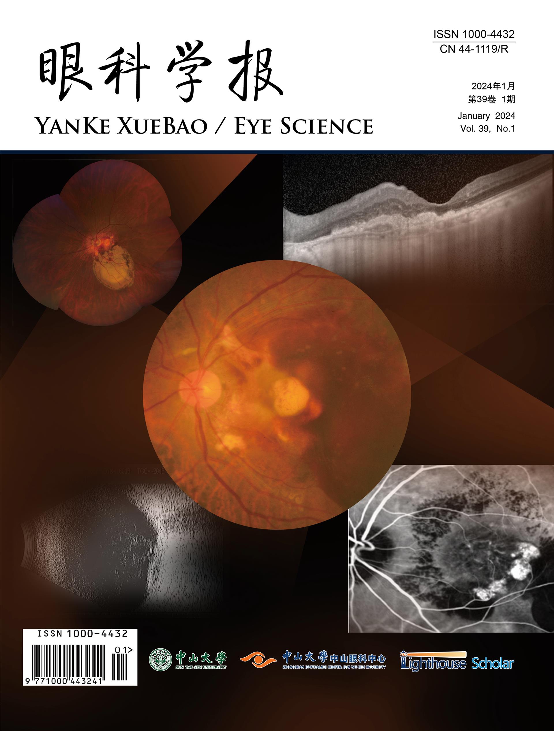
方法:收集2020年1月1日至2022年4月30日就诊于东莞东华医院、横沥医院及东莞市寮步镇社区卫生服务中心并诊断为糖尿病的患者,采集其一般信息并拍摄以黄斑为中心、图片边缘距离视盘中心超过1PD的50°眼底彩照。眼底彩照由人工智能诊断系统分析获得视盘及血管检测参数,由3-4名眼底专家阅片后分为DON(+)、DON(-)两组并作糖尿病视网膜病变(diabetic retinopathy,DR)分期诊断。比较两组间视盘、血管检测参数的差异性,并分析各项参数以及DR分期与DON发病的相关性。
结果:研究共纳入糖尿病患者526人(945眼),其中男性335人,女性191人;平均年龄为51.58±12.21岁,平均病程为5.51±5.20年。所有入组病例中,DON(+)组205眼,DON(-)740眼;根据专科医师判读结果,无DR 723眼,轻度非增殖期糖尿病视网膜病变(non-proliferrative diabetic retinopathy,NPDR)7眼,中度NPDR 184眼,重度NPDR 24眼,增殖期糖尿病视网膜病变(proliferrative diabetic retinopathy,PDR)7眼。AI检测的视盘及血管参数中,水平视杯直径、垂直视杯直径、水平杯盘比、垂直杯盘比、B区视网膜静脉血管当量、B区视网膜动静脉比值在有或无DON组间存在显著差异;水平视盘直径、垂直视盘直径、弧形斑和视盘面积比、B区视网膜动脉当量在两组之间无显著差异。相关性分析发现,水平视杯直径、垂直视杯直径、水平杯盘比、垂直杯盘比、B区视网膜动静脉比值与DON患病呈负相关;B区视网膜静脉血管当量、DR分期则与其呈正相关。
结论:DON患者的视杯直径、杯盘比、B区视网膜静脉血管当量等基于眼底彩照的人工智能检测参数有显著改变;DON的发病与DR病变严重程度有关。
Objective: To explore the application value of artificial intelligence (AI) in the diagnosis of diabetic optic neuropathy (DON) by analyzing the parameter characteristics of artificial intelligence (AI) based on fundus color photos.
Methods: From January 1, 2020 to April 30, 2022, patients diagnosed with diabetes were collected in Dongguan Donghua Hospital, Hengli Hospital of Dongguan and Community Healthcare Center of Dongguan Liaobu. General information was collected and 50°field vision fundus images(centered on macula and the edge of the images were more than 1PD away from the center of the optic disc) were taken. All the images were divided into DON(+) and DON(-) groups by 3-4 ophthalmologists. All the parameters were detected and analyzed by AI system, and their differences between the two groups were compared. The correlation between each parameter and DR stage with the incidence of DON was analyzed as well.
Results: A total of 526 diabetic patients (945 eyes) were included in this study, including 335 males and 191 females. The mean age was 51.58±12.21 years, and the mean disease duration was 5.51±5.20 years. All the enrolled cases were divided into DON (+) group (205 eyes) and DON (-) group (740 eyes) . According to ophthalmologists’ interpretation, 723 eyes had no DR, 7 eyes had mild nonproliferrative diabetic retinopathy (NPDR), 184 eyes had moderate NPDR, 24 eyes had severe NPDR, 7 eyes had Proliferrative diabetic retinopathy (PDR). Among the parameters detected by AI, there were significant differences in horizontal and vertical optic cup diameter, horizontal and vertical C/D, retinal vein equivalent(RVE) in zone B, and retinal arteriole-to venule ratio(AVR) in zone B between DON(+) and DON(-) groups. There were no significant differences between the two groups in horizontal and vertical optic disc diameter, arc-shaped spot-to-disc area ratio, and retinal artery equivalent(RAE) in zone B. In the analysis of risk factors, horizontal and vertical optic cup diameter, horizontal and vertical C/D, and AVR in zone B were negatively correlated with the diagnosis of DON. RVE in zone B and the severity of DR were positively correlated with the diagnosis of DON.
Conclusions: The AI detection parameters based on fundus color photography have significant changes in the diameter of optic cup, C/D and RVE in zone B in DON patients. The incidence of DON is related to the severity of DR.