您的位置: 首页 > 2023年3月 第38卷 第3期 > 文字全文
2023年7月 第38卷 第7期11
目录

单中心神经眼科住院患者疾病谱及流行病学分析

Analysis of disease spectrum and epidemiology of inpatients with neuroophthalmic disease from single center

来源期刊: 眼科学报 | 2023年3月 第38卷 第3期 190-198 发布时间: 收稿时间:2023/3/27 16:27:35 阅读量:6544
作者:
关键词:
神经眼科视神经病变视神经炎非动脉炎性前部缺血性视神经病变传出神经系统
neuro-ophthalmology optic neuropathy optic neuritis non-arteritic anterior ischemic optic neuropathy efferent nervous system
DOI:
10.12419/j.issn.1000-4432.2023.03.03
收稿时间:
 
修订日期:
 
接收日期:
 
目的:分析单中心神经眼科疾病谱及流行病学特点,为指导神经眼科疾病诊断和治疗提供基础。方法:纳入2010年1月1日—2021年12月31日中国人民解放军总医院神经眼科病区收治的神经眼科疾病患者,从电子病例系统检索和记录所有纳入病例的年龄、性别、地区分布及病种亚型分析。结果:共计7245例神经眼科患者纳入统计,其中男性3331例(46.0%)、女性3914例(54.0%),男女比例1∶1.2;年龄(38.2±17.5)岁。83.25%(6031/7245)为传入神经系统疾病,9.92%(719/7245)为传出神经系统疾病和眼眶疾病,6.83%(495/7245)未归类。病种分析显示,占比最高的是脱髓鞘性视神经炎(demyelinating optic neuritis,DON),为40.17%(2910/7245);占比第二的是非动脉炎性前部缺血性视神经病变(nonarteritic anterior ischemic optic neuropathy,NAION),为11.37%(824/7245);占比第三的是外伤性视神经病变5.15%(373/7245),其中7.85%(569/7245)表现为不明原因视神经萎缩。从年龄分布来看,DON和外伤性视神经病变患者中18~40岁者占比最高(分别为48.63%和44.24%),NAION患者中41~60岁者占比最高(66.14%),小于18岁的未成年患者在遗传性视神经病变中占比最高,比例为48.58%。在2226例DON患者中,视神经脊髓炎(neuromyelitis optica,NMO)/视神经脊髓炎谱系疾病(neuromyelitis optica spectrum disorder,NMOSD)比例最高,为60.02%;髓鞘少突胶质细胞糖蛋白抗体(myelinoligodendrocyte glycoprotein antibody,MOG-IgG)阳性视神经炎比例为11.68%;多发性硬化(multiple sclerosis,MS)和MS相关性视神经炎和慢性复发性炎性视神经病变(chronic recurrent inflammatory optic neuropathy,CRION)占比较低,分别是1.8%和2.25%。DON整体患者中,男女比例为1∶3.08;在NMO/NMOSD患者中男女比例为1∶8;MOG阳性视神经炎患者中,男女比例为4∶5;在非典型视神经炎患者中,男性比例高于女性,为1.28∶1;DON患者中,81.79%患者为中青年,MOG阳性视神经炎未成年患者可达41.15%。结论:DON和NAION是神经眼科传入系统疾病最常见两大病种。
Objective: To analyze the spectrum and epidemiological characteristics of neuro-ophthalmic diseases from single center, and to provide basis for guiding the diagnosis and treatment of neuro-ophthalmic diseases. Methods: Patients with neuro-ophthalmic diseases admitted to the neuro-ophthalmology ward of Chinese PLA General Hospital from January 1, 2010 to December 31, 2021 were enrolled. The age, gender, regional distribution and disease subtypes of all included patients were retrieved and recorded from the electronic case system. Results: A total of 7245 patients with neuro-ophthalmic diseases were enrolled, including 3331 males(46.0%)and 3914 females(54.0%), with a male to female ratio of 1:1.2. The average age was 38.2±17.5 years. 83.25%(6031/7245)were afferent nervous system diseases, 9.92% (719/7245)were efferent nervous system diseases and orbital diseases, and 6.83%(495/7245)were not classified. The ratio of demyelinating optic neuritis(DON)was the highest(40.17%,2910/7245), followed by nonarteritic anterior ischemic optic neuropathy(NAION)(11.37%,824/7245)and traumatic optic neuropathy(TON) (5.15%,373/7245). The ratio of optic nerve atrophy with unknown causes was 7.85%(569/7245). Characteristics of age distribution, the DON and TON were more common in 18-40 age group(the proportion were 48.63% and 44.24%,respectively), the NAION was common in 41-60 age group(66.14%), and the hereditary optic neuropathy was common in younger 18 age group (48.58%). In 2226 DON patients, the proportion of neuromyelitis optica(NMO)/neuromyelitis optica spectrum disorder(NMOSD)-optic neuritis(ON)was the highest(60.02%)and myelinoligodendrocyte glycoprotein antibody(MOG-IgG)ON was 11.68%, while multiple sclerosis(MS)-ON and chronic recurrent inflammatory optic neuropathy(CRION)were relatively low(1.8% and 2.25%,respectively). In DON patients, the male to female ratio was 1:3.08. In NMO/NMOSD-ON patients, the ratio of male to female was 1:8, and that of MOG-ON was 4:5. In atypical ON, the ratio of male to female was higher than that of female(1.28:1). In DON patients, 81.79% of patients were young and middle-aged, and the proportion of children with MOG-ON(less than 18 years old)was 41.15%.Conclusions: DON and NAION are the two most common diseases of neuro-ophthalmic afferent system.
神经眼科是一门新兴的眼科亚专科,与全身系统性疾病有着非常密切和广泛的联系。如视神经炎(optic neuritis,ON)可能是某些全身多系统疾病的眼部表现,其中特发性视神经炎可再被分为多发性硬化相关性视神经炎(multiple sclerosis-optic neuritis,MS-ON)、视神经脊髓炎相关性视神经炎(neuromyelitis optica-optic neuritis, NMO-ON)、血清髓鞘少突胶质细胞糖蛋白抗体(myelinoligodendrocyte glycoprotein antibody,MOG-Ab)介导的ON、其他中枢神经系统脱髓鞘疾病相关性视神经炎和孤立性神经炎。其中孤立性神经炎仅表现为视神经的脱髓鞘改变,其治疗与糖皮质激素(激素)相关,且和神经内科关系密切。有原因的视神经炎包括感染相关和自身免疫相关。感染相关包括眶周、筛窦、蝶窦、上颌窦的感染性炎症,梅毒、艾滋病、乙型肝炎等传染性疾病,此外脑炎也可引起ON。自身免疫相关疾病包括干燥综合征、系统性红斑狼疮等。神经眼科作为一个多学科的交叉领域,涉及眼科学、神经病学、神经外科学、神经影像学等,各相关专业学科紧密结合、发挥所长,共同解决交叉、边缘学科中的医学难题[1-2]
神经眼科疾病可以分为传入性与传出性两大类,前者通常指视觉通路的各种病变引起的视力下降和(或)视野缺损;后者主要与复视和眼肌麻痹相关[3]。神经影像学检查手段和专业的神经眼科查体有助确定病变部位、范围和与邻近组织的毗邻关系,进而为明确病变的性质提供线索[4-5]。神经眼科疾病的诊疗思路与手段具有特殊性,临床医生从每个日常的病种、病例中汲取经验和教训,善于积累和总结,具备基本的神经眼科相关知识技能和诊疗思维,对于疾病正确诊断与及时治疗具有非常重大意义。
本文将神经眼科常见疾病的分类做一归纳,为临床诊疗工作提供借鉴。本文对笔者近年收治的神经眼科疾病患者的来源及分布作一分析,并按照传入障碍与传出障碍进行具体分类,旨在为我国神经眼科疾病研究提供临床流行病学数据。

1 对象与方法

1.1 研究对象

收集2010年1月1日—2021年12月31日在中国人民解放军总医院神经眼科住院患者,分析患者的疾病分类、人口统计学和病因学的分布概况。本研究已通过解放军总医院伦理委员会的批准(S2019-111-01号)。

1.2 研究方法

纳入标准[6]:有明确的神经眼科症状和体征:1)单眼或双眼的急性、亚急性、慢性视力下降,伴或不伴有眼痛、眼球转动痛、色觉异常和亮度、对比敏感度异常等;2)各种类型视野缺损;3)复视、眼球转动异常、上睑下垂等;4 )瞳孔大小、光反射、近反射异常或相对瞳孔传入障碍阳性;5)与眼科相关的神经系统异常如头痛、恶心、反复呃逆、手脚麻痹、截瘫、共济失调;6 )非器质性视力下降:(因临床容易误诊为视神经疾病,需要排除诊断,故将其纳入)。
排除标准[6]:多次住院的患者按首次明确诊断住院资料录入,其后院资料排除。
从电子病例系统记录所有纳入病例的病历信息,并填写Excel表格。收集的人口统计和临床数据包括患者年龄、性别、地区分布、神经系统疾病、受累等信息。
由经过神经眼科培训的高年资医生审核纳入患者的诊断,包括传入性与传出性神经眼科疾病两大类。传入性神经眼科疾病包括脱髓鞘性视神经炎(demyelinating optic neuritis,DON)、其他视神经炎、非动脉炎性前部缺血性视神经病变(nonarteritic anterior ischemic optic neuropathy,NAION)、外伤、遗传性视神经病变、视乳头水肿、其他视神经病变、视神经萎缩、视交叉及以上部位病变和视交叉及后视路病变等疾病。其中,DON包括:NMO/视神经脊髓炎谱系疾病(neuromyelitis optica spectrum disorder,NMOSD)、MOG-ON、M S和MS-ON、特发性脱髓鞘性视神经病变(idiopathic demyelinating optic neuropathy,IDON)(包含特发性、儿童、复发性、慢性复发性)、非典型(双眼视神经炎和非典型视神经炎)和慢性复发性炎性视神经病变(chronic recurrent inflammatory optic neuropathy,CRION)[1, 6];其他视神经炎分为感染性和免疫性;遗传性视神经病变包括:Leber遗传性视神经病变、显性遗传性视神经病变、线粒体相关和其他。传出性神经眼科疾病包括眼眶和传出两大类。

1.3 统计学处理

使用SPSS19.0进行统计分析,各种疾病以例(%)表示,平均年龄等正态分布的计量资料用x±s表示,患者年龄分为小于18岁、18~40岁、41~60岁及大于60岁共4个组别。

2 结果

2.1 人口学特点及地域分布

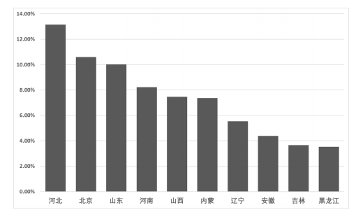
本研究共纳入7245例神经眼科患者,其中男性3331例(46.0%),女性3914例(54.0%),男女比例1—1.2,年龄(38.2±17.5)岁。7245例患者来自全国共计31个省、自治区、直辖市(图1),其中,排名前10位的依次为河北953例(13.15%)、北京768例(10.60%)、山东313例(10.02%)、河南596例(8.23%)、山西541例(7.47%)、内蒙古534例(7.37%)、辽宁401例(5 .53%)、安徽317例(4.38%)、吉林265例(3.66%)和黑龙江256例(3.53%)。
20230328091441_2020.png
图 1 单中心神经眼科疾病患者地域分布特点
Figure 1 Geographical distribution characteristics of patients with nerve ophthalmic diseases from single center

2.2 神经眼科疾病病种分析及疾病性别构成比特点

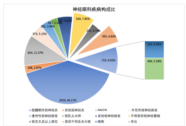
在7245例神经眼科疾病患者中,83.25%(6031/7245)为传入神经系统疾病9.92% (719/7245)为传出神经系统疾病和眼眶疾病,6.83%(495/7245)未归类(包含误诊为神经眼科疾病的视网膜疾病、正常眼压性青光眼等)。病种分析显示:脱髓鞘性视神经炎占比最高,为40.17%(2910/7245),然后依次是NAION 11.37%(824/7245)、外伤性视神经病变5.15%(373/7245),其中7.85%(569/7245)表现为不明原因视神经萎缩(图2)。
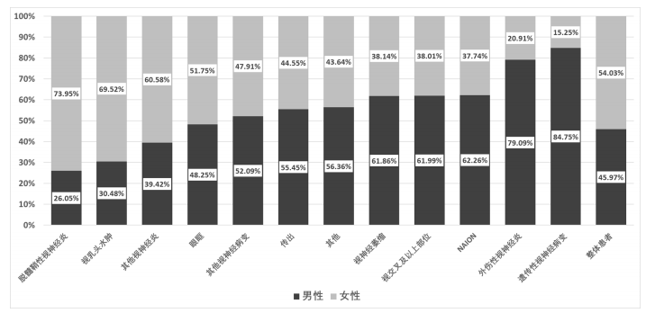
性别分析结果可见脱髓鞘性视神经炎、其他类型视神经疾病、视乳头水肿女性发病为主,比例分别为73.95%、60.58%和69.52%,NAION、外伤性视神经病变、遗传性视神经病变、不明原因视神经萎缩、视交叉及以上后视路病变男性发病为主,比例分别是62.26%、79.09%、84.75%、61.86%和61.99%,其余病种男女比例相当(图3)。
20230328091554_7817.png
图 2 单中心神经眼科疾病住院患者疾病构成比
Figure 2 Disease composition ratio of inpatients with neuroophthalmic disease from single center


图 3 单中心神经眼科住院患者不同病种性别分布
Figure 3 Gender distribution of inpatients with neuroophthalmic disease from single center

2.3 神经眼科疾病患者的年龄分布特点

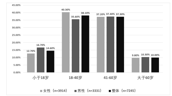
在整体神经眼科疾病患者中,小于18岁者占14.56%(1055/7245),中青年患者占比较高,18~40岁者占比最高,达38.11%(2761/7245),41~60岁者占37.29%(2702/7245),两者共占75.4%,大于60岁者占10.03%(727/7245)。在男性神经眼科疾病患者中,41~60岁者占比最高,达17.18%(1245/7245);女性神经眼科疾病患者中,18~40岁者占比最高,达21.75%(1576/7245)。见图4。
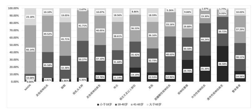
在DON患者中,18~40岁者占比最高(48.63%),NAION患者中41~60岁者占比最高(66.14%)。在外伤性视神经病变和遗传性视神经病变患者中,占比最高的分别为18~40岁者(44.24%)和小于18岁者(48.58%);视交叉及以上后视路病变中41~60岁者占比最高(36.53%)。在传出系统疾病和眼眶疾病患者中,均是41~60岁者占比最高(分别为38.61%和45.71%),其余占比见图5。
20230328091726_3924.png
图 4 单中心神经眼科疾病住院患者不同性别年龄分布
Figure 4 Gender and age distribution of inpatients with neuroophthalmic disease from single center


图 5 单中心神经眼科住院患者不同病种年龄分布
Figure 5 Age distribution of patients with neuroophthalmic disease from single center

2.4 脱髓鞘性视神经炎疾病患者的临床亚型、年龄及性别分布特点

本中心在2015年开始对脱髓鞘性视神经炎患者检测MOG抗体,因此本研究统计了2015年1月1日至2021年2226例DON患者的不同亚型比例。
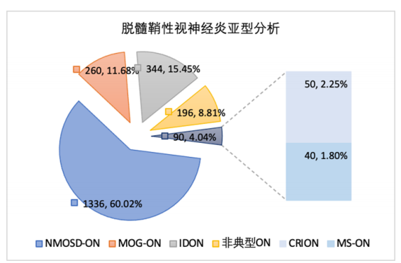
在DON患者中(图6),NMOSD-ON是DON最常见的亚型,比例最高为60.02%(1336/2226),IDON占15.45%(344/2226),MOG-ON占11.68%(260/2226),8.81%(196/2226)患者表现为非典型ON,MS-ON和CRION占比较低,分别是1.80%(40/2226)和2.24%(50/2226)。
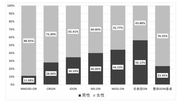
DON整体患者中,76.55%(1704/2226)为女性,男女比例为1 ∶ 3.26。在NMOSD患者中,女性比例最高(88.92%),男女比为1 ∶ 8;MOG-ON男女比例相当,55.77%为女性,男女比例为4 ∶ 5。在非典型ON患者中,男性比例高于女性,为56.12%,男女比为1.28 ∶ 1,其他神经眼科疾病患者的性别构成见图7。
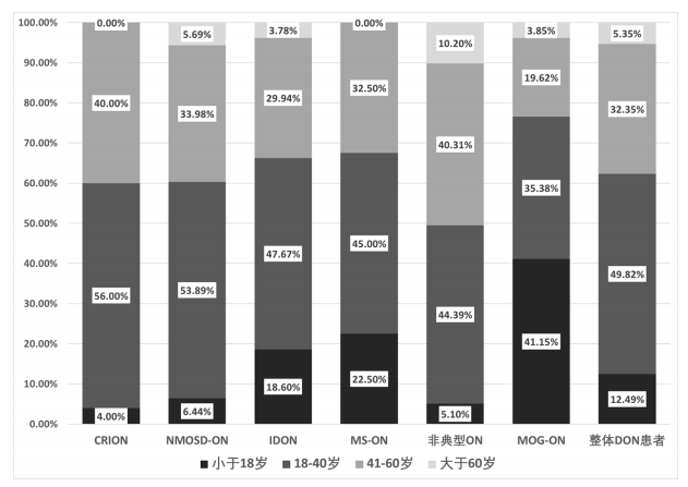
DON患者中,81.79%的患者为中青年,18~40岁者和41~60岁者占比分别为48.63%和33.16%,小于18岁未成年患者占比为12.96%,大于60岁者占比最低,为5.25%。其他亚型视神经炎年龄比例不同,NMOSD-ON患者18~40岁者占比为53.89%,MOG-ON未成年患者可达41.15%,其余神经眼科疾病患者的年龄分布见图8。
20230328091915_8616.png
图 6 DON 患者中的临床亚型及比例分布
Figure 6 Clinical subtypes and proportion distribution of DON patients

图 7 单中心脱髓鞘性视神经炎患者不同性别构成
Figure 7 Gender composition of patients with demyelinating optic neuritis from single center

图 8 单中心脱髓鞘性视神经炎患者不同亚型年龄构成
Figure 8 Age composition of different subtypes in patients with demyelinating optic neuritis from single center

3 讨论

本研究统计了单中心神经眼科住院患者近10年的病种分析,其样本量较大、涵盖病种较为全面。分析了不同病种年龄与性别分布特点,着重介绍了第一大病种脱髓鞘性视神经炎的亚型分析和人口学特点。患者来源于全国共计31个省、自治区、直辖市,其中,排名前10位为北京及其周边省份,这与笔者团队在2016年的报道相似,提示患者来源很大原因和地域交通相关[6]
本研究对神经眼科疾病病种分析结果显示,以传入神经系统疾病为主(83.25%),也是神经眼科疾病的主要医治病种,而这些疾病也是目前我们国家主要的致盲性眼病[7]。本研究还显示,9.92%(719/7245)为传出神经系统疾病和眼眶疾病,6.83%(495/7245)为误诊为视神经疾病的视网膜疾病、正常眼压性青光眼、或非器质性视力下降,因此未能归类。病种分析显示,脱髓鞘性视神经炎占比最高,为40.17%(2910/7245),第二是NAION 11.37%(824/7245),第三是外伤性视神经病变5.15%(373/7245),其中7.85%(569/7245)表现为不明原因视神经萎缩,近几年,随着基因检测技术和影像学的发展,神经眼科疾病的确诊率明显提高,但仍有部分患者应用现有手段未能明确疾病性质[8-9],此类患者目前也是眼科医生临床诊治比较棘手和关注的对象,本次研究对其进行单独统计,与既往研究不同。
从年龄分布看,神经眼科住院患者以18~40岁和41~60岁中青年患者为主(75.40%),年龄分布和神经眼科病种发病特点有关,DON和外伤性视神经病变患者,18~40岁者占比最高(分别是48.63%和44.24%),NAION患者中41~60岁者占比最高(66.14%),小于18岁儿童患者在遗传性视神经病变中占比者最高是48.58%。该结果与既往研究结果一致[10]。不同神经眼科疾病的性别分布不同,DON、其他类型视神经疾病、视乳头水肿女性发病为主,比例分别为73.95%、60.58%和69.52%,与既往文献报道一致[11-12],视乳头水肿多为良性颅高压和CVST为主,既往文献报道也是女性多发[13-14];NAION、外伤性视神经病变、遗传性视神经病变,男性高发,占比分别为62.26%、79.09%、84.75%、61.86%和61.99%,其和疾病发病特点相关,NAION主要累及中老年男性,外伤性视神经病变因从事工作性质及本身易冲动性,男性发病率高于女性,遗传性视神经病变多为Leber遗传性视神经病变,主要累及儿童及青年男性[15-16]。本研究发现不明原因视神经萎缩、视交叉及以上后视路病变男性多发,既往研究及本团队前期研究并未关注此类患者[17]
DON在全球的发病率约为每年(0.94~ 2.18)/100000[18]。美国约翰霍普金斯大学公共卫生学院报告,在临床上小于50岁的患者所患视神经损害性疾病中,脱髓鞘性视神经炎的发病率排第二位[19-20]。本研究结果提示,脱髓鞘性视神经炎是神经眼科疾病最主要病种。研究结果提示,NMO/NMOSD-ON是ION最常见的亚型(60.02%,1336/2226),后依次为IDON(15.45%,344/2226)和MOG-ON(11.68%,260/2226)。MS-ON和CRION占比较低,分别是1.8%(40/226)和2.25% (50/2226)。8.81%(196/2226)患者表现为非典型ON,此类视神经炎预后较差,激素治疗不敏感[21-22],需要进一步研究其发病机制及临床转归。本研究中,DON患者中男女比例为1 ∶ 3.26;其亚型性别倾向不同,在NMO/NMOSD患者中,男女比为1 ∶ 8,MOG-ON男女比例为4 ∶ 5,在非典型ON中,男性比例高于女性为1.28 ∶ 1。ION患者以中青年为主(81.79%),18~40岁者和41~60岁者占比分别为48.63%和33.16%,小于18岁未成年患者占比为12.96%,大于60岁者最低,为5.25%。其他亚型视神经炎年龄比例不同,NMOSD-ON患者中18~40岁者占比最高,为53.89%,MOG-ON儿童多发,小于18岁的未成年人占比可达41.15%。
从以上分析可以看出,本中心神经眼科收治的病例以传入神经系统疾病为主,主要包含DON、NAION、外伤和遗传性视神经病变;此四类疾病是神经眼科最主要的致盲性眼病,这些疾病应作为神经眼科医生今后防盲治盲工作的重中之重。同时,尽管目前视神经疾病治疗水平明显提高,但仍有一部分视神经萎缩患者未能查到病因[23-24],要加强对此类视神经疾病的早期初筛,做到早发现、早诊断、早治疗,减少致盲率[25],进一步提高全国神经眼科疾病诊治水平。本研究可为神经眼科疾病的流行病学和临床研究提供基础。

开放获取声明

本文适用于知识共享许可协议 (Creative Commons),允许第三方用户按照署名(BY)-非商业性使用(NC)-禁止演绎(ND)(CCBY-NC-ND)的方式共享,即允许第三方对本刊发表的文章进行复制、发行、展览、表演、放映、广播或通过信息网络向公众传播,但在这些过程中必须保留作者署名、仅限于非商业性目的、不得进行演绎创作。详情请访问:https://creativecommons.org/licenses/by-nc-nd/4.0/。

1、宋宏鲁, 魏世辉, 孙明明, 等. 《中国脱髓鞘性视神经炎诊断和治 疗循证指南(2021年)》解读[ J]. 中华眼底病杂志, 2021(10): 753-757.
Song HL, Wei SH, Sun MM, et al. Interpretation of Evidence-Based Guidelines for Diagnosis and Treatment of demyelinating optic neuritis in China(2021)[ J]. Chinese Journal of fundus Diseases, 2021(10): 753-757.
宋宏鲁, 魏世辉, 孙明明, 等. 《中国脱髓鞘性视神经炎诊断和治 疗循证指南(2021年)》解读[ J]. 中华眼底病杂志, 2021(10): 753-757.
Song HL, Wei SH, Sun MM, et al. Interpretation of Evidence-Based Guidelines for Diagnosis and Treatment of demyelinating optic neuritis in China(2021)[ J]. Chinese Journal of fundus Diseases, 2021(10): 753-757.
2、Stunkel L. Big data in neuro-ophthalmology: international classification of diseases codes[ J]. J Neuroophthalmol, 2022, 42(1): 1-5.Stunkel L. Big data in neuro-ophthalmology: international classification of diseases codes[ J]. J Neuroophthalmol, 2022, 42(1): 1-5.
3、Cherayil NR, Tamhankar MA. Neuro-ophthalmology for internists[ J]. Med Clin North Am, 2021, 105(3): 511-529.Cherayil NR, Tamhankar MA. Neuro-ophthalmology for internists[ J]. Med Clin North Am, 2021, 105(3): 511-529.
4、Rucker JC. Neuro-ophthalmology of systemic disease[ J]. Semin Neurol, 2009, 29(2): 111-123.Rucker JC. Neuro-ophthalmology of systemic disease[ J]. Semin Neurol, 2009, 29(2): 111-123.
5、Weber KP, Straumann D. Neuro-ophthalmology update[ J]. J Neurol, 2014, 261(7): 1251-1256.Weber KP, Straumann D. Neuro-ophthalmology update[ J]. J Neurol, 2014, 261(7): 1251-1256.
6、王雪琼, 李军, 周欢粉, 等. 单中心神经眼科住院患者疾病构成比 分析[ J]. 眼科, 2018, 27(5): 381-385.
Wang XQ, Li J, Zhou HF, et al. Analysis of disease composition ratio of inpatients from nerve ophthalmology of single center [ J]. Ophthalmology, 2018, 27(5): 381-385.
王雪琼, 李军, 周欢粉, 等. 单中心神经眼科住院患者疾病构成比 分析[ J]. 眼科, 2018, 27(5): 381-385.
Wang XQ, Li J, Zhou HF, et al. Analysis of disease composition ratio of inpatients from nerve ophthalmology of single center [ J]. Ophthalmology, 2018, 27(5): 381-385.
7、Song HL, Xu QG, Wei SH. Progress in the treatment of neuromyelitis optica spectrum disorders related optic neuritis[ J]. Zhonghua Yan Ke Za Zhi, 2020, 56(7): 539-543.Song HL, Xu QG, Wei SH. Progress in the treatment of neuromyelitis optica spectrum disorders related optic neuritis[ J]. Zhonghua Yan Ke Za Zhi, 2020, 56(7): 539-543.
8、Winter A, Chwalisz B. MRI characteristics of NMO, MOG and MS related optic neuritis[ J]. Semin Ophthalmol, 2020, 35(7/8): 333-342.Winter A, Chwalisz B. MRI characteristics of NMO, MOG and MS related optic neuritis[ J]. Semin Ophthalmol, 2020, 35(7/8): 333-342.
9、Costello F, Scott JN. Imaging in neuro-ophthalmology[ J]. Continuum (Minneap Minn), 2019, 25(5): 1438-1490.Costello F, Scott JN. Imaging in neuro-ophthalmology[ J]. Continuum (Minneap Minn), 2019, 25(5): 1438-1490.
10、李星仪, 张秀兰, 王伟, 等. 儿童及青少年神经眼科住院患者病因 分析[ J]. 中国实用眼科杂志, 2013, 31(10): 1281-1286.
Li XY, Zhang XL, Wang W, et al. Etiology analysis of pediatric and adolescent inpatients with neuroophthalmology [ J]. Chinese Journal of Practical Ophthalmology, 2013, 31(10): 1281-1286.
李星仪, 张秀兰, 王伟, 等. 儿童及青少年神经眼科住院患者病因 分析[ J]. 中国实用眼科杂志, 2013, 31(10): 1281-1286.
Li XY, Zhang XL, Wang W, et al. Etiology analysis of pediatric and adolescent inpatients with neuroophthalmology [ J]. Chinese Journal of Practical Ophthalmology, 2013, 31(10): 1281-1286.
11、Dohlman JC, Cestari DM, Freitag SK. Orbital disease in neuroophthalmology[ J]. Curr Opin Ophthalmol, 2020, 31(6): 469-474.Dohlman JC, Cestari DM, Freitag SK. Orbital disease in neuroophthalmology[ J]. Curr Opin Ophthalmol, 2020, 31(6): 469-474.
12、Levin LA, Sengupta M, Balcer LJ, et al. Report from the national eye institute workshop on neuro-ophthalmic disease clinical trial endpoints: optic neuropathies[ J]. Invest Ophthalmol Vis Sci, 2021, 62(14): 30.Levin LA, Sengupta M, Balcer LJ, et al. Report from the national eye institute workshop on neuro-ophthalmic disease clinical trial endpoints: optic neuropathies[ J]. Invest Ophthalmol Vis Sci, 2021, 62(14): 30.
13、Yan Y, Liao YJ. Updates on ophthalmic imaging features of optic disc drusen, papilledema, and optic disc edema[ J]. Curr Opin Neurol, 2021, 34(1): 108-115.Yan Y, Liao YJ. Updates on ophthalmic imaging features of optic disc drusen, papilledema, and optic disc edema[ J]. Curr Opin Neurol, 2021, 34(1): 108-115.
14、Sibony PA , Kupersmith MJ, Kardon RH. Optical coherence tomography neuro-toolbox for the diagnosis and management of papilledema, optic disc edema, and pseudopapilledema[ J]. J Neuroophthalmol, 2021, 41(1): 77-92.Sibony PA , Kupersmith MJ, Kardon RH. Optical coherence tomography neuro-toolbox for the diagnosis and management of papilledema, optic disc edema, and pseudopapilledema[ J]. J Neuroophthalmol, 2021, 41(1): 77-92.
15、Kim US, Jurkute N, Yu-Wai-Man P. Leber hereditary optic neuropathy-light at the end of the tunnel?[ J]. Asia Pac J Ophthalmol (Phila), 2018, 7(4): 242-245.Kim US, Jurkute N, Yu-Wai-Man P. Leber hereditary optic neuropathy-light at the end of the tunnel?[ J]. Asia Pac J Ophthalmol (Phila), 2018, 7(4): 242-245.
16、Zuccarelli M, Vella-Szijj J, Serracino-Inglott A, et al. Treatment of Leber 's hereditary optic neuropathy: an overview of recent developments[ J]. Eur J Ophthalmol, 2020, 30(6): 1220-1227.Zuccarelli M, Vella-Szijj J, Serracino-Inglott A, et al. Treatment of Leber 's hereditary optic neuropathy: an overview of recent developments[ J]. Eur J Ophthalmol, 2020, 30(6): 1220-1227.
17、庄淑流, 刘利娟, 陈积斌, 等. 378例视神经疾病病因分析[ J]. 中 国中医眼科杂志, 2009, 19(3): 145-147.
Zhuang SL, Liu LJ, Chen JB, et al. Etiological analysis of 378 cases of optic nerve disease[ J]. Chinese Journal of Traditional Chinese Ophthalmology, 2009, 19(3): 145-147.
庄淑流, 刘利娟, 陈积斌, 等. 378例视神经疾病病因分析[ J]. 中 国中医眼科杂志, 2009, 19(3): 145-147.
Zhuang SL, Liu LJ, Chen JB, et al. Etiological analysis of 378 cases of optic nerve disease[ J]. Chinese Journal of Traditional Chinese Ophthalmology, 2009, 19(3): 145-147.
18、Toosy AT, Mason DF, Miller DH. Optic neuritis[ J]. Lancet Neurol, 2014, 13(1): 83-99.Toosy AT, Mason DF, Miller DH. Optic neuritis[ J]. Lancet Neurol, 2014, 13(1): 83-99.
19、Levin MH. Demyelinating optic neuritis and its subtypes[ J]. Int Ophthalmol Clin, 2019, 59(3): 23-37.Levin MH. Demyelinating optic neuritis and its subtypes[ J]. Int Ophthalmol Clin, 2019, 59(3): 23-37.
20、García Ortega A, Monta?ez Campos FJ, Mu?oz S, et al. Autoimmune and demyelinating optic neuritis[ J]. Arch Soc Esp Oftalmol (Engl Ed), 2020, 95(8): 386-395.García Ortega A, Monta?ez Campos FJ, Mu?oz S, et al. Autoimmune and demyelinating optic neuritis[ J]. Arch Soc Esp Oftalmol (Engl Ed), 2020, 95(8): 386-395.
21、Sarkar P, Mehtani A, Gandhi H C, et al. Atypical optic neuritis: an overview[ J]. Indian J Ophthalmol, 2021, 69(1): 27-35.Sarkar P, Mehtani A, Gandhi H C, et al. Atypical optic neuritis: an overview[ J]. Indian J Ophthalmol, 2021, 69(1): 27-35.
22、Abel A, McClelland C, Lee MS. Critical review: typical and atypical optic neuritis[ J]. Surv Ophthalmol, 2019, 64(6): 770-779.Abel A, McClelland C, Lee MS. Critical review: typical and atypical optic neuritis[ J]. Surv Ophthalmol, 2019, 64(6): 770-779.
23、Shrestha P, Sitaula S, Sharma AK, et al. Clinical assessment and etiological evaluation of optic nerve atrophy[ J]. Nepal J Ophthalmol, 2021, 13(25): 73-81.Shrestha P, Sitaula S, Sharma AK, et al. Clinical assessment and etiological evaluation of optic nerve atrophy[ J]. Nepal J Ophthalmol, 2021, 13(25): 73-81.
24、Jones R, Al-Hayouti H, Oladiwura D, et al. Optic atrophy in children: current causes and diagnostic approach[ J]. Eur J Ophthalmol, 2020, 30(6): 1499-1505.Jones R, Al-Hayouti H, Oladiwura D, et al. Optic atrophy in children: current causes and diagnostic approach[ J]. Eur J Ophthalmol, 2020, 30(6): 1499-1505.
25、滕克禹, 吕丽萍. 视神经萎缩病因分析[ J]. 中国中医眼科杂志, 2013, 23(6): 428-430.
Teng KY, Lv LP. Etiology analysis of optic atrophy [ J]. Chinese Journal of Traditional Chinese Medicine Ophthalmology, 2013, 23(6): 428-430.
滕克禹, 吕丽萍. 视神经萎缩病因分析[ J]. 中国中医眼科杂志, 2013, 23(6): 428-430.
Teng KY, Lv LP. Etiology analysis of optic atrophy [ J]. Chinese Journal of Traditional Chinese Medicine Ophthalmology, 2013, 23(6): 428-430.
1、国家自然科学基金青年项目 (82101110)。
This work was supported by Youth Program of National Natural Science Foundation of China(82101110)()
上一篇
下一篇
其他期刊
  • 眼科学报

    主管:中华人民共和国教育部
    主办:中山大学
    承办:中山大学中山眼科中心
    主编:林浩添
    主管:中华人民共和国教育部
    主办:中山大学
    浏览
  • Eye Science

    主管:中华人民共和国教育部
    主办:中山大学
    承办:中山大学中山眼科中心
    主编:林浩添
    主管:中华人民共和国教育部
    主办:中山大学
    浏览
推荐阅读
出版者信息
目录