您的位置: 首页 > 2022年7月 第37卷 第7期 > 文字全文
2023年7月 第38卷 第7期11
目录

周边遮盖对成年视皮层双眼优势平衡的作用

Effect of peripheral patching on binocular dominance in adult visual cortex

来源期刊: 眼科学报 | 2022年7月 第37卷 第7期 527-536 发布时间: 收稿时间:2022/12/1 15:15:29 阅读量:4008
作者:
关键词:
周边遮盖双眼竞争双眼平衡可塑性反向遮盖
peripheral patching binocular rivalry binocular balance plasticity inverse patching
DOI:
10.3978/j.issn.1000-4432.2022.06.08
收稿时间:
 
修订日期:
 
接收日期:
 
目的:探究短期周边遮盖对成年视皮层双眼优势平衡的作用。方法:对12名正常成年人的各眼 (24只眼)分别进行单眼短期周边遮盖。遮盖方式为单眼佩戴90 min的环形、半透明的塑料遮盖板,遮盖板仅能透光,中央留有1 0°~15°视野范围的圆孔,从而实现周边遮盖。受试者在周边遮盖前、遮盖后的0~3、3~6、6~9、9~12、12~15、30、60和90 min均完成双眼竞争任务 (binocular rivalry task)。记录并分析各时间段中各眼的占优时间、双眼竞争在眼别间切换周期数和各眼占优概率随时间改变的特点等。每位受试者左右眼测试间隔1周进行。结果:在遮盖前,12名正常成年受试者被遮盖眼的占优时间与非遮盖眼的差异无统计学意义(92.78±6.33 s vs 87.22±6.23 s,P>0.05),提示眼优势平衡。遮盖去除后的0~3 min,被遮盖眼占优比例显著增加至 0.721±0.11(P<0.001),该效应在遮盖去除后的3~30 min均存在(P<0.05),直至60 min(P=0.445)双眼基本恢复优势平衡。双眼优势转换周期在周边遮盖前后差异无统计学意义(P=0.064)。主导眼在去除周边遮盖后的0~3 min遮盖眼占优时间比例相对基线的改变幅度与遮盖非主导眼的差异无统 计学意义(P=0.835)。结论:短期的周边遮盖可改变成年双眼优势平衡,有望应用于视觉关键期后的弱视治疗中。视觉关键期后双眼视功能仍保留有一定的可塑性。

Objective: To study the effect of short-term peripheral patching on binocular dominance in adult visual cortex. Methods: Monocular short-term peripheral patching was performed on each eye (24 eyes) of 12 normal adults. The patching was achieved by monocularly wearing a ring-shaped, translucent and plastic patch for 90 minutes. The patch could only transmit light, but not pattern, and there was a circular hole with a visual field of 10°–15°, so as to achieve peripheral patching. Participants completed the binocular rivalry task at baseline and 0–3, 3–6, 6–9, 9–12, 12–15, 30, 60 and 90 min after peripheral patching. The dominance duration of each eye and the number of dominance switches between eyes were recorded. The probability of perceiving stimulus of each eye was calculated in each time period. Each participant’s both left and right eyes performed peripheral patching one week apart. Results: Before patching, the dominance duration of the patched eye was not significantly different from the non-patched eye (92.78±6.33 s vs 87.22±6.23 s, P>0.05), which suggests that the eye dominance was balanced. At 0–3 min after the removal of the patch, the dominance duration of the patched eye was increased significantly (P<0.001), and this effect existed until 30 min after the removal of the patch (P<0.05). The dominance duration of the patched eye at post-60 min was not significantly different from the baseline (P=0.445). There was no significant difference in the dominance switches among baseline and each period after patching (P=0.064). After the removal of patch on the dominant eye, the amplitude of change in the dominance duration of the patched eye at 0–3 min was not significantly different from that after the removal of patch on the non-dominant eyes (P=0.835). Conclusion: Short-term peripheral patching can also change the binocular dominance in adults, and it has the potential to be applied in treatment of adult amblyopia. After the critical period for visual development, binocular vision function still retains plasticity.
      弱视是在视觉发育期内由于单眼或双眼视觉输入不足、双眼间异常相互作用,导致视觉系统异常发育的眼病,其确切发病机制仍不清楚[1]。经典的遮盖治疗通过遮盖非弱视眼,消除双眼间的竞争性抑制,促进弱视眼的使用,期望恢复双眼间优势平衡,从而提高弱视眼视功能[2]。作为目前弱视的标准治疗方法之一,遮盖治疗在低龄患儿中具有较好的效果[3-6],但对于视觉关键期后的弱视患者则效果有限[7]。寻找新的弱视治疗策略对改 善大龄弱视患者视功能具有重要意义。
      近年来研究[8-10]发现:成年人在短期单眼完 全遮盖后双眼优势平衡发生明显改变,但与传统 弱视理论不同的是,被遮盖眼在去遮盖后反而占优。有学者[8]基于此提出了完全遮盖弱视眼以改变双眼优势平衡的反向遮盖治疗方法(inverse patching),并在部分成年弱视患者中应用,取得了 一定的效果。但全视野的反向遮盖治疗与传统遮 盖的理念相冲突,尤其是对弱视眼中央视觉的遮盖可能加重弱视眼的视功能损害,因此在国内外引起了关于其伦理、有效性等问题的争议[11]。 
      周边视觉占视野大部分区域,不少研究发现其对中央视觉的正常发育具有重要的作用[12]。一 个非常有意思的问题是,在保留中央视力的情况下,短期的单眼周边遮盖能否代替完全遮盖,并改变关键期后的双眼优势平衡?因此,本研究拟 研究短期周边遮盖对发育关键期后视觉系统双眼 平衡的改变作用及其随时间的变化趋势,旨在为大龄弱视治疗提供新的治疗策略,也为成年视觉 系统的可塑性提供新证据。

1 对象与方法

1.1 对象

      纳入12名来自中山大学的成年学生作为受试者。纳入标准:1 )年龄≥1 8岁;2 )各眼等效球镜 在?6.00~+3.00 D,其中柱镜度数在?2.00~+2.00 D, 双眼间等效球镜差≤±1.50 D;3 )左右眼最佳矫 正视力等于或优于1.0;4)近立体视优于40″。排 除标准:1 )存在双眼视功能异常、单眼抑制、眼 位或眼动异常;2 )眼部结构存在病理性改变,如 角膜疾病、视网膜疾病等;3 )患有糖尿病、高血 压、心脏病、精神疾病等全身疾病史;4 )曾有眼 部手术、外伤史;5 )无法配合完成本研究的临床 检查和视觉任务者。本研究已通过中山眼科中心 医学伦理委员会审查(审批号:2020KYPJ006),受 试者均已签署知情同意书。

1.2 仪器

      所有刺激均在计算机中运行的Matlab(美 国 MathWorks公 司)和Psychtoolbox(3.0版)生成和 控制,呈现于伽马校正过的3 D显示器上(A SUS VG278HE;刷新率144 Hz;分辨率1 920×1 080像 素;背景平均亮度54 cd/m2 )。受试者通过3D快门眼 镜(NVIDIA 3D Shuttle Goggle)接受双眼分视刺激[13]。 在实验期间,受试者距离屏幕57 cm,并以下巴额 托固定头部位置。所有实验均在光线昏暗的房间 (<5 lx)中进行。

1.3 方法

1.3.1 一般资料采集

      收集受试者姓名、年龄、性别、疾病史等。

1.3.2 基本视功能评价

      受试者在足度矫正屈光度下进行下述视功能检测及视觉任务。通过标准对数视力E字表检测各眼远距离最佳矫正视力;通过非接触眼压计检 测各眼眼压;通过裂隙灯、直接检眼镜检测各眼眼前段和眼后段病理性改变;采用四孔灯、线状镜、随机点立体视本检测双眼视功能;通过卡洞法确定受试者的主导眼[14]

1.3.3 双眼眼优势平衡的测量

       双眼竞争任务是衡量双眼优势平衡的经典心理物理学方法,在研究中被广泛使用[15-16]。该任务通过在左右眼呈现不相容的一对刺激(一般为正交朝向),使两眼的视觉输入相互竞争视觉意识。受试者通过按键反馈知觉到的视觉刺激特征(如不同 朝向),以各眼占优势时间或占优势次数来衡量双 眼优势平衡情况。
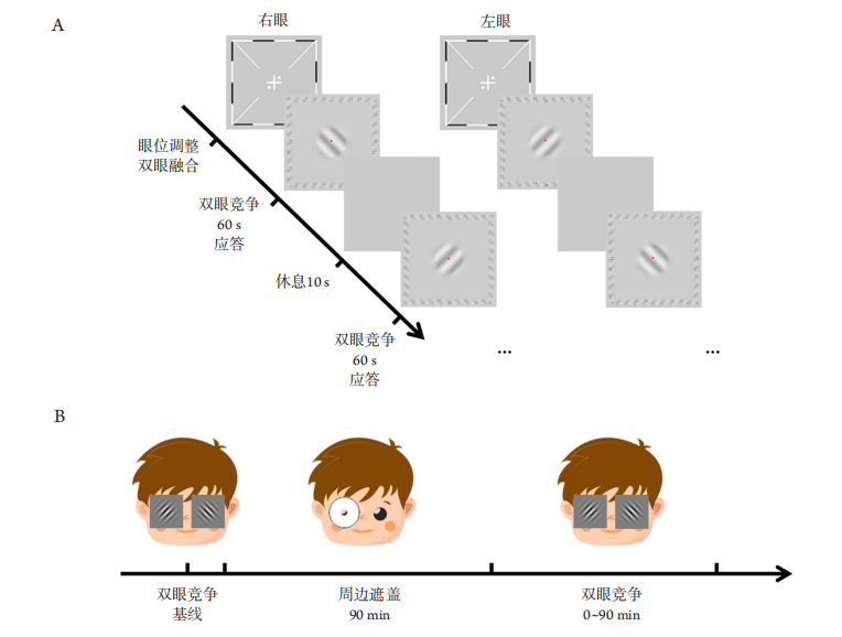
       本研究中使用的视觉刺激为1对40%对比度、空 间频率为4c/d(cycle/degree)的正交正弦光栅(±45°), 大小均为3°×3°,边缘有0.25°的半高斯渐变处理, 使光栅的最大对比度部分大小约为2.5° ×2.5°。为帮助受试者在分视状态下融合,光栅刺激的周边还呈现了由黑白小方块组成的高对比度框架。在视觉刺激的中央始终有1个红色的中心固定点(亮度为56.6 cd/m2 )帮助受试者固视中央(图1A)。 
       双眼竞争任务在每轮次(block)进行3个试次 (trial)。在每轮次前均需进行双眼融合调整:通过 双眼分视技术在左右眼呈现十字与黑白框刺激, 以及互补的圆点,调整图像相对位置直至左右眼画面中的十字和黑白框刺激相互融合,且四个象限中的圆点对称分布,记录此时图像的相对位置并用于后续双眼竞争任务。在每个试次中,受试者在分视下各眼分别接受持续60 s的正交朝向光栅 刺激(±45°,每试次在眼间随机),并通过左右按键报告他们所感知到的光栅刺激朝向(图1A)。记录 所有按键的时间点,记录“左”与“右”按键的切换次数。

1.3.4 实验设计

      本研究中的周边遮盖方式为单眼佩戴环形、半透明塑料遮盖板(仅透过20%光,无形觉信息)。 遮盖板总直径为50 mm,遮盖板中央有约5mm的圆孔,其对应的视野范围为中央10°~15°,可实现对单眼中央15 °以外的形觉信息进行遮盖。每次 (session)遮盖时间为90 min(图1B)。 
20230210151822_3233.png

图1 视觉刺激、双眼竞争任务和实验设计
Figure 1 Visual stimuli, binocular rivalry task and experimental design
(A)受试者在每次双眼竞争任务前均通过调整图像相对位置确保双眼融合,后在各眼呈现持续的60 s光栅刺激,受试者进行应答,休息10 s后进入下一个试次,直至结束;(B)受试者首先进行双眼竞争任务获得双眼平衡的基线状态,后进行单眼周边遮盖90 min,再次完成多次双眼竞争任务以评价双眼优势随时间的变化特性。
(A) Prior to each block of binocular rivalry task, participants adjusted the position of dichoptic images of the frames and fixation crosses to ensure binocular fusion. After that, two orthogonal gratings were presented to the two eyes dichoptically. Participants viewed the dichoptic images for 60 s and reported their dominant percept using keyboard. After 10 s for resting, the next trial was initiated. (B) Participants first performed the binocular rivalry task to obtain the baseline of binocular balance, and then performed the peripheral patching of one eye for 90 min, and subsequently completed the binocular rivalry tasks at each timepoint, to evaluate the changes in binocular dominance over time.

      对于每名受试者,主导眼和非主导眼均接受1次周边遮盖(顺序随机),时间间隔为1周。在1次周边遮盖实验中,受试者首先进行1 min的双眼竞争任务演示练习,然后进行3个轮次(共3 min)的眼优势平衡测量以确定遮盖前的眼优势指数;受试者随后通过磨砂遮盖板进行90 min周边遮盖。为使被遮盖眼的中央视野保持在遮盖板孔中,固定受试者的头部在下颌托中,调整头位和遮盖板至合适位置,进行阅读或观看电影等。在撤去遮盖板后的0~3、3~6、6~9、9~12、12~15、3 0、 60和90 min分别完成3个轮次(共3 min)的双眼竞争任务。

1.4 统计学处理

      在每轮次的测试中(3 min,180 s),计算所有受试者在单个时刻中报告遮盖眼和报告非遮盖眼的概率,获得遮盖眼和非遮盖眼平均占优概率随时间的变化图。计算各眼占优时间在总测试时间中的比例,即各眼的占优时间比例。为更好地描 述周边遮盖对眼优势平衡的改变作用,我们进一步将各时间段中遮盖眼的占优时间比例除以各受试者在基线时的遮盖眼占优时间比例,进而获得各时间段中遮盖眼标准化后的占优时间比例。按键切换次数计作眼优势转换周期数。 
      采用SPSS 20.0统计学软件进行数据分析。计量资料满足正态分布及方差齐性条件,采用配对t检验,以均数±标准差(x±s)表示。利用单因素重复测量方差分析(ANOVA)分析不同时间点下眼优势指数、遮盖眼占优时间、眼优势转换周期的改变,以Shapiro-Wilk检验正态性,以Mauchly’s 球形假设检验球形度,若不服从球形假设则以 Greenhouse & Geisser法进行校正,并采用事后 LSD-t检验分析不同时间点的改变。以Bonferroni方法对各组均数间多重检验进行校正。P<0.05为差异有统计学意义。

2 结果

2.1 受试者基本资料

      本研究共纳入12名成年受试者,其中女7名, 男5名,年龄18~26(25.25±2.48)岁。主导眼视力为 1.01±0.05,非主导眼为1.01±0.06,眼间视力差为 0.01±0.08,差异无统计学意义(P>0.05)。等效球镜 为(-4.06 ±1.59) D,范围为(-5.50~?0.50) D,眼间等效球镜差为(-0.04±0.43) D。具体临床数据见表1。

2.2 正常成年人双眼平衡

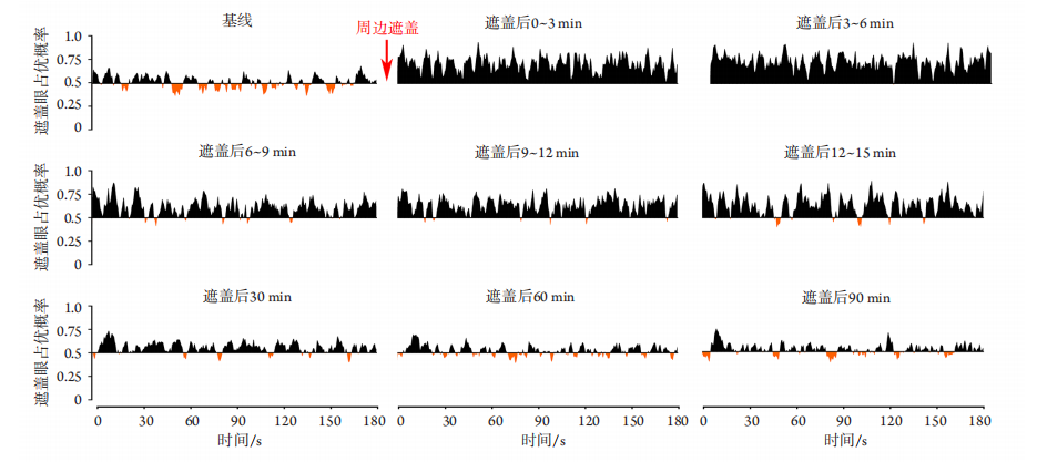
      12名受试者在遮盖前基线处,遮盖眼和非遮盖眼平均占优概率基本平衡(水平轴上方和下方的曲线下面积基本相同;图2 )。其中,被遮盖眼在每个试次中的占优时间为(92.78±6.33) s,非遮盖眼占优时间为(87.22±6.23) s,两者差异无统计学意义(t 23=1.952,P >0.05),占优时间比为 0.515±0.04,提示受试者在遮盖前双眼基本平衡。 双眼优势转换周期次数为(18.99±3.21)次。

表1 受试者临床数据
Table 1 Clinical data of subjects

20230210152003_0481.png
20230210152059_9613.png

图2 受试者对呈现在被遮盖眼视觉刺激的应答概率在周边遮盖前后的变化图
Figure 2 Changes in the average proportion of times participants reported seeing the stimulus presented to the patched eye before and after peripheral patching
在基线处,水平轴(X轴)上下方曲线数量和曲线下面积基本相等,即对被遮盖眼的应答概率与随机概率相近,提示双眼基 本平衡。周边遮盖后,X轴上方曲线下面积明显增加,提示受试者更有可能看到呈现在被遮盖眼的视觉刺激。60、90 min 时双眼基本恢复平衡。X轴以上代表被遮盖眼占优,X轴以下代表非遮盖眼占优。
At the baseline, the number of curves and the area under curve (AUC) above and below the X-axis were basically similar, indicated that the probability of reporting the stimuli presented to the patched eye was similar to the random probability, so the dominance of the two eyes were balanced. After the removal of peripheral patching, the number of curves and the AUC above the X-axis increased significantly, suggesting that participants were more likely to report the stimuli presented to the patched eye. The dominance of the two eyes did not rebalance until post-60 min. Curves above the X-axis represents the predominance of the patched eye, while those below the X-axis represents the predominance of the non-patched eye.

2.3 正常成年人周边遮盖后双眼优势平衡改变

      图2展示了12名受试者在周边遮盖(遮盖主导眼和遮盖非主导眼)90 min前后,遮盖眼和非遮盖眼平均占优概率随时间的变化情况,可见遮盖后遮盖眼的平均占优概率即刻增加(水平轴上方的曲线数量和曲线下面积均比轴下方的增多)。随着时间推移(3~30 min),遮盖眼占优效应持续存在但逐渐减弱,非遮盖眼在双眼竞争任务中的比重逐渐恢复(水平轴下方的曲线数量和曲线下面积逐渐增多)。遮盖后的60 min和90 min各眼占优比例已基本恢复平衡,与基线情况基本相同。 
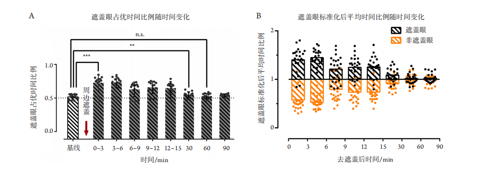
      进一步通过单因素重复测量ANOVA分析不同时间段间遮盖眼占优时间的改变(图3 ),经箱线图判断,数据无异常值;经Shapiro-Wilk检验,各组数据服从正态分布(P>0.05);经过Mauchly’s 球形假设检验,因变量的方差协方差矩阵不相等,χ 2 (35)=84.517,P<0.001,通过Greenhouse & Geisser方法校正ε =0.576。去遮盖前后各时间段间(基线、0~3 min、3~6 min 、6~9 min、 9~12 min 、 12~15 min 、30 min 、60 min 、90 min)发生显著改变[校正后F(4.607,105.95)=28.211,P<0.001,偏ω2 =0.502]。通过事后LSD-t检验可发现直至去遮盖后的30 min,遮盖眼的占优时间仍显著高于基线(基线 vs 0~3 min,基线 vs 3~6 min, 基线 vs 6~9 min,基线 vs 9~12 min,基线 vs 12~ 15 min,均P<0.001;基线 vs 30 min,P=0.002,小于多重检验校正后的P值(0.05/8=0.006),提示周边遮盖显著改变了中央视觉的双眼优势平衡,且使双眼平衡向被遮盖眼倾斜。随着去遮盖后的时间延长,遮盖眼的占优时间逐渐减少,60、90 min与基线差异均无统计学意义(基线 vs 60 min,P=0.445;基线 vs 90 min,P=0.196)。
20230210152230_0688.png

图3 周边遮盖对双眼平衡的改变
Figure 3 Peripheral patching changes binocular balance
(A)去遮盖后,遮盖眼占优时间比例在0~3 min、3~6 min、6~9 min、9~12 min、12~15 min与30 min均显著高于基线,而在60 min、90 min与基线差异无统计学意义;(B)遮盖眼与非遮盖眼标准化后的平均占优时间(各时间段的周期内占优时间与基线的比值)在去遮盖后随时间的变化图。若时间比值为1,代表占优时间与基线水平相同;越高于1则占优时间越多,最多为2;越低于1则占优时间越少,最少为0。周边遮盖后,被遮盖眼的占优时间明显增多,而相应的非遮盖眼占优时间减少,直至30 min。在60 min时,各眼占优时间基本恢复至基线水平。
(A) The proportion of the patched eye dominance durations at 0–3 min, 3–6 min, 6–9 min, 9–12 min, 12–15 min and 30 min after the removal of the patch were significantly higher than the baseline. Neither the proportion of the patched eye dominance duration at the 60 min nor that at 90 min was significantly different from the baseline. B. The normalized mean dominance duration for each eye (the ratio of dominance duration at each timepoint to that at baseline) after the removal of patch. The ratio of 1 means that the dominance duration is similar to the baseline, while the ratio more or less than 1 means that the dominance duration is above or below the baseline, respectively. Until 30 min after the removal of peripheral patching, the dominance duration of the patched eye increased significantly, while the corresponding non-patched eye dominance duration decreased. At 60 min, the dominance duration of each eye basically returned to the baseline level.

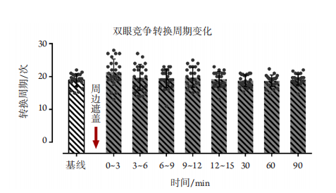
      各时间段的眼优势转换周期变化如图4所示。 单因素重复测量ANOVA未发现各时间段间转换周期差异有统计学意义[校正F(5.227,120.225)=2.127, P =0.064,偏ω2 =0.040]。尽管未发现各时间段间的眼优势转换周期差异,配对t检验发现去遮盖后的3 min内转换周期显著大于基线(P =0.020), 而其余时间段间差异均无统计学意义 ( 基 线 v s 3~6 min,基线 vs 6~9 min,基线 vs 9~12 min, 基 线 vs 12~15 min,基线 vs 30 min,基线 vs 60 min,基线 vs 90 min,均P>0.05)。 同一受试者均在不同时间 (间隔1周) 完成了主导眼和非主导眼的单眼周边遮盖。分别对主导/非主导眼进行单因素重复测量ANOVA分析发现,不论是遮盖主导眼 [ F(8,88)=13.067 , P < 0.001 , 偏 ω 2 =0.472] ,还是遮盖非主导眼[校正后F(3.76,41.363)=14.676 ,P < 0.001 ,偏 ω2 =0.503],遮盖眼占优时间比例均发生显著改变。但通过事后 LSD- t 检验发现,遮盖主导眼在30 min的遮盖眼占优时间比例与基线差异无统计学意义(基线 vs 30 min,P =0.132); 除6 ~ 9 min(P=0.008,多重比较校正大于多重检验校正后的P值(0.05/8=0.006)外,0~3 min(P<0.001)、 3~6 min( P < 0.001) 、 9~12 min( P < 0.001) 、 12~15 min( P < 0.001) 仍显著高于基线。除 6~9 min(P=0.037)和9~12 min(P=0.009)外,遮盖非主导眼所引起的遮盖眼占优时间比例在30 min 内的各时间段与基线比较,差异均有统计学意义(基线 vs 0~3 min,P<0.001;基线 vs 3~6 min, P<0.001;基线 vs 12~15 min,P=0.001;基线 vs 30 min,P=0.004,多重比较校正小于多重检验校正后的P值(0.05/8=0.006)。进一步比较去遮盖后的0~3 min遮盖眼占优时间比例相对基线的改变幅度,发现遮盖主导眼引起的占优时间比例改变 幅度与遮盖非主导眼间差异无统计学意义(配 对 t 11=0.214,P=0.835)
20230210152344_3358.png

图4 双眼竞争转换周期变化图示:周边遮盖前后双眼竞争转换周期无显著改变
Figure 4 Dominance switch between the two eyes shows that there was no significant change in the dominance switch among baseline and each timepoint after peripheral patching

3 讨论

    本研究发现短期周边遮盖改变了正常成年人的双眼优势平衡,显著地提高了被遮盖眼在双眼平衡中的占优时间,使双眼优势平衡向被遮盖眼倾斜,而眼优势转换周期没有发生明显改变。周边遮盖所产生的效应及其持续时间与过往完全遮 盖的研究结果基本相同。
    初级视皮层V1区形成眼优势柱均衡依赖于平衡的双眼视觉信息输入[17]。当眼部发生单眼形觉剥夺、斜视、屈光不正或屈光参差等因素引起双眼视觉输入出现不平衡时,视皮层发生竞争性抑制,使双眼优势平衡向优势眼倾斜[1,17-19]。传统观点认为若双眼视觉输入不平衡在视觉发育关键期内长期存在,眼优势柱在结构和功能上均发生明显改变,最后形成持久、难以逆转的双眼优势不平衡,即弱视。而基于这一观点,临床医生[2]认为在视觉发育关键期内,及时遮盖非弱视眼可改变双眼视觉输入平衡,从而改善这种双眼优势不平衡。作为弱视治疗的标准方法,遮盖应用于临床长达250年,但在临床实践中仍然存在一些问题[20]:15%~50%患者仍无法获得视力提高[21]、单眼遮盖的接受度较低、影响日常生活、治疗周期长[21]、依从性差[22]、易复发[3]等。许多临床医生和学者[2,7,17,19]还发现:非弱视眼遮盖对关键期以外的弱视患者效果较差,这可能与关键期后视皮层可塑性下降有关。
    传统观点[23-24]认为:视觉发育关键期后的成年视觉系统可塑性减弱。但国内外越来越多的研究发现成年视觉系统仍保留有一定的可塑性,尤其是在双眼视功能上。Lunghi等[10]发现:短期的完全遮盖可显著改变正常成年人的双眼优势平衡,使被遮盖眼在平衡中占据显著的优势,其持续时间约为30 min。基于此,有学者提出完全遮盖弱视眼以改变双眼优势平衡的反向遮盖疗法[9]。 Lunghi等[25]进一步发现,弱视眼通过短期的反向遮盖结合运动4周后,视功能获得显著的提高。 Zhou等[9]通过基于相位的双眼间平衡测量方法,发现弱视眼在接受反向遮盖后能显著减少来源于非弱视眼的视觉抑制;而进行约2个月的反向遮盖后,弱视患者也获得了一定的视功能提高。但由于对弱视眼进行完全遮盖与传统的遮盖非弱视眼治疗理念(遮盖非弱视眼而强迫弱视眼使用)不同, 而且对弱视眼中央视觉的完全遮盖存在加重弱视眼视功能损害的风险,国内外关于反向遮盖的伦理、有效性等问题仍存在争议[11]。 
    本研究提出新的周边遮盖方法(遮盖周边形觉而完全保留中央视觉),以替代反向遮盖(对非弱视眼中央和周边的完全遮盖),并利用经典的双眼竞争任务对正常成年人的双眼视平衡进行连续观察,为改良弱视眼反向遮盖方法以适应未来的临床应用提供了可能。周边遮盖的提出源于大量研究中发现的周边视觉对中央视觉的功能和发育具有一定的作用。Smith等[12]通过对发育期的恒河猴进行黄斑中心凹激光消融后发现眼球的中央眼轴仍然出现增长,提示中心凹的视觉信号不是导致眼球和视觉改变的必要因素,周边视觉对眼球发育和中央视觉起到十分重要的作用。Hussain等[26] 通过对正常成年人的周边拥挤进行训练,发现其训练效果能转化到中央视觉。本研究也发现短期 的遮盖单眼周边形觉可明显改变视觉关键期后视觉系统的双眼优势平衡,被遮盖眼在双眼平衡中占优时间明显增加,且该效应可维持约60min,与短期的完全遮盖效果相似。此外,本研究还发现周边遮盖对成年双眼平衡的作用具有一定的个体差异。大部分受试者可获得较为明显的双眼平衡向遮盖眼倾斜,而少部分受试者则没有出现明显改变,甚至出现非遮盖眼占优时间提高。这可能与不同个体间视觉可塑性、双眼视平衡程度、注意力和视觉任务范式等差异有关。但本研究仅在正常成年受试者中进行,而关于弱视患者的弱视眼在进行周边遮盖后能否出现双眼平衡向弱视眼倾斜,并产生与反向遮盖相似的双眼视功能提高效果、其应用的安全性及其内在机制等问题仍有待进一步深入研究。
    综上所述,短期周边遮盖可改变正常成年人中央视觉的双眼优势平衡,使被遮盖眼在短期内占优,为大龄弱视治疗提供一种新的治疗策略。但周边遮盖对弱视单眼和双眼视功能效果、长期应用的安全性及其内在机制,仍需要借助客观检测手段和更大样本量的研究。

开放获取声明

    本文适用于知识共享许可协议 (Creative Commons),允许第三方用户按照署名(BY)-非商业性使用(NC)-禁止演绎(ND)(CC BY-NC-ND)的方式共享,即允许第三方对本刊发表的文章进行复制、发行、展览、表演、放映、广播或通过信息网络向公众传播,但在这些过程中必须保留作者署名、仅限于非商业性目的、不得进行演绎创作。详情请访问:https://creativecommons.org/licenses/by-nc-nd/4.0/
1、Holmes JM, Clarke MP. Amblyopia[ J]. Lancet, 2006, 367(9519):1343-1351.Holmes JM, Clarke MP. Amblyopia[ J]. Lancet, 2006, 367(9519):1343-1351.
2、Holmes JM, Repka MX , Kraker RT, et al. The treatment of amblyopia[ J]. Strabismus, 2006, 14(1): 37-42.Holmes JM, Repka MX , Kraker RT, et al. The treatment of amblyopia[ J]. Strabismus, 2006, 14(1): 37-42.
3、Holmes JM, Kraker RT, Beck RW, et al. A randomized trial of prescribed patching regimens for treatment of severe amblyopia in children[ J]. Ophthalmology, 2003, 110(11): 2075-2087.Holmes JM, Kraker RT, Beck RW, et al. A randomized trial of prescribed patching regimens for treatment of severe amblyopia in children[ J]. Ophthalmology, 2003, 110(11): 2075-2087.
4、Wallace DK, Lazar EL, Holmes JM, et al. A randomized trial of increasing patching for amblyopia[ J]. Ophthalmology, 2013, 120(11):2270-2277.Wallace DK, Lazar EL, Holmes JM, et al. A randomized trial of increasing patching for amblyopia[ J]. Ophthalmology, 2013, 120(11):2270-2277.
5、Wang S, Wen W, Zhu WQ, et al. Effect of combined atropine and patching vs patching alone for treatment of severe amblyopia in children aged 3 to 12 years a randomized clinical trial[ J]. JAMA Ophthalmol,2021, 139(9): 990-996.Wang S, Wen W, Zhu WQ, et al. Effect of combined atropine and patching vs patching alone for treatment of severe amblyopia in children aged 3 to 12 years a randomized clinical trial[ J]. JAMA Ophthalmol,2021, 139(9): 990-996.
6、Li TJ, Qureshi R , Taylor K . Conventional occlusion versus pharmacologic penalization for amblyopia[ J]. Cochrane Database Syst Rev, 2019, 8(8): CD006460.Li TJ, Qureshi R , Taylor K . Conventional occlusion versus pharmacologic penalization for amblyopia[ J]. Cochrane Database Syst Rev, 2019, 8(8): CD006460.
7、Holmes JM, Levi DM. Treatment of amblyopia as a function of age[ J]. Vis Neurosci, 2018, 35: E015.Holmes JM, Levi DM. Treatment of amblyopia as a function of age[ J]. Vis Neurosci, 2018, 35: E015.
8、Zhou JW, Baker DH, Simard M, et al. Short-term monocular patching boosts the patched eye's response in visual cortex[ J]. Restor Neurol Neurosci, 2015, 33(3): 381-387.Zhou JW, Baker DH, Simard M, et al. Short-term monocular patching boosts the patched eye's response in visual cortex[ J]. Restor Neurol Neurosci, 2015, 33(3): 381-387.
9、Zhou JW, He ZF, Wu YD, et al. Inverse occlusion: a binocularly motivated treatment for amblyopia[ J]. Neural Plast, 2019, 2019: 5157628.Zhou JW, He ZF, Wu YD, et al. Inverse occlusion: a binocularly motivated treatment for amblyopia[ J]. Neural Plast, 2019, 2019: 5157628.
10、Lunghi C, Burr DC, Morrone C. Brief periods of monocular deprivation disrupt ocular balance in human adult visual cortex[ J]. Curr Biol, 2011,21(14): R538-R539.Lunghi C, Burr DC, Morrone C. Brief periods of monocular deprivation disrupt ocular balance in human adult visual cortex[ J]. Curr Biol, 2011,21(14): R538-R539.
11、Zurevinsky J. Eccentric fixation and inverse occlusion: renewing our interest?[ J]. J Binocul Vis Ocul Motil, 2019, 69(4): 136-140.Zurevinsky J. Eccentric fixation and inverse occlusion: renewing our interest?[ J]. J Binocul Vis Ocul Motil, 2019, 69(4): 136-140.
12、Smith EL, Ramamirtham R, Qiao-Grider Y, et al. Effects of foveal ablation on emmetropization and form-deprivation myopia[ J]. Invest Ophthalmol Vis Sci, 2007, 48(9): 3914-3922.Smith EL, Ramamirtham R, Qiao-Grider Y, et al. Effects of foveal ablation on emmetropization and form-deprivation myopia[ J]. Invest Ophthalmol Vis Sci, 2007, 48(9): 3914-3922.
13、Liu ZT, Chen ZD, Gao L, et al. A new dichoptic training strategy leads to better cooperation between the two eyes in amblyopia[ J]. Front Neurosci, 2020, 14: 593119.Liu ZT, Chen ZD, Gao L, et al. A new dichoptic training strategy leads to better cooperation between the two eyes in amblyopia[ J]. Front Neurosci, 2020, 14: 593119.
14、Rice ML, Leske DA, Smestad CE, et al. Results of ocular dominance testing depend on assessment method[ J]. J AAPOS, 2008, 12(4):365-369.Rice ML, Leske DA, Smestad CE, et al. Results of ocular dominance testing depend on assessment method[ J]. J AAPOS, 2008, 12(4):365-369.
15、Zhao W, Jia WL, Chen G, et al. A complete investigation of monocular and binocular functions in clinically treated amblyopia[ J]. Sci Rep,2017, 7(1): 10682.Zhao W, Jia WL, Chen G, et al. A complete investigation of monocular and binocular functions in clinically treated amblyopia[ J]. Sci Rep,2017, 7(1): 10682.
16、Knapen T, Brascamp J, Adams WJ, et al. The spatial scale of perceptual memory in ambiguous figure perception[ J]. J Vis, 2009, 9(13): 1-12.Knapen T, Brascamp J, Adams WJ, et al. The spatial scale of perceptual memory in ambiguous figure perception[ J]. J Vis, 2009, 9(13): 1-12.
17、 Kiorpes L, Daw N. Cortical correlates of amblyopia[ J]. Vis Neurosci,2018, 35: E016. Kiorpes L, Daw N. Cortical correlates of amblyopia[ J]. Vis Neurosci,2018, 35: E016.
18、Wiesel TN, Hubel DH. Comparison of the effects of unilateral and bilateral eye closure on cortical unit responses in kittens[ J]. J Neurophysiol, 1965, 28(6): 1029-1040.Wiesel TN, Hubel DH. Comparison of the effects of unilateral and bilateral eye closure on cortical unit responses in kittens[ J]. J Neurophysiol, 1965, 28(6): 1029-1040.
19、 Birch EE. Amblyopia and binocular vision[ J]. Prog Retin Eye Res,2013, 33: 67-84. Birch EE. Amblyopia and binocular vision[ J]. Prog Retin Eye Res,2013, 33: 67-84.
20、Meier K , Tarczy-Hornoch K . Recent treatment advances in amblyopia[ J]. Annu Rev Vis Sci, 2022, Epub ahead of print. doi: 10.1146/annurev-vision-100720-022550.Meier K , Tarczy-Hornoch K . Recent treatment advances in amblyopia[ J]. Annu Rev Vis Sci, 2022, Epub ahead of print. doi: 10.1146/annurev-vision-100720-022550.
21、Birch EE, Stager DR . Long-term motor and sensory outcomes after early surgery for infantile esotropia[ J]. J AAPOS, 2006,10(5): 409-413.Birch EE, Stager DR . Long-term motor and sensory outcomes after early surgery for infantile esotropia[ J]. J AAPOS, 2006,10(5): 409-413.
22、Stewart CE, Moseley MJ, Stephens DA, et al. Treatment dose-response in amblyopia therapy: the Monitored Occlusion Treatment of Amblyopia Study (MOTAS)[ J]. Invest Ophthalmol Vis Sci, 2004,45(9): 3048-3054.Stewart CE, Moseley MJ, Stephens DA, et al. Treatment dose-response in amblyopia therapy: the Monitored Occlusion Treatment of Amblyopia Study (MOTAS)[ J]. Invest Ophthalmol Vis Sci, 2004,45(9): 3048-3054.
23、Consorti A, Sansevero G, Torelli C, et al. Visual perceptual learning induces long-lasting recovery of visual acuity, visual depth perception abilities and binocular matching in adult amblyopic rats[ J]. Front Cell Neurosci, 2022, 16: 840708.Consorti A, Sansevero G, Torelli C, et al. Visual perceptual learning induces long-lasting recovery of visual acuity, visual depth perception abilities and binocular matching in adult amblyopic rats[ J]. Front Cell Neurosci, 2022, 16: 840708.
24、Mitchell DE, Maurer D. Critical periods in vision revisited[ J]. Annu Rev Vis Sci, 2022, Epub ahead of print. doi:10.1146/annurev-vision-090721-110411.Mitchell DE, Maurer D. Critical periods in vision revisited[ J]. Annu Rev Vis Sci, 2022, Epub ahead of print. doi:10.1146/annurev-vision-090721-110411.
25、Lunghi C, Sframeli AT, Lepri A, et al. A new counterintuitive training for adult amblyopia[ J]. Ann Clin Transl Neurol, 2019, 6(2): 274-284.Lunghi C, Sframeli AT, Lepri A, et al. A new counterintuitive training for adult amblyopia[ J]. Ann Clin Transl Neurol, 2019, 6(2): 274-284.
26、Hussain Z, Webb BS, Astle AT, et al. Perceptual learning reduces crowding in amblyopia and in the normal periphery[ J]. J Neurosci,2012, 32(2): 474-480.Hussain Z, Webb BS, Astle AT, et al. Perceptual learning reduces crowding in amblyopia and in the normal periphery[ J]. J Neurosci,2012, 32(2): 474-480.
1、邹捷粮,李迎红,文业千等.青少年弱视康复治疗研究进展[J].眼科学报,2023,38(01):52-62.ZOU Jieliang, LI Yinghong, WEN Yeqian, et al. Research progress on rehabilitation treatment of adolescent amblyopia[J]. Eye Sci, 2023, 38(1): 52-62.
1、广东省自然科学基金 (2015A030312016);广东省基础与应用基础研究基金 (2021A1515110479)。This work was supported by the Natural Science Foundation of GuangdongProvince (2015A030312016) and the Guangdong Basic and Applied Basic Research Foundation (2021A1515110479), China()
上一篇
下一篇
其他期刊
  • 眼科学报

    主管:中华人民共和国教育部
    主办:中山大学
    承办:中山大学中山眼科中心
    主编:林浩添
    主管:中华人民共和国教育部
    主办:中山大学
    浏览
  • Eye Science

    主管:中华人民共和国教育部
    主办:中山大学
    承办:中山大学中山眼科中心
    主编:林浩添
    主管:中华人民共和国教育部
    主办:中山大学
    浏览
推荐阅读
出版者信息
目录