您的位置: 首页 > 2023年12月 第38卷 第12期 > 文字全文
2023年7月 第38卷 第7期11
目录

准分子激光原位角膜磨镶术后白内障患者术后远视漂移一例

A case of hyperopic drift after cataract surgery in a patient with previous laser in situ keratomileusis

来源期刊: 眼科学报 | 2023年12月 第38卷 第12期 874-879 发布时间:2023-11-15 收稿时间:2024/3/11 17:20:22 阅读量:4060
作者:
关键词:
白内障准分子激光原位角膜磨镶术人工晶状体远视漂移
cataract laser in situ keratomileusis intraocular lens hyperopic drif
DOI:
10.12419/2307240005
收稿时间:
 
修订日期:
 
接收日期:
 
该文报道了一例40岁女性患者,因“双眼渐进性视物模糊3个月”就诊。患者既往于2005年因高度近视行双眼准分子激光原位角膜磨镶术 (LASIK)。最佳矫正视力OD:0.2 (–11.00 DS/ –1.25 DC×170 °),OS:0.7 (–4.00 DS/ –0.75 DC×25 °)。双眼角膜透明,前房中深,晶状体混浊,豹纹状眼底伴后巩膜葡萄肿。诊断为双眼并发性白内障,并行右眼白内障超声乳化联合人工晶状体 (IOL) 植入术,术中植入+14.0 D IOL一枚,目标屈光度为–0.5 D。术后1周裸眼视力0.3,验光结果示右眼屈光度+2.75 DS,最佳矫正视力0.7。术后2周行右眼IOL置换术,由+14.0 D置换为+17.0 D。右眼术后1周裸眼视力0.8,验光结果示右眼屈光度–0.75 DC×15 °。

It is reported in this article that a 40-year-old female patient presented with "progressive blurred vision of both eyes for 3 months". The patient underwent bilateral laser in situ keratomileusis (LASIK) because of high myopia in 2005. It was recorded that her best corrected visual acuity was 0.2 (–11.00 DS/ –1.25 DC×170 °) in the right eye and 0.7 (–4.00 DS/ –0.75 DC×25 °) in the left, and clear cornea, normal anterior chamber, cloudy lens, tessellated fundus with posterior staphyloma in both eyes. The patient was diagnosed with bilateral complicated cataract. Phacoemulsification combined with intraocular lens (IOL, +14.0 diopter (D)) implantation was performed on the right eye, with the target –0.5D refractive diopter . One week after surgery, it was recorded that the uncorrected visual acuity of the right eye was 0.3, and the best corrected visual acuity was 0.7 (+2.75 DS). IOL replacement of the right eye was performed two weeks after surgery, the +14.0 D IOL was replaced by +17.0 D IOL. One week after surgery, the uncorrected visual acuity of the right eye was 0.8 (–0.75 DC×15 °).
准分子激光原位角膜磨镶术(laser in situ keratomileusis,LASIK)是一种常用的激光角膜屈光手术,发展至今已有二十多年。随着时间推移,既往选择角膜屈光手术的患者如今已步入老年,需要行白内障手术改善屈光间质混浊。角膜屈光手术改变了角膜屈光状态,给人工晶状体(intraocular lens,IOL)目标屈光度计算带来较大难度,导致白内障术后发生近视或远视漂移的概率增加。现将1例LASIK术后白内障患者IOL植入术后发生远视漂移的病例报告如下,供临床同道参考。

1 病例资料

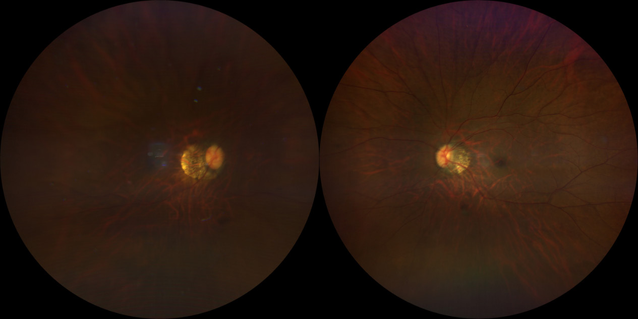
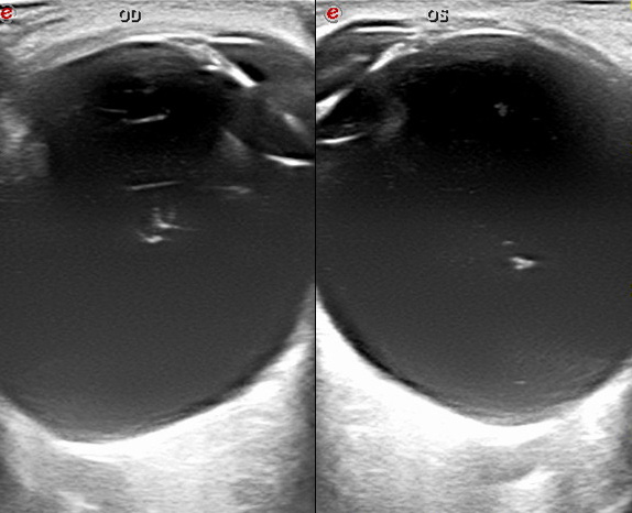
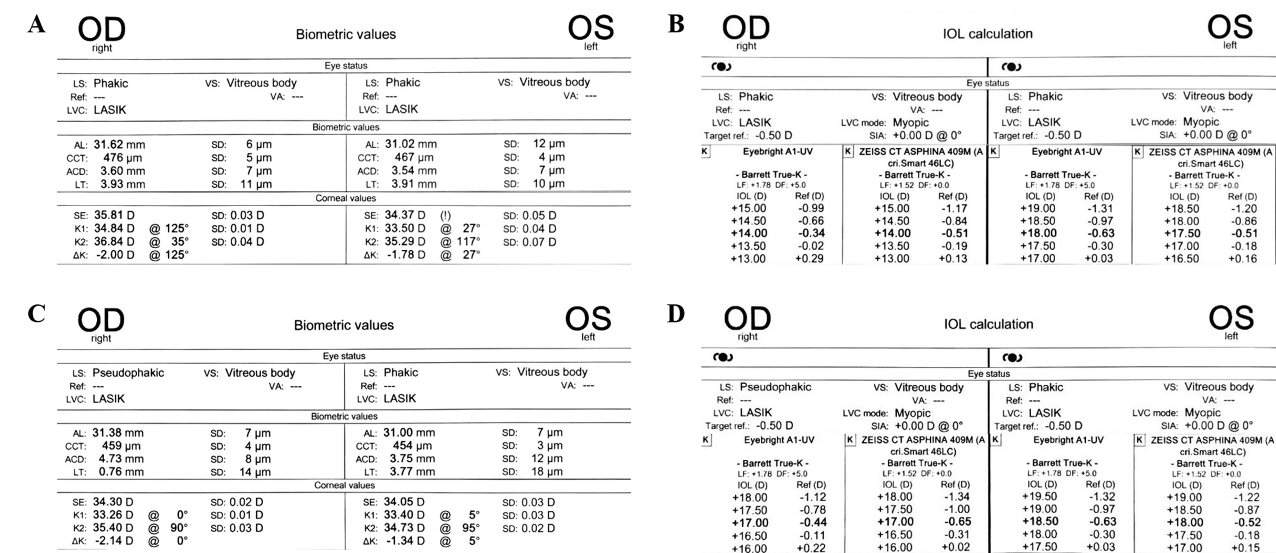
患者女性,4 0岁,因“双眼渐进性视物模糊3月”于2023年2月2日就诊。患者既往于2005年因高度近视行双眼LASIK手术。术前检查:最佳矫正视力OD:0.2 (–11.00 DS/ –1.25 DC×170 °),OS:0.7 (–4.00 DS/ –0.75 DC×25 °)。眼压右眼11 mmHg(1 mmHg=0.133 kPa),左眼12 mmHg。双眼结膜无充血水肿,角膜透明,前房中深,虹膜纹理清,瞳孔圆,直径约3 mm,对光反射灵敏,晶状体皮质混浊,核Ⅲ级,眼底检查见双眼豹纹状眼底伴后巩膜葡萄肿,视网膜平伏在位,视盘旁可见萎缩弧(图1)。B超检查示双眼玻璃体混浊,后巩膜葡萄肿(图2)。使用IOL Master700光学生物测量仪获取眼球生物学参数,双眼中央角膜厚度(central corneal thickness,CCT)、角膜曲率(keratometry,K)、前房深度(anterior chamber depth,ACD)、晶状体厚度(lens thickness,LT)、眼轴长度(axial length, AL)分别为OD:CCT 476 μm,K1 34.84 D,K2 36.84 D,ACD 3.60 mm,LT 3.93 mm,AL 31.62 mm;OS:CCT 467 μm,K1 33.50 D,K2 35.29 D,ACD 3.54 mm,LT 3.91 mm,AL 31.02 mm(图3A)。采用Barrett True-K公式计算IOL目标屈光度,右眼若植入A=117.7、+14.0 D后房型IOL,术后残余屈光度–0.51 D;左眼若植入A=117.7、+17.5 D后房型IOL,术后残余屈光度–0.51 D(图3B)。
20240313103252_9716.png
图1 眼底检查结果
Figure 1 Fundus examination results
眼底相示双眼豹纹状眼底伴后巩膜葡萄肿,视网膜平伏在位,视盘旁可见萎缩弧。
Fundus image showed tessellated fundus, posterior staphyloma and peri-papillary atrophy in both eyes.
20240313103321_5214.png
图2 B超检查结果
Figure 2 B-mode ultrasound examination results
B超示双眼玻璃体混浊,后巩膜葡萄肿。
B-mode ultrasound showed vitreous opacity and posterior staphyloma in both eyes.
20240313103407_1159.png
图3 眼球生物学参数及IOL目标屈光度计算结果
Figure 3 Ocular biological parameters and IOL power calculation results
(A)第一次术前眼球生物学参数; (B)第一次术前IOL目标屈光度计算结果; (C)第二次术前眼球生物学参数; (D)第二次术前IOL
目标屈光度计算结果。
(A) The first pre-operative results of ocular biological parameters. (B) The first pre-operative results of IOL power calculation. (C) The second
pre-operative results of ocular biological parameters. (D) The second pre-operative results of IOL power calculation
患者初步诊断为双眼并发性白内障,双眼角膜屈光术后(LASIK术后),双眼高度近视。患者诊断明确,为进一步治疗,于2023年2月22日行右眼白内障超声乳化联合IOL植入术,手术顺利。术中植入+14.0 D ZEISS CT ASPHINA 409M (A=117.7) IOL一枚,目标屈光度为–0.5 D。右眼术后1周(2023年3月2日)裸眼视力0.3,验光结果示右眼屈光度+2.75 DS,最佳矫正视力0.7。排除引起视力下降的眼前节、眼底及视神经病变等因素后,再次使用IOLMaster700光学生物测量仪行生物学测量,结果示OD:CCT 459 μm,K1 33.26 D,K2 35.40 D,ACD 4.73 mm,LT 0.76 mm,AL 31.38 mm;OS:CCT 454 μm,K1 33.40 D,K2 34.73 D,ACD 3.75 mm,LT 3.77 mm,AL 31.00 mm(图3C)。采用Barrett True-K公式重新计算IOL目标屈光度,右眼若植入A=117.7、+17.0 D后房型IOL,术后残余屈光度–0.65 D;左眼若植入A=117.7、+18.0 D后房型IOL,术后残余屈光度–0.52 D(图3D)。患者于2023年3月8日行右眼IOL置换术,由原ZEISS CT ASPHINA 409M (A=117.7) +14.0 D置换为+17.0 D。右眼术后1周(2023年3月16日)裸眼视力0.8,验光结果示右眼屈光度–0.75 DC×15 °。患者于2023年3月29日行左眼白内障超声乳化联合IOL植入术,手术顺利。术中植入+18.0 D ZEISS CT ASPHINA 409M (A=117.7) IOL一枚,目标屈光度为–0.5 D。右眼术后4周、左眼术后1周(2023年4月6日)裸眼视力OD:1.0 (+0.50 DS),OS:0.7 (–1.00 DS/ –0.50 DC×20 °)。

2 讨论

高度近视角膜屈光术后的患者,其眼球形态及角膜形态等的改变容易引发IOL度数测算误差,使术后实际屈光度与术前目标屈光度相距较大,患者对术后视力不满意。该病例中右眼术后出现约2.75 D的远视漂移,分析误差产生的原因可能有以下几个方面。

2.1 角膜屈光力误差

该误差可能为计算误差或测量误差。计算误差指IOL度数计算使用的角膜屈光力多为SimK,是通过设备测得的角膜前表面曲率半径,利用公式K= (n–1) /r计算得到的角膜屈光力,并非真实角膜屈光力。其中K为角膜屈光力,n为角膜屈光指数(多为1.337 5),r为角膜前表面曲率半径[1]。公式成立前提之一是角膜前后表面曲率半径比值约为0.82,角膜厚度为500 μm。而LASIK术后,角膜变薄(本病例中右眼术前中央角膜厚度为476 μm),且前表面形态显著改变,后表面形态基本不变,导致前后表面曲率半径比值改变,因而使用公式K= (n-1) /r得到的SimK易产生计算误差。然而本病例使用的公式为Barrett True-K,能计算出角膜屈光术后的修正K值[2],使用K、AL、ACD、LT、白到白距离(white to white,WTW)等多个参数计算IOL度数。因此暂不考虑将SimK代入Barrett True-K公式计算导致的计算误差是远视漂移的主要原因。另一个可能原因为测量误差。右眼第一次术前角膜曲率为K1 34.84 D, K2 36.84 D,平均角膜曲率35.81 D;右眼第二次术前角膜曲率为K1 33.26 D, K2 35.40 D,平均角膜曲率34.30 D,两次角膜曲率相差约1.5 D(左眼两次角膜曲率测量相差约0.3~0.4 D),而1.00 D的角膜屈光力测量误差又会导致1.3~1.6 D的IOL度数计算误差[3]。两次角膜曲率相差较大的原因可能为第一次生物学测量时,患者在高度近视(–11.00 D)及白内障的影响下视物模糊,注视固视灯不佳,导致略微偏中心测量,第二次生物学测量时,患者屈光不正及屈光间质混浊已得到较大改善,能更好地注视固视灯,因而得到更加准确的角膜曲率。此外两次角膜曲率相差较大的原因还可能为白内障术中手术切口引起的术源性散光(surgically induced astigmatism,SIA)、术后角膜轻度水肿、术后泪膜不稳定等。针对该病例而言,测量误差在角膜屈光力误差中占主要影响。

2.2 眼轴测量误差

右眼第一次术前AL为31.62 mm,第二次术前AL为31.38 mm,相较于第一次缩短了0.24 mm(左眼两次AL测量仅相差0.02 mm),而1 mm的AL测量误差又会导致2.5~3.5 D的IOL屈光度计算误差[4]。有研究提出,高度近视白内障术后可能出现AL缩短导致远视漂移,原因可能为高度近视后巩膜葡萄肿使得巩膜性质改变,在晶状体摘除后眼内容物减少,眼压降低使其收缩从而导致AL变短[5]。此外测量误差也不能排除。尽管AL测量方面,IOLMaster光学生物测量仪较传统A型超声更为精确(IOLMaster结果可精确至0.01 mm),但高度近视伴有后巩膜葡萄肿患者,眼球后凸程度加深,后极部球壁由平滑变为陡峭,即使眼球仅为轻微偏离固视灯注视也可能造成较大的眼轴测量误差,因而不排除第一次因固视不佳导致AL测量偏长。

2.3 IOL屈光度计算公式误差

有效IOL位置(effective lens position,ELP)为角膜前表面顶点到IOL光学中心的垂直距离。目前广泛使用的第三代公式HolladayⅠ、Hoer Q、SRK/T是应用角膜屈光力预测ELP[1],而激光角膜屈光术后角膜屈光力的不准确测量会导致ELP乃至IOL度数计算的误差。研究表明1 mm ELP误差可产生1.44 D屈光误差[6]。基于此误差来源,目前已有20余种用于计算角膜屈光术后IOL度数的方法,主要分为两类:一类需要角膜屈光术前资料,例如临床病史法[7],该方法是公认的评估角膜屈光术后角膜屈光力的金标准;另一类不需要角膜屈光术前数据,包括Barrett True-K公式[2]、Haigis-L公式[8]、Shammas公式[9]、Wang-Koch-Maloney公式[10]、基于光学相干断层扫描技术(optical coherence tomography,OCT)的公式[11]、基于Scheimpflug成像技术的Potvin-Hill公式[12]、基于光线追踪技术的Olsen公式[13]以及基于人工智能(artificial intelligence,AI)技术的Hill-RBF公式[14]、Kane公式[15]等。本例使用的公式为Barre True-K公式,是基于第五代IOL度数计算公式Barrett UniversalⅡ[16]推演得到的,对于角膜屈光术后IOL度数计算的准确性得到广泛认可[2,17],被认为是目前角膜屈光术后IOL度数计算最准确的公式之一。为初步验证本病例中因Barrett True-K公式计算误差导致术后屈光误差的可能性,将患者右眼两次术前生物学参数代入美国白内障与屈光手术学会网站(http://iolcalc.ascrs.org/),使用Barrett True-K,Haigis-L及Shammas公式计算ZEISS CT ASPHINA 409M (A=117.7),目标屈光度为–0.5 D情况下的IOL度数,结果为:第一次术前Barrett True-K 13.99 D,Haigis-L 13.64 D,Shammas 13.73 D;第二次术前Barrett True-K 16.60 D,Haigis-L 17.20 D,Shammas 16.32 D。可以看出,三个公式使用第一次术前生物学参数计算出的IOL度数较为接近,使用第二次术前生物学参数计算出的IOL度数略有些差别,原因可能为右眼在第二次术前已经是IOL眼状态,代入的ACD及LT略超出正常值范围。由此也验证了这三个用于角膜屈光术后IOL度数计算的公式具有相似的精确性,推测因公式误差导致本病例右眼IOL度数计算误差的可能性较小。此外,左眼前后两次使用Barrett True-K公式计算出的IOL度数均接近左眼最终植入且无较大屈光误差的+18.0 D(第一次计算为+17.5 D,术后残余屈光度–0.51 D,第二次计算为+18.0 D,术后残余屈光度–0.52 D),进一步验证了公式误差的可能性较小。
综上,LASIK术后IOL目标屈光度精确计算具有较大挑战性,可通过重复多次生物学测量取平均值、选择合适的IOL度数计算公式、使用多种仪器(IOLMaster光学生物测量仪、Pentacam三维眼前节分析仪、前节OCT等)行生物学测量并借助仪器配套公式计算IOL目标屈光度,最后进行结果比较等以减少术后屈光误差的发生。

利益冲突

所有作者均声明不存在利益冲突

开放获取声明

本文适用于知识共享许可协议(Creative Commons),允许第三方用户按照署名(BY)-非商业性使用(NC)-禁止演绎(ND)(CC BY-NC-ND)的方式共享,即允许第三方对本刊发表的文章进行复制、发行、展览、表演、放映、广播或通过信息网络向公众传播,但在这些过程中必须保留作者署名、仅限于非商业性目的、不得进行演绎创作。详情请访问:https://creativecommons.org/licenses/by-nc-nd/4.0/。
1、Savini G, Hoffer KJ. Intraocular lens power calculation in eyes with previous corneal refractive surgery[ J]. Eye Vis, 2018, 5: 18.Savini G, Hoffer KJ. Intraocular lens power calculation in eyes with previous corneal refractive surgery[ J]. Eye Vis, 2018, 5: 18.
2、Abulafia A, Hill WE, Koch DD, et al. Accuracy of the Barrett True-K formula for intraocular lens power prediction after laser in situ keratomileusis or photorefractive keratectomy for myopia[ J]. J Cataract Refract Surg, 2016, 42(3): 363-369.Abulafia A, Hill WE, Koch DD, et al. Accuracy of the Barrett True-K formula for intraocular lens power prediction after laser in situ keratomileusis or photorefractive keratectomy for myopia[ J]. J Cataract Refract Surg, 2016, 42(3): 363-369.
3、Shammas HJ. Intraocular IOL power calculations (Illustrated ed)[M]. Thorofare, NJ: SLACK Incorporated, 2004.Shammas HJ. Intraocular IOL power calculations (Illustrated ed)[M]. Thorofare, NJ: SLACK Incorporated, 2004.
4、Olsen T. Sources of error in intraocular lens power calculation[ J]. J Cataract Refract Surg, 1992, 18(2): 125-129.Olsen T. Sources of error in intraocular lens power calculation[ J]. J Cataract Refract Surg, 1992, 18(2): 125-129.
5、Kanthan GL, Mitchell P, Rochtchina E, et al. Myopia and the long-term incidence of cataract and cataract surgery: the Blue Mountains Eye Study[ J]. Clin Exp Ophthalmol, 2014, 42(4): 347-353.Kanthan GL, Mitchell P, Rochtchina E, et al. Myopia and the long-term incidence of cataract and cataract surgery: the Blue Mountains Eye Study[ J]. Clin Exp Ophthalmol, 2014, 42(4): 347-353.
6、李修远, 常平骏, 赵云娥. 人工晶状体屈光度计算公式中有效晶 状体位置的预测及其影响因素[ J]. 国际眼科纵览, 2021, 45(5): 409-414.
Li XY, Chang PJ, Zhao YE. Prediction of effective lens position in intraocular lens calculation formula and its influencing factors[ J]. Int Rev Ophthalmol, 2021, 45(5): 409-414.
李修远, 常平骏, 赵云娥. 人工晶状体屈光度计算公式中有效晶 状体位置的预测及其影响因素[ J]. 国际眼科纵览, 2021, 45(5): 409-414.
Li XY, Chang PJ, Zhao YE. Prediction of effective lens position in intraocular lens calculation formula and its influencing factors[ J]. Int Rev Ophthalmol, 2021, 45(5): 409-414.
7、Hoffer KJ. Intraocular lens power calculation for eyes after refractive keratotomy[ J]. J Refract Surg, 1995, 11(6): 490-493.Hoffer KJ. Intraocular lens power calculation for eyes after refractive keratotomy[ J]. J Refract Surg, 1995, 11(6): 490-493.
8、Haigis W. Intraocular lens calculation after refractive surgery for myopia: Haigis-L formula[ J]. J Cataract Refract Surg, 2008, 34(10): 1658-1663.Haigis W. Intraocular lens calculation after refractive surgery for myopia: Haigis-L formula[ J]. J Cataract Refract Surg, 2008, 34(10): 1658-1663.
9、Shammas HJ, Shammas MC. No-history method of intraocular lens power calculation for cataract surgery after myopic laser in situ keratomileusis[ J]. J Cataract Refract Surg, 2007, 33(1): 31-36.Shammas HJ, Shammas MC. No-history method of intraocular lens power calculation for cataract surgery after myopic laser in situ keratomileusis[ J]. J Cataract Refract Surg, 2007, 33(1): 31-36.
10、Wang L, Booth MA, Koch DD. Comparison of intraocular lens power calculation methods in eyes that have undergone LASIK[ J]. Ophthalmology, 2004, 111(10): 1825-1831.Wang L, Booth MA, Koch DD. Comparison of intraocular lens power calculation methods in eyes that have undergone LASIK[ J]. Ophthalmology, 2004, 111(10): 1825-1831.
11、Huang D, Tang M, Wang L, et al. Optical coherence tomography-based corneal power measurement and intraocular lens power calculation following laser vision correction (an American Ophthalmological Society thesis)[ J]. Trans Am Ophthalmol Soc, 2013, 111: 34-45.Huang D, Tang M, Wang L, et al. Optical coherence tomography-based corneal power measurement and intraocular lens power calculation following laser vision correction (an American Ophthalmological Society thesis)[ J]. Trans Am Ophthalmol Soc, 2013, 111: 34-45.
12、Potvin R, Hill W. New algorithm for intraocular lens power calculations after myopic laser in situ keratomileusis based on rotating Scheimpflug camera data[ J]. J Cataract Refract Surg, 2015, 41(2): 339-347.Potvin R, Hill W. New algorithm for intraocular lens power calculations after myopic laser in situ keratomileusis based on rotating Scheimpflug camera data[ J]. J Cataract Refract Surg, 2015, 41(2): 339-347.
13、Olsen T, Hoffmann P. C constant: new concept for ray tracing-assisted intraocular lens power calculation[ J]. J Cataract Refract Surg, 2014, 40(5): 764-773.Olsen T, Hoffmann P. C constant: new concept for ray tracing-assisted intraocular lens power calculation[ J]. J Cataract Refract Surg, 2014, 40(5): 764-773.
14、Kane JX, Van Heerden A, Atik A, et al. Accuracy of 3 new methods for intraocular lens power selection[ J]. J Cataract Refract Surg, 2017, 43(3): 333-339.Kane JX, Van Heerden A, Atik A, et al. Accuracy of 3 new methods for intraocular lens power selection[ J]. J Cataract Refract Surg, 2017, 43(3): 333-339.
15、Connell BJ, Kane JX. Comparison of the Kane formula with existing formulas for intraocular lens power selection[ J]. BMJ Open Ophthalmol, 2019, 4(1): e000251.Connell BJ, Kane JX. Comparison of the Kane formula with existing formulas for intraocular lens power selection[ J]. BMJ Open Ophthalmol, 2019, 4(1): e000251.
16、Barrett GD. An improved universal theoretical formula for intraocular lens power prediction[ J]. J Cataract Refract Surg, 1993, 19(6): 713- 720.Barrett GD. An improved universal theoretical formula for intraocular lens power prediction[ J]. J Cataract Refract Surg, 1993, 19(6): 713- 720.
17、Wang L, Tang M, Huang D, et al. Comparison of newer intraocular lens power calculation methods for eyes after corneal refractive surgery[ J]. Ophthalmology, 2015, 122(12): 2443-2449.Wang L, Tang M, Huang D, et al. Comparison of newer intraocular lens power calculation methods for eyes after corneal refractive surgery[ J]. Ophthalmology, 2015, 122(12): 2443-2449.
上一篇
下一篇
其他期刊
  • 眼科学报

    主管:中华人民共和国教育部
    主办:中山大学
    承办:中山大学中山眼科中心
    主编:林浩添
    主管:中华人民共和国教育部
    主办:中山大学
    浏览
  • Eye Science

    主管:中华人民共和国教育部
    主办:中山大学
    承办:中山大学中山眼科中心
    主编:林浩添
    主管:中华人民共和国教育部
    主办:中山大学
    浏览
推荐阅读
出版者信息
目录