您的位置: 首页 > 2023年11月 第38卷 第11期 > 文字全文
2023年7月 第38卷 第7期11
目录

双眼瞳孔残膜切除联合有晶状体眼后房型环曲面人工晶状体植入治疗先天性视力不佳一例

Bilateral pupil residual membrane resection combined with TICL implantation in the treatment of congenital poor vision: a case report

来源期刊: 眼科学报 | 2023年11月 第38卷 第11期 770-774 发布时间: 收稿时间:2024/3/13 16:30:57 阅读量:3052
作者:
关键词:
瞳孔残膜后房型环曲面人工晶状体屈光不正晶状体
pupil residual membrane toric implantable collamer lens ametropia lens
DOI:
10.12419/2312140003
收稿时间:
 
修订日期:
 
接收日期:
 
该文报道一例30岁的男性患者因“双眼自幼视力不佳,强光下视物模糊加重4年余”就诊,经过眼部检查评估,诊断为双眼瞳孔残膜、双眼屈光不正。患者接受一期双眼瞳孔残膜切除、二期双眼行有晶状体眼后房型环曲面人工晶状体(toric implantable collamer lens,TICL)植入手术,术后视力恢复良好。文章回顾了该例患者的诊治过程,为临床屈光不正同时伴有瞳孔残膜患者的诊治提供参考。
A 30-year-old male patient presented at our institution with a history of poor vision in both eyes since childhood, exacerbated by blurriness under bright light for over four years. Following a comprehensive ophthalmic examination, the patient was diagnosed with bilateral pupillary membrane remnants and refractive errors. The patient underwent a two-stage surgical intervention, starting with the removal of the pupillary membrane remnants, followed by the implantation of toric implantable collamer lenses (TICL) in the posterior chamber of the lensless eyes. Postoperative outcomes were favorable, with significant improvement in visual acuity. This article reviews the therapeutic journey of the patient, offering insights into the diagnosis and management of individuals with concurrent refractive anomalies and pupillary membrane remnants, thereby contributing to the clinical discourse on the subject.
原始眼动脉在胎儿发育的第3周发展成一个围绕晶状体的血管网络,为晶状体和其他眼部组织提供营养。至第6周末,这个血管网络与间充质在瞳孔区形成一层膜状结构,即瞳孔膜。瞳孔膜在胚胎7个月大时开始收缩,形成瞳孔,并在第8~9个月完全消失[1]。然而,在某些胎儿中,瞳孔膜的收缩不完全,导致出生后瞳孔区还残留着一部分组织,形成先天性瞳孔残膜(persistent pupillary membrane,PPM)。PPM在新生儿中相对常见[2],大多数PPM在出生后的前几周内会自然消退,但如果1岁时仍未完全消失,则通常不会自然消退。在临床上,这种异常情况的发生率约为10%[3],大多数情况下患儿并没有明确的病因。过了视觉发育关键期的较大年龄儿童和成人通常认为治疗效果不大。本例患者经PPM切除联合有晶状体眼后房型环曲面人工晶状体(toric implantable collamer lens,TICL)植入治疗获得良好的效果,为临床PPM患者恢复视功能提供新的治疗思路,现报道如下。

1 临床资料

1.1 病史

患者男性,30岁,因“双眼自幼视力不佳,强光下视物模糊加重4年余”就诊于当地医院,当地医院诊断为“双眼PPM,双眼先天性白内障,双眼屈光不正”,建议行PPM切除联合晶状体摘除及人工晶状体植入手术。为求进一步诊疗,2022年10月11日来我院就诊,患者否认相关疾病家族史,否认早产病史、手术史等。

1.2 眼部检查及辅助检查

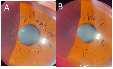
裸眼视力:右眼0.08,左眼0.06;矫正视力:右眼+0.75 DS/-5.50 DC×15→0.6,左眼-3.50 DS/-1.50 DC×180→0.8,非接触式眼压:右眼18 mmHg(1 mmHg=0.133 kPa),左眼18 mmHg,双眼结膜无充血(-),角膜透明,前房深,房水清,瞳孔对光反射存在,晶状体前囊中央可见色素膜附着并与周边虹膜呈丝状连接,扩瞳后见晶状体周边点状混浊,玻璃体轻度液化混浊,眼底黄斑中心凹反光可见,视网膜平伏。诊断:双眼PPM,双眼屈光不正。双眼眼前节裂隙灯照片见图1。角膜内皮计数:右眼2 420/mm2,左眼2 559/mm2;双眼眼轴:右眼23.47 mm,左眼24.52 mm;前房深度:右眼3.88 mm,左眼3.97 mm;晶状体厚度:右眼3.80 mm,左眼3.77 mm;角膜水平直径:右眼11.4 mm,左眼11.4 mm;角膜曲率:右眼44.08D@13,48.41D@103,左眼44.98D@15,46.30D@105;明室瞳孔大小:右眼2.29 mm,左眼2.59 mm;暗室下瞳孔:右眼4.5 mm,左眼4.6 mm;双眼眼部B超见轻度玻璃体混浊;双眼光学相干断层扫描(optical coherence tomography,OCT)检查未见明显异常。
20240315101120_7051.png
图1 PPM切除术前双眼眼前段裂隙灯照片
Figure1 Slit lamp photograph of anterior segment of both eyes before pupil remnant membrane resection
A:右眼;B:左眼。
A: Right eye; B: Left eye.

1.3 诊断、治疗和随访

术前和患者充分沟通,制定诊疗计划,建议先行PPM切除,但术中可能存在囊膜损伤或术源性白内障可能,因此术中预备“Phaco+IOL植入术”,且术后视力因为存在屈光不正可能不会提高,需进一步配镜或行屈光手术进一步改善。患者签署知情同意书后,分别于2022年10月12日和2022年10月31日分别行右眼和左眼PPM切除术。术前扩瞳,术中在角膜陡峭轴制作角膜缘2.4 mm切口,在PPM下方注入粘弹剂,利用虹膜恢复器于残膜较致密侧轻轻将PPM和晶状体前囊粘连处剥离,囊膜剪将PPM虹膜粘连端剪断,利用无齿镊取出游离残膜,检查见晶状体前膜靠上方瞳孔缘残留少量虹膜色素,未给予处理,清除前房粘弹剂,水密主切口。术后半年随访,于2023年4月先后行双眼TICL植入术。
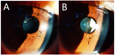
右眼PPM切除术后第一天裸眼视力0.4,矫正视力+0.50 DS/-4.75 DC×15→0.6,左眼PPM切除术后裸眼视力0.4,矫正视力-3.75 DS/-1.25 DC×7→1.0,术后半年复诊,眼前段照相见图2。晶状体未见明显进行性混浊。
20240315101343_1625.png
图2 PPM切除术后双眼眼前段裂隙灯照片
Figure2 Slit lamp photograph of anterior segment of both eyes after pupil remnant membrane resection
A:右眼;B:左眼。
A: Right eye; B: Left eye.
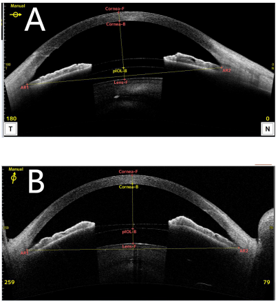
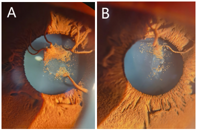
右眼TICL植入术后第1天裸眼视力0.8,左眼TICL植入术后裸眼视力1.0,非接触式眼压右眼16 mmHg,左眼17 mmHg,TICL植入术后1个月,右眼裸眼视力1.0,左眼裸眼视力1.0,综合验光:右眼+0.25 DS→1.0,左眼+0.25 DS/-0.25 DC×5→1.0。复查角膜内皮计数:右眼2 351 /mm2,左眼2 452 /mm2,双眼TICL植入术后眼前段照片见图3;眼前段OCT(CASIA2)测量右眼拱高681 μm,左眼拱高861 μm,见图4。术后双眼视觉质量分析检查结果见图5。
20240315101422_3046.png
图3 TICL植入术后双眼眼前段裂隙灯照片
Figure3 Slit lamp photograph of anterior segment of both eyes after TICL implantation
A:右眼;B:左眼。
A: Right eye; B: Left eye.
20240315101744_4598.png
图4 TICL植入术后双眼眼前节OCT检查结果
Figure4 OCT examination of the anterior segment of both eyes after TICL implantation
A:右眼;B:左眼。
A: Right eye; B: Left eye.
20240315101818_5790.png
图5 TICL植入术后双眼视觉质量分析(iTrace)结果
Figure5 Results of binocular visual quality analysis (iTrace) after TICL implantation
A:右眼;B:左眼。
A: Right eye; B: Left eye.

2 讨论

先天性PPM亦称永存瞳孔膜,是虹膜的一种先天性异常,属于血管系统发育异常的一部分。Goldberg[4]在1997年将PPM和永存原始玻璃体增生症(persistent hyperplasia of primary vitreous,PHPV)归类为永存性胚胎血管,其中PHPV与晶状体血管膜后部的不完全消退有关,而PPM则与前部的不完全消退相关。他将这类血管系统异常重新命名为持续性胎儿血管症(persistent fetal vasculature,PFV)。PPM通常不影响视力,但若膜较致密可能导致视力障碍、弱视或斜视。对于成年人和大龄儿童,治疗通常被认为意义不大,除非为美容或视力问题。然而,研究显示[5-6]即使在成人中,去除遮挡物可以提高视力,特别是伴有其他眼疾如白内障或青光眼时。例如,Ramappa等[7]报道了一例通过保留晶状体进行PPM膜剥离手术的12岁患者,术后视力有所提高,且未见晶状体混浊。
既往研究[8-9]认为,激光治疗PPM是安全、有效的,但多适用于丝状或较小的膜状残膜,且残膜与晶状体无粘连,因此可以通过激光将上方的残膜条索打断从而暴露出瞳孔,但创伤性白内障、前房出血、色素播散致继发青光眼等并发症发生风险相应增加。本例患者因瞳孔区残膜与晶状体形成膜状附着,因此只能考虑应用手术剥离。术中采用与Hu等[10]改良的残膜切除术式类似方法进行残膜的切除,为了进一步减少术中对晶状体的损伤,充分扩瞳后,术者在处理晶状体中央残膜的时候采用钝性的虹膜恢复器进行缓慢并尽量一次性完整剥离残膜组织,术中应用冲洗针头在切口处进行前房粘弹剂的清除,术后主切口自闭良好,未给予缝线,因此避免术后较严重术源性散光的出现。在残膜切除的第二天,患者右眼虽然仍然存在-4.75 D散光,但裸眼视力提升至0.4,与既往研究结果(即使在成人中,去除PPM遮挡仍可以提高视力)一致[5-6],这也让患者对左眼的手术有了更强的治疗信心,同样左眼术后裸眼视力也达到了0.4。因此,提示对于成人较大的PPM也可以积极给予治疗,残膜的切除对于视力的提升可能具有潜在价值。另外,患者双眼手术后没有立即进一步行屈光手术矫正屈光不正,随访观察半年,发现患者双眼晶状体未见明显进行性混浊,与Ramappa等[7]随访16个月的结果一致,提示单纯晶状体前囊残膜剥离不会引起晶状体前囊下混浊。
患者在PPM剥离半年后自愿双眼植入TICL来矫正屈光不正,获得双眼良好的术后效果,相对于Gupta等[11]采用手术切除PPM,并进行晶状体摘除联合后房型IOL植入手术治疗方式,虽然术后视力也能提高,但保留自然晶状体的TICL植入术对于患者术后视功能的恢复拥有更大的优势,既往研究[12]认为,TICL植入术后较IOL植入术后拥有更好的视力,更高的满意度和脱镜率,且无后发性白内障等并发症。
综上所述,对于成年人较明显的瞳孔区的PPM,仍然可以考虑一期行PPM切除,对于术后拥有良好的矫正视力的患者,择期再行角膜或者眼内屈光手术,可以为患者提供良好的术后视功能。

利益冲突

所有作者均声明不存在利益冲突

开放获取声明

本文适用于知识共享许可协议(Creative Commons),允许第三方用户按照署名(BY)-非商业性使用(NC)-禁止演绎(ND)(CC BY-NC-ND)的方式共享,即允许第三方对本刊发表的文章进行复制、发行、展览、表演、放映、广播或通过信息网络向公众传播,但在这些过程中必须保留作者署名、仅限于非商业性目的、不得进行演绎创作。详情请访问:https://creativecommons.org/licenses/by-nc-nd/4.0/。
1、Banigallapati S, Potti S, Marthala H. A rare case of persistent pupillary membrane: case-based approach and management[ J]. Indian J Ophthalmol, 2018, 66(10): 1480-1483.Banigallapati S, Potti S, Marthala H. A rare case of persistent pupillary membrane: case-based approach and management[ J]. Indian J Ophthalmol, 2018, 66(10): 1480-1483.
2、Wang YJ, Ke M, Yan M. Wide-field digital imaging system for assessing ocular anterior segment development in very preterm infants[ J]. Indian J Ophthalmol, 2023, 71(11): 3484-3488.Wang YJ, Ke M, Yan M. Wide-field digital imaging system for assessing ocular anterior segment development in very preterm infants[ J]. Indian J Ophthalmol, 2023, 71(11): 3484-3488.
3、Wang YJ, Ke M, Yan M. The ocular anterior segment examination of perinatal newborns by wide-field digital imaging system: a cross-sectional study[ J]. BMC Ophthalmol, 2023, 23(1): 411.Wang YJ, Ke M, Yan M. The ocular anterior segment examination of perinatal newborns by wide-field digital imaging system: a cross-sectional study[ J]. BMC Ophthalmol, 2023, 23(1): 411.
4、Goldberg MF. Persistent fetal vasculature (PFV): an integrated interpretation of signs and symptoms associated with persistent hyperplastic primary vitreous (PHPV) LIV Edward Jackson memorial lecture[ J]. Am J Ophthalmol, 1997, 124(5): 587-626.Goldberg MF. Persistent fetal vasculature (PFV): an integrated interpretation of signs and symptoms associated with persistent hyperplastic primary vitreous (PHPV) LIV Edward Jackson memorial lecture[ J]. Am J Ophthalmol, 1997, 124(5): 587-626.
5、Gavri%C5%9F%20M%2C%20Horge%20I%2C%20Avram%20E%2C%20et%20al.%20Persistent%20pupillary%20membrane%20or%20%0Aaccessory%20iris%20membrane%3F%5B%20J%5D.%20Rom%20J%20Ophthalmol%2C%202015%2C%2059(3)%3A%20184-%0A187.Gavri%C5%9F%20M%2C%20Horge%20I%2C%20Avram%20E%2C%20et%20al.%20Persistent%20pupillary%20membrane%20or%20%0Aaccessory%20iris%20membrane%3F%5B%20J%5D.%20Rom%20J%20Ophthalmol%2C%202015%2C%2059(3)%3A%20184-%0A187.
6、Gokhale V, Agarkar S. Persistent pupillary membrane[ J]. N Engl J Med, 2017, 376(6): 561.Gokhale V, Agarkar S. Persistent pupillary membrane[ J]. N Engl J Med, 2017, 376(6): 561.
7、Ramappa M, Murthy SI, Chaurasia S, et al. Lens-preserving excision of congenital hyperplastic pupillary membranes with clinicopathological correlation[ J]. J AAPOS, 2012, 16(2): 201-203.Ramappa M, Murthy SI, Chaurasia S, et al. Lens-preserving excision of congenital hyperplastic pupillary membranes with clinicopathological correlation[ J]. J AAPOS, 2012, 16(2): 201-203.
8、Wang JK, Wu CY, Lai PC. Sequential argon-YAG laser membranectomy and phacoemulsification for treatment of persistent pupillary membrane and associated cataract[ J]. J Cataract Refract Surg, 2005, 31(8): 1661-1663.Wang JK, Wu CY, Lai PC. Sequential argon-YAG laser membranectomy and phacoemulsification for treatment of persistent pupillary membrane and associated cataract[ J]. J Cataract Refract Surg, 2005, 31(8): 1661-1663.
9、Mansour AM, Hamade I, Antonios RS. Sequential argon-YAG laser membranotomy of extensive persistent pupillary membrane with visual loss[ J]. BMJ Case Rep, 2015, 2015: bcr2015210140.Mansour AM, Hamade I, Antonios RS. Sequential argon-YAG laser membranotomy of extensive persistent pupillary membrane with visual loss[ J]. BMJ Case Rep, 2015, 2015: bcr2015210140.
10、Hu H, Nie D, Zou Y, et al. Surgical membranectomy with modified incision and capsulotomy microscissors for persistent pupillary membrane[ J]. Ann Palliat Med, 2021, 10(5): 5619-5626.Hu H, Nie D, Zou Y, et al. Surgical membranectomy with modified incision and capsulotomy microscissors for persistent pupillary membrane[ J]. Ann Palliat Med, 2021, 10(5): 5619-5626.
11、Gupta Y, Gupta M, Sambhav K, et al. A rare case of persistent pupillary membrane associated with high myopia and amblyopia[ J]. Nepal J Ophthalmol, 2010, 2(1): 68-70.Gupta Y, Gupta M, Sambhav K, et al. A rare case of persistent pupillary membrane associated with high myopia and amblyopia[ J]. Nepal J Ophthalmol, 2010, 2(1): 68-70.
12、李玉雯, 吴保华, 吴鹏程, 等. 探讨矫正高度近视的两种眼内屈光 手术的临床效果[ J]. 中国医学创新, 2014, 11(12): 74-76.
Li YW, Wu BH, Wu PC, et al. Clinical eect of two kinds of intraocular refractive operation to correct high myopia[ J]. Med Innov China, 2014, 11(12): 74-76.
李玉雯, 吴保华, 吴鹏程, 等. 探讨矫正高度近视的两种眼内屈光 手术的临床效果[ J]. 中国医学创新, 2014, 11(12): 74-76.
Li YW, Wu BH, Wu PC, et al. Clinical eect of two kinds of intraocular refractive operation to correct high myopia[ J]. Med Innov China, 2014, 11(12): 74-76.
上一篇
下一篇
其他期刊
  • 眼科学报

    主管:中华人民共和国教育部
    主办:中山大学
    承办:中山大学中山眼科中心
    主编:林浩添
    主管:中华人民共和国教育部
    主办:中山大学
    浏览
  • Eye Science

    主管:中华人民共和国教育部
    主办:中山大学
    承办:中山大学中山眼科中心
    主编:林浩添
    主管:中华人民共和国教育部
    主办:中山大学
    浏览
推荐阅读
出版者信息
目录