您的位置: 首页 > 2024年10月 第39卷 第10期 > 文字全文
2023年7月 第38卷 第7期11
目录

广角 SS-OCT En Face 结构投射图揭示未成年人玻璃体早期液化特征

Wide-angle SS-OCT en face structural projection images reveal early vitreous liquefaction characteristics in minors

来源期刊: 眼科学报 | 2024年10月 第39卷 第10期 533-540 发布时间:2024-10-28 收稿时间:2024/11/1 14:42:37 阅读量:164
作者:
关键词:
糖扫频源光学相干断层扫描成像 en face结构投射图玻璃体液化液化池玻璃体后脱离
SS-OCTen facevitreous liquefactioncistern PVD
DOI:
10.12419/24090305
收稿时间:
2024-09-03 
修订日期:
2024-09-30 
接收日期:
2024-10-19 
目的:应用广角扫频源光学相干断层扫描成像(swept-source optical coherence tomography, SS-OCT)的en face结构投射图研究玻璃体早期液化特征。方法:使用SS-OCT进行18 mm×18 mm 的容积(Cube)扫描,创建并分析健康未成年人(年龄5~18岁)70眼的系列玻璃体en face结构投射图。 结果:在未成年人中,视网膜前的玻璃体包含4种液化结构,分别为后皮质前玻璃体囊袋(posterior precortical vitreous pocket, PPVP)、视盘前Martegiani区(the area of Martegiani, AM)、血管前液化裂隙(prevascular vitreous fissures,PVF)和液化池(cistern)。所有研究眼均能检出PPVP、AM和PVF,其中22眼(31.4%)的PPVP和AM连通。41眼(58.6%)可检出液化池,且其年龄大于未检出液化池的个体(=0.01),液化池的发生与年龄呈正相关(rs=0.315,=0.008)。液化池的象限空间分布频率依次为颞上(90.2%)、鼻上(58.5%)、颞下(36.6%)、鼻下(24.4%),最常累及颞上象限(P<0.001)。 结论:PPVP、AM和PVF是健康人群视网膜前玻璃体早期液化过程中均出现的特征。液化池的发生与年龄呈正相关,最常出现在颞上象限,可能是年龄相关性玻璃体液化变性的结果。
Objective: To investigate the early vitreous liquefaction characteristics using en face structural projection images obtained by wide-angle swept-source optical coherence tomography (SS-OCT). Methods: SS-OCT was employed to perform 18*18mm volumetric (Cube) scans. A series of en face structural projection images of the vitreous were created and analyzed for 70 eyes from healthy minors aged between 5 and 18 years. Results: In minors, four types of vitreous liquefaction structures were identified anterior to the retina: pre-posterior vitreous pocket (PPVP), the preoptic area of Martegiani (AM), pre-vascular liquefaction fissures (PVF), and cisterns. PPVP, AM, and PVF were detectable in all studied eyes, with PPVP and AM being interconnected in 22 eyes (31.4%). Cisterns were observed in 41 eyes (58.6%), and the mean age of individuals with cisterns was higher than those without (=0.01). The occurrence of cisterns positively correlated with age (r=0.315; P=0.008). The frequency of cistern quadrant distribution was highest in the superotemporal quadrant (90.2%), followed by the superonasal quadrant (58.5%), inferotemporal quadrant (36.6%), and inferonasal quadrant (24.4%). The superotemporal quadrant was the most frequently affected (<0.001). Conclusion: PPVP, AM, and PVF are features consistently observed in the early vitreous liquefaction process anterior to the retina in healthy individuals. The occurrence of cisterns positively correlates with age and is most common in the superotemporal quadrant, possibly representing the result of age-related vitreous liquefaction degeneration. These findings provide a theoretical foundation for studying the pathogenesis of vitreoretinal interface diseases. 

文章亮点

1. 关键发现

 • 视网膜前的玻璃体包含 4 种液化结构,分别为 PPVP、视盘前 Martegiani 区、PVF 和液化池。
 • 其中,液化池的发生与年龄呈正相关,可能是年龄相关性玻璃体液化变性的结果。
 • 早期液化池最常出现在颞上象限,与早期 PVD 的上方起源一致。

2. 已知与发现

 • 在玻璃体液化的基础上,玻璃体视网膜粘连减弱可导致 PVD,这个过程可能导致多种玻璃体视网膜界面疾病的发生。

3. 意义与改变

 • 探索早期玻璃体液化特征及其与 PVD 之间的密切联系有助于更好地理解玻璃体视网膜界面疾病的发病机制。

       正常人眼玻璃体是由99%的水、透明质酸和少量胶原组成的三维网状透明凝胶结构,具有屈光功能和支持眼球的作用。在老龄化生理或病理情况下,胶原纤维支架塌陷浓缩,水分析出,玻璃体凝胶状态破坏变为液体。在玻璃体液化的基础上,玻璃体视网膜粘连减弱可导致玻璃体后脱离(posterior vitreous detachment, PVD),这个过程可能导致黄斑裂孔、黄斑前膜、玻璃体积血和视网膜脱离等玻璃体视网膜界面(vitreoretinal interface, VRI)疾病的发生[1-2]。鉴于此,探索玻璃体液化特征及其与PVD之间的密切联系有助于更好地理解VRI疾病的发病机制。 
       虽然玻璃体占据了眼睛80%的体积,对其正常解剖特征和生理病理变化的理解却相对落后。这是因为玻璃体主要由水组成,近乎光学透明的结构使玻璃体的研究特别具有挑战性。接触式B超可以提供玻璃体的低分辨率图像,但不能显示精细的解剖细节。20世纪70年代至90年代,多项组织学研究证实了存在于黄斑前的玻璃体液化腔隙[3-6],并最终由Kishi命名为后皮质前玻璃体囊袋 (posterior precortical vitreous pocket, PPVP )。Cloquet管是原始玻璃体的残余,从视盘前Martegiani区(the area of Martegiani, AM)发出并连接至晶状体后极部的Berger区。然而,关于其他玻璃体液化结构的组织学研究较少,相关结论也存在诸多争议。
       最近,高分辨率扫频源光学相干断层扫描成像(swep-source optical coherence tomography , SS-OCT)的出现有助于阐明玻璃体腔中复杂的液化结构。2013年,Itakura等[7]使用SS-OCT首次在青年人群中观察到PPVP及其与Cloquet 管的连接。Li 等[8]通过SS-OCT成像观察儿童后玻璃体并证明PPVP是人类眼睛几乎普遍的特征。之后,其他玻璃体液化结构包括黄斑上囊(supramacular bursa)、液化池(cistern)、液化腔(lacunae)、退行性劈裂(degenerative cleavages)等也陆续被观察到[9-10]。2015年Pang 等[11]使用SS-OCT 5线交叉扫描模式识别出血管前玻璃体液化裂隙(prevascular vitreous fissures,PVF)和液化池,并提出PVF可能是液化池的前体。2020年Leong 等[12]通过12 mm×12 mm SS-OCT en face成像第一次清晰显示了PVF沿着视网膜浅层血管分布的连续行径,并观察到液化池与PVF相连续。这些研究结果揭开了玻璃体液化的神秘面纱。   
       相较于断层扫描,SS-OCT en face图像可以清楚呈现后玻璃体液化的全景图,因此有助于获取新的信息。通过分析健康成年人群29*25mm超广角SS-OCT en-face图像,我们发现后玻璃体液化遵循刻板模式,均发生在玻璃体视网膜紧密粘连处,且所有的PVF和液化池均沿着视网膜浅层血管分布。基于先前的证据,本研究拟使用18 mm×18 mm广角SS-OCT Z-Scan en face结构投射图分析未成年人群后玻璃体液化特征,以进一步探索玻璃体液化的早期起源。

1  对象与方法

1.1 对象

       本研究为观察性横断面研究。从2024年4月—2024年7月,中山眼科中心连续招募了46例未成年参与者(年龄在5~18岁),共纳入70只眼睛。纳入标 准:1)等效球镜度数为−3. 00 ~ +3.00 D,最佳矫正视力为−0.1 ~ +0.1(LogMAR); 2) 裂隙灯、间接检眼镜检查未见明显异常。排除标准:配合不良或玻璃体显示不清者。 本研究经中山大学中山眼科中心伦理委员会审核批准(批件号:2024KYPJ036),所有参与者及监护人均签署知情同意书。 

1.2 方法

       受检者取坐位,将头部置于下颌托上,双眼平视。玻璃体图像通过VG200 SS-OCT(Intalaight Imaging,洛阳,中国)采集,扫描模式为经过黄斑中心凹的容积扫描(Cube Scan,1 024×1 024),扫描线长度为18 mm,扫描深度为6.3 mm;扫描激光波长为1 050 nm;扫描速度为200 000次/秒。在扫描采集过程中,通过将视网膜和脉络膜定位在扫描窗口的下方,从而增强了玻璃体结构的可视化。
创建玻璃体en face结构投射图的方法:选择结构投射图的玻璃体层面,设置下边界为内界膜0 μ m,上边界为内界膜前−100 μ m创建玻璃体en face结构投射图,通过手动调整en face板层厚度(范围50~100 μ m)及其与视网膜表面的距离,对后玻璃体结构进行动态en face图像评估。玻璃体各种液化结构在SS-OCT en face图像上表现为低信号腔隙。使用Intalight 200软件的内置卡尺测量视网膜附近(内界膜前−100 μ m)PPVP的水平直径。由2名技术娴熟眼科医师进行独立分析和测量,测量结果偏差较大或有争议时由高年资医师进行裁决。

1.3 统计学分析

       采用SPSS 27.0统计学软件对采集的数据进行分析。分类变量包括眼数、性别、PPVP和AM连接、液化池象限分布、液化池累及象限数,采用例数(n)和百分率(%)描述,通过Pearson χ 2检验进行组间比较。定量变量包括年龄、等效球镜度数、PPVP宽度,经K-S检验符合正态分布,以± 表示,采用独立样本检验进行比较。年龄与液化池发生的相关性使用Spearman检验。 P < 0. 05为差异有统计学意义。  

2  结果

2.1 研究对象的一般资料

       本研究共纳入46例未成年人70眼,其中男31例48眼、女15例22眼,年龄5~18岁,年龄为(10.83±4.05)岁,等效球镜度数为(0.11±1.46)D。 根据年龄将受检者分为儿童组(5~11岁)和少年组(12~18岁),两组人群特征见表1。

表 1 儿童组与少年组一般资料比较
Table 1 Comparison of general information between children and young adolescents

项目

儿童组(5~11岁)

少年组(12~18岁)

/ χ² 

眼数

41

29


年龄(x±s)/ 岁

7.85±1.78

15.03±2.13

-15.31

<0.001*

男性[ (%)]

2868.3

2069.0

-

0.952

等效球镜度数(x±s)/D

0.72±1.25

-0.79±1.31

4.88

<0.001*

PPVP宽度(x±s)/ μm

5 325.2±875.0

5 266.1±710.5

0.30

0.765

液化池[ n (%)]

19 (46.3)

22 (75.9)

-

0.014*

2.2 后玻璃体液化结构

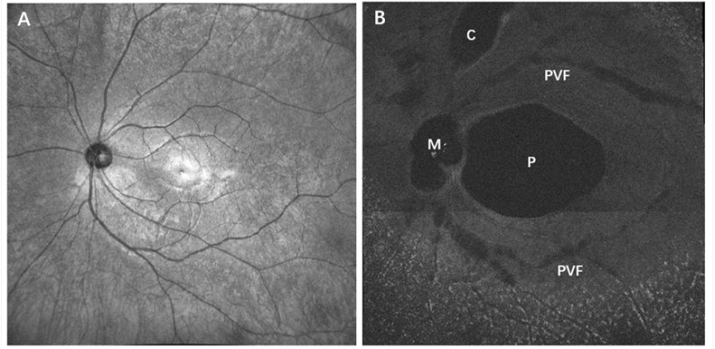
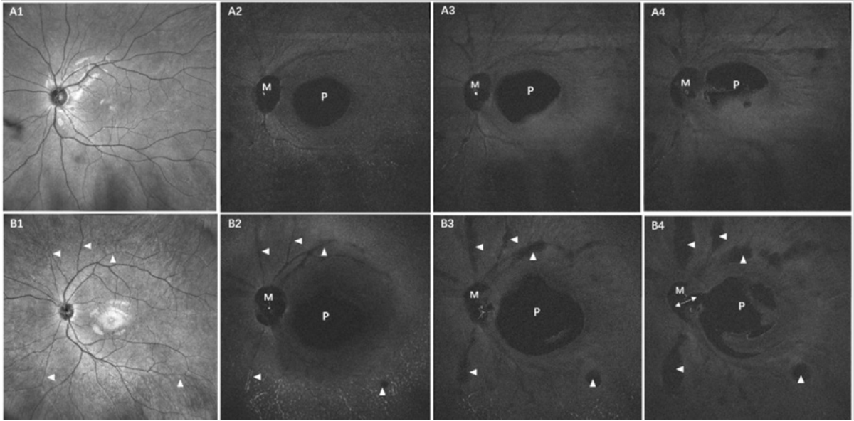
       在未成年人中,视网膜前的玻璃体包含4种液化结构,分别是PPVP、AM、PVF和液化池。其中,PPVP被定义为黄斑前的低信号腔隙, AM被定义为视盘前的低信号腔隙,PVF被定义为覆盖视网膜浅层血管的细长低信号腔隙,而液化池是比PVF更宽大的低信号腔隙,其位置与PVF一致,见图1。本研究中,所有眼睛均能检出PPVP、AM和PVF。PPVP位于颞侧血管弓内,直径为(5 318.9±809.6)μ m,且儿童组的(5 325.2±875.0)μ m和少年组的(5 266.1±710.5)μ mPPVP的宽度比较差异无统计学意义(=0.765)。动态en face投射图(图2)显示其逐渐从椭圆形变成新月形或不规则形,22眼(31.4%)的PPVP和AM在视盘前上方连通。PVF主要位于后部玻璃体并围绕PPVP和AM区域分布,en face图像能够显示PVF沿着视网膜浅表血管走行的连续行径,见图2A2~A4。与PVF位置一样,液化池也沿着视网膜浅层血管分布,且液化池向上逐渐扩大形成更大的液化池,通过动态en face图像评估可以很容易地区分出来,见图2B2~B4。

图 1 En Face结构投射图显示四种玻璃体液化结构
Figure 1 En Face vitreous images showing four types of preretinal vitreous liquefaction structures
(A)IR参照图;(B)玻璃体en face结构投射图。各种液化结构均显示为低信号腔隙:PPVP是黄斑前的低信号腔隙, AM是视盘前的低信号腔隙,PVF是覆盖视网膜浅层血管的细长低信号腔隙,液化池是比PVF更宽大的低信号腔隙,其位置与PVF一致。P:PPVP, M:AM,C: Cistern。
(A) Infrared reference image; (B) En Face vitreous images. Various liquefaction structures are displayed as low signal cavities: PPVP is a low signal cavity overlying the macular area; AM is a low signal cavity over the optic disc; PVFs are slender, hyporeflective vitreous cavities overlying retinal superficial vessels; cistern are wider spaces than PVFs, and the position is consistent with PVFs. P: PPVP, M: AM, C: Cistern.

图 2 动态en face图像评估后玻璃体液化特征
Figure 2 Dynamic en face assessment of posterior vitreous liquefaction
(A1)8岁远视(+1.5 D)男性的18*18mm IR图,内界膜前(A2)−100μ m、(A3)−250μ m和(A4)−450μ m en face投射图显示PVF沿着视网膜浅层血管走行的连续行径,未检出明显的液化池。(B1)17岁近视(−1.5 D)男性的18 mm×18mm IR图,内界面前(B2)−100μ m、(B3)−250μ m、(B4)−450μ m en face投射图显示PVF和液化池(三角形)均沿着视网膜浅层血管分布,且液化池逐渐向上扩大。PPVP(P)位于颞侧血管弓内,在系列en face图像上逐渐从椭圆形变成半圆形或不规则形状。B4显示PPVP和AM(M)在视盘前上方的连接通道(双箭头)。
(A1)is the 18 × 18mm IR image of an 8-year-old hyperopic (+1.5 D) male, dynamic en face scans at (A2) −100 μ m, (A3) −250 μ m, and (A4) −450 μ m anterior to the retinal surface demonstrating the continuous course of PVFs along retinal superficial vessels, but cisterns were not detected. (B1) is the 18 mm × 18 mm IR image of a 17-year-old myopic (−1.5 D) male, dynamic en face scans at (B2) −100 μ m, (B3) −250 μ m, and (B4) −450 μ m anterior to the retinal surface demonstrating that both PVFs and cisterns (triangle) distributed along retinal superficial vessels, and cisterns gradually expanded more anteriorly. PPVP (P) is located within the temporal vascular arch and gradually changes from an elliptical shape to a semi-circular or irregular shape on the series of en face images. B4 shows the connecting channel (double arrow) between PPVP and AM (M) in front of of the optic disc.

2.3液化池的发生跟年龄呈正相关

       在两个年龄组中,儿童组19眼(46.3%)检出液化池,且最年轻的检出者为5岁,少年组22眼(75.9%)检出液化池,少年组液化池的发生率高于儿童组(=0.014),但两组间的年龄和等效球镜度数均具有显着差异(<0.001),见表1。进一步分析液化池发生的相关因素,根据en face图像是否检出液化池将所有未成年受检者分为液化池(+)组和液化池(–)组。在所有70只研究眼中,41眼(58.6%)可检出液化池,29眼(41.4%)未检出液化池。液化池 (+)组的年龄为(11.73±4.14)岁,液化池 (−)组的年龄为(9.55±3.61)岁,组间的年龄比较差异有统计学意义 (=0.01),见表2。液化池的发生跟年龄增加呈正相关(rs=0.315,=0.008),可能是年龄相关性玻璃体液化的结果。两组间的性别( P =0.952)、等效球镜度数(=0.052)、PPVP宽度(P =0.856)、PPVP和 AM连通(P=0.269)差异无统计学意义,见表2。

表 2 未成年人中液化池(+)组与液化池(−)组一般资料比较
Table 2 Comparison of general information between the cistern (+) and the cistern (−) groups among minors

项目

化池(+)组

液化池()组

/ χ² 

眼数[ n (%)]

41 (58.6)

29 (41.4)


性别(男)[ n (%)]

28(68.3)

20(69.0)

0.952

年龄(x±s)/ 岁

11.73±4.14

9.55±3.61

2.67

0.010

等效球镜度数(x±s)/D)

−0.18±1.41

0.51±1.45


-1.98

0.052

PPVP宽度(x±s)/μ m

5333.9±847.3

5297.7±767.4

0.18

0.856

PPVP和AM连接[ n (%)]

15 (36.6)

7 (24.1)

0.269


2.4 早期液化池最常发生于颞上象限

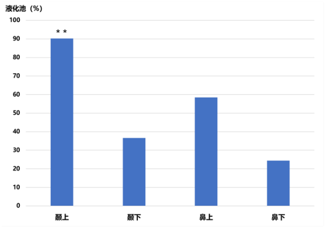
       在所有检出液化池的41只眼中,15眼在1个象限检出液化池,14眼在2个象限检出液化池,6眼在3个象限检出液化池,6眼在4个象限检出液化池。总体而言,未成年阶段液化池最常累及1~2个象限(29眼,70.7%,P<0.001)。值得注意的是,液化池的空间分布频率依次为颞上(90.2%)、鼻上(58.5%)、颞下(36.6%)、鼻下(24.4%),颞上象限最常见 (P <0.001),见图3。此外,在所有仅检出1个象限液化池的15只眼睛中,13眼(86.7%)位于颞上象限,2眼(13.3%)位于颞下象限,也是颞上象限最常见(P <0.001)。

图 3 液化池出现的象限频率
Figure 3 Geographic distribution of cisterns
依次为颞上(90.2%)>鼻上(58.5%)>颞下(36.6%)>鼻下(24.4%),早期液化池最常累及颞上象限(**P<0.001)。
Incidence of cisterns was in the order of supertemporal (90.2%)> supranasal (58.5%)> infratemporal (36.6%)> infranasal (24.4%), and early cisterns predominately involved the supertemporal quadrant (* * P<0.001).

3  讨论

       因为玻璃体液化的复杂性和广泛性,相较于传统的OCT B-Scan,SS-OCT Z-Scan可视技术更有助于完整显示整个后玻璃体的液化结构及空间分布。利用这种方法,本研究显示在生命早期视网膜前玻璃体包含4种液化结构,分别是PPVP、AM、PVF和液化池,与笔者团队先前在健康成年人中的研究结果一致。其中,PPVP、AM和PVF是在所有研究眼中一致性出现的玻璃体早期液化结构,其生理作用值得进一步探讨。
       眼睛每天约扫视10万次,眼球承受扫视运动高角速度和加速度的机械应力[13]。神经视网膜组织持续暴露于极端的剪切应力中,却能稳定生成高分辨率的图像可能得益于玻璃体的2个主要特征:黏弹性[14-15]和有益于减少黄斑区应力载荷的解剖结构。基于SS-OCT动态聚焦技术观察后玻璃体解剖结构,Spaide[16]首先提出PPVP在眼球扫视运动过程中可能发挥着能量阻尼的作用,有助于将黄斑区玻璃体的机械应力转移到黄斑以外区域。最近有研究者通过创建数学模型进行玻璃体动力学研究[17],模拟了0.17 s内跨越50 °的眼球水平扫视运动,结果显示PPVP可显著减少其下方黄斑区的壁剪切应力;相反,增加黄斑邻近区域的壁剪切应力。如此,PPVP对黄斑区玻璃体视网膜界面的保护作用在一定程度上似乎可以解释早期PVD均起源于PPVP以外区域这一现象[18-19],反之亦然。
       PPVP位于颞侧血管弓内,在玻璃体腔中继续向上向前延伸,因此在en face图像上逐渐从椭圆形变成新月形或不规则形。同样地,AM也继续向上延伸形成Cloquet管,并在Berger区的晶状体后极部截止。Itakura 等 [20]观察到成人中93. 1%的PPVP与 AM间存在连接通道,Park 等[21]报道3~4岁的受检者无连接通道,而11岁受检者中50%存在连接通道,金波等[22]报道8~63岁人群中约71.7%的受检者存在连接通道。本研究中仅在31.4%未成年人中观察到连接通道,可能是因为入选人群年龄较小,也可能因为en face结构图像不容易发现早期较细小或不规则的连接通道。个体发生理论认为Cloquet管代表胎儿时期初级玻璃体的残余物,Worst和Los[23]假设Cloquet管是玻璃体凝胶内的液体通道系统,作为眼球前后段离子、细胞和分子的生物通讯方式。由此推测PPVP中的液体可能为房水,而房水中高浓度的维生素C可能有助于中和黄斑区产生的氧化自由基继而保护黄斑[8,24]。同样地,前葡萄膜炎房水中的炎症因子也可能通过这个通道到达眼后段,从而导致黄斑水肿[25]。 
       在本研究中,约58.6%的未成年人可以检出液化池,最小检出者年龄仅为5岁。与既往研究一致[11-12],本研究显示,生命早期液化池的发生与年龄呈正相关,可能是年龄相关性玻璃体液化变性的结果。结合所有PVF与液化池均沿着视网膜浅层血管分布,推测液化池可能起源于PVF,随着进行性玻璃体液化逐渐扩大形成更大的液化池。近年一系列OCT研究表明与年龄相关的PVD可能不是急性事件,而是一种经过多年演变缓慢进展的过程,甚至从生命的第一个十年开始就可以观察到早期PVD,且早期PVD最常发生在上方象限[18-19]。有趣的是,早期液化池也最常出现在颞上象限,恰与早期PVD的上方起源一致,这也进一步证实了玻璃体液化与PVD之间的密切关系。Pang等[11]认为这种现象可能至少部分是由于直立状态下的重力作用导致高密度的玻璃体凝胶向下移动,而上方低密度的玻璃体凝胶更容易脱水液化。 
       这项研究的局限性在于缺乏纵向研究来观察玻璃体液化随着时间推移的演变进程。尽管广角SS-OCT en face图像有助于可视化后玻璃体结构,进一步改进方法通过3D成像使玻璃体液化的定量分析成为可能。此外,OCT成像的未来进展将提供更宽和更深范围区域的玻璃体成像。 
       总之,SS-OCT Z-Scan可视技术有助于更好地理解复杂的后玻璃体液化结构。通过分析未成年人广角SS-OCT en face玻璃体结构投射图,我们发现PPVP、AM区和PVF是恒定性出现的玻璃体早期液化结构。液化池的发生与年龄呈正相关,可能是年龄相关性玻璃体液化变性的结果。与早期PVD起源一致,早期液化池最常出现在颞上象限。本研究有助于阐明后部玻璃体年龄相关性变化及各种VRI疾病的机制。 

利益冲突

所有作者均声明不存在利益冲突。

开放获取声明

本文适用于知识共享许可协议(Creative Commons),允许第三方用户按照署名(BY)-非商业性使用(NC)-禁止演绎(ND)(CC BY-NC-ND)的方式共享,即允许第三方对本刊发表的文章进行复制、发行、展览、表演、放映、广播或通过信息网络向公众传播,但在这些过程中必须保留作者署名、仅限于非商业性目的、不得进行演绎创作。


1、Duker JS, Kaiser PK, Binder S, et al. The International Vitreomacular Traction Study Group classification of vitreomacular adhesion, traction, and macular hole[ J]. Ophthalmology, 2013, 120(12): 2611-2619. DOI: 10.1016/j.ophtha.2013.07.042.Duker JS, Kaiser PK, Binder S, et al. The International Vitreomacular Traction Study Group classification of vitreomacular adhesion, traction, and macular hole[ J]. Ophthalmology, 2013, 120(12): 2611-2619. DOI: 10.1016/j.ophtha.2013.07.042.
2、Sebag J. Age-related changes in human vitreous structure[ J]. Albrecht Von Graefes Arch Fur Klin Und Exp Ophthalmol, 1987, 225(2): 89-93. DOI: 10.1007/BF02160337.Sebag J. Age-related changes in human vitreous structure[ J]. Albrecht Von Graefes Arch Fur Klin Und Exp Ophthalmol, 1987, 225(2): 89-93. DOI: 10.1007/BF02160337.
3、Eisner G. Biomicroscopy of the peripheral fundus[ J]. Surv Ophthalmol, 1972, 17(1): 1-28.Eisner G. Biomicroscopy of the peripheral fundus[ J]. Surv Ophthalmol, 1972, 17(1): 1-28.
4、Jongebloed WL, Worst JF. The cisternal anatomy of the vitreous body[ J]. Doc Ophthalmol, 1987, 67(1-2): 183-196. DOI: 10.1007/ BF00142712.Jongebloed WL, Worst JF. The cisternal anatomy of the vitreous body[ J]. Doc Ophthalmol, 1987, 67(1-2): 183-196. DOI: 10.1007/ BF00142712.
5、Worst JG. Cisternal systems of the fully developed vitreous body in the young adult[ J]. Trans Ophthalmol Soc U K, 1977, 97(4): 550-554.Worst JG. Cisternal systems of the fully developed vitreous body in the young adult[ J]. Trans Ophthalmol Soc U K, 1977, 97(4): 550-554.
6、Kishi S. Posterior precortical vitreous pocket[ J]. Arch Ophthalmol, 1990, 108(7): 979. DOI: 10.1001/archopht.1990.01070090081044.Kishi S. Posterior precortical vitreous pocket[ J]. Arch Ophthalmol, 1990, 108(7): 979. DOI: 10.1001/archopht.1990.01070090081044.
7、Itakura H, Kishi S, Li D, etal. Observation of posterior precortical vitreous pocket using swept-source optical coherence tomography[ J]. Invest Ophthalmol Vis Sci, 2013, 54(5): 3102-3107. DOI: 10.1167/ iovs.13-11769.Itakura H, Kishi S, Li D, etal. Observation of posterior precortical vitreous pocket using swept-source optical coherence tomography[ J]. Invest Ophthalmol Vis Sci, 2013, 54(5): 3102-3107. DOI: 10.1167/ iovs.13-11769.
8、Li D, Kishi S, Itakura H, et al. Posterior precortical vitreous pockets and connecting channels in children on swept-source optical coherence tomography[ J]. Invest Ophthalmol Vis Sci, 2014, 55(4): 2412-2416. DOI: 10.1167/iovs.14-13967.Li D, Kishi S, Itakura H, et al. Posterior precortical vitreous pockets and connecting channels in children on swept-source optical coherence tomography[ J]. Invest Ophthalmol Vis Sci, 2014, 55(4): 2412-2416. DOI: 10.1167/iovs.14-13967.
9、Schaal KB, Pang CE, Pozzoni MC, et al. The premacular bursa's shape revealed in vivo by swept-source optical coherence tomography[ J]. Ophthalmology, 2014, 121(5): 1020-1028. DOI: 10.1016/ j.ophtha.2013.11.030.Schaal KB, Pang CE, Pozzoni MC, et al. The premacular bursa's shape revealed in vivo by swept-source optical coherence tomography[ J]. Ophthalmology, 2014, 121(5): 1020-1028. DOI: 10.1016/ j.ophtha.2013.11.030.
10、Gal-Or O, Ghadiali Q, Dolz-Marco R, et al. In vivo imaging of the fibrillar architecture of the posterior vitreous and its relationship to the premacular bursa, Cloquet’s canal, prevascular vitreous fissures, and cisterns[ J]. Albrecht Von Graefes Arch Fur Klin Und Exp Ophthalmol, 2019, 257(4): 709-714. DOI: 10.1007/s00417-018-04221-x.Gal-Or O, Ghadiali Q, Dolz-Marco R, et al. In vivo imaging of the fibrillar architecture of the posterior vitreous and its relationship to the premacular bursa, Cloquet’s canal, prevascular vitreous fissures, and cisterns[ J]. Albrecht Von Graefes Arch Fur Klin Und Exp Ophthalmol, 2019, 257(4): 709-714. DOI: 10.1007/s00417-018-04221-x.
11、Pang CE, Schaal KB, Engelbert M. Association of prevascular vitreous fissures and cisterns with vitreous degeneration as assessed by swept source optical coherence tomography[ J]. Retina, 2015, 35(9): 1875- 1882. DOI: 10.1097/IAE.0000000000000540.Pang CE, Schaal KB, Engelbert M. Association of prevascular vitreous fissures and cisterns with vitreous degeneration as assessed by swept source optical coherence tomography[ J]. Retina, 2015, 35(9): 1875- 1882. DOI: 10.1097/IAE.0000000000000540.
12、Leong BCS, Fragiotta S, Kaden TR, et al. OCT en face analysis of the posterior vitreous reveals topographic relationships among premacular bursa, prevascular fissures, and cisterns[ J]. Ophthalmol Retina, 2020, 4(1): 84-89. DOI: 10.1016/j.oret.2019.09.002.Leong BCS, Fragiotta S, Kaden TR, et al. OCT en face analysis of the posterior vitreous reveals topographic relationships among premacular bursa, prevascular fissures, and cisterns[ J]. Ophthalmol Retina, 2020, 4(1): 84-89. DOI: 10.1016/j.oret.2019.09.002.
13、Ibbotson M, Krekelberg B. Visual perception and saccadic eye movements[ J]. Curr Opin Neurobiol, 2011, 21(4): 553-558. DOI: 10.1016/j.conb.2011.05.012.Ibbotson M, Krekelberg B. Visual perception and saccadic eye movements[ J]. Curr Opin Neurobiol, 2011, 21(4): 553-558. DOI: 10.1016/j.conb.2011.05.012.
14、张鹏程, 严宏. 玻璃体液化生物力学特性及临床研究进展 [ J]. 国际眼科杂志, 2017, 17(8): 1485-1488. DOI: 10.3980/ j.issn.1672-5123.2017.8.21.
Zhang PC, Yan H. Research advances in biomechanical properties and its clinical significance of vitreous liquefaction[ J]. Int Eye Sci, 2017, 17(8): 1485-1488. DOI: 10.3980/j.issn.1672-5123.2017.8.21.
15、Filas BA, Zhang Q, Okamoto RJ, etal. Enzymatic degradation identifies components responsible for the structural properties of the vitreous body[ J]. Invest Ophthalmol Vis Sci, 2014, 55(1): 55-63. DOI: 10.1167/iovs.13-13026.Filas BA, Zhang Q, Okamoto RJ, etal. Enzymatic degradation identifies components responsible for the structural properties of the vitreous body[ J]. Invest Ophthalmol Vis Sci, 2014, 55(1): 55-63. DOI: 10.1167/iovs.13-13026.
16、Spaide RF. Visualization of the posterior vitreous with dynamic focusing and windowed averaging swept source optical coherence tomography[ J]. Am J Ophthalmol, 2014, 158(6): 1267-1274. DOI: 10.1016/j.ajo.2014.08.035.Spaide RF. Visualization of the posterior vitreous with dynamic focusing and windowed averaging swept source optical coherence tomography[ J]. Am J Ophthalmol, 2014, 158(6): 1267-1274. DOI: 10.1016/j.ajo.2014.08.035.
17、Rossi%20T%2C%20Badas%20MG%2C%20Querzoli%20G%2C%20et%20al.%20Does%20the%20Bursa%20Pre-Macularis%20%0Aprotect%20the%20fovea%20from%20shear%20stress%3F%20A%20possible%20mechanical%20role%5B%20J%5D.%20Exp%20%0AEye%20Res%2C%202018%2C%20175%3A%20159-165.%20DOI%3A%2010.1016%2Fj.exer.2018.06.022.Rossi%20T%2C%20Badas%20MG%2C%20Querzoli%20G%2C%20et%20al.%20Does%20the%20Bursa%20Pre-Macularis%20%0Aprotect%20the%20fovea%20from%20shear%20stress%3F%20A%20possible%20mechanical%20role%5B%20J%5D.%20Exp%20%0AEye%20Res%2C%202018%2C%20175%3A%20159-165.%20DOI%3A%2010.1016%2Fj.exer.2018.06.022.
18、Tsukahara M, Mori K, Gehlbach PL, et al. Posterior vitreous detachment as observed by wide-angle OCT imaging[ J]. Ophthalmology, 2018, 125(9): 1372-1383. DOI: 10.1016/j.ophtha.2018.02.039.Tsukahara M, Mori K, Gehlbach PL, et al. Posterior vitreous detachment as observed by wide-angle OCT imaging[ J]. Ophthalmology, 2018, 125(9): 1372-1383. DOI: 10.1016/j.ophtha.2018.02.039.
19、Hayashi A, Ito Y, Takatsudo Y, etal. Posterior vitreous detachment in normal healthy subjects younger than age twenty[ J]. Invest Ophthalmol Vis Sci, 2021, 62(13): 19. DOI: 10.1167/iovs.62.13.19.Hayashi A, Ito Y, Takatsudo Y, etal. Posterior vitreous detachment in normal healthy subjects younger than age twenty[ J]. Invest Ophthalmol Vis Sci, 2021, 62(13): 19. DOI: 10.1167/iovs.62.13.19.
20、Itakura H, Kishi S, Li D, et al. En face imaging of posterior precortical vitreous pockets using swept-source optical coherence tomography[ J]. Invest Ophthalmol Vis Sci, 2015, 56(5): 2898-2900. DOI: 10.1167/ iovs.15-16451.Itakura H, Kishi S, Li D, et al. En face imaging of posterior precortical vitreous pockets using swept-source optical coherence tomography[ J]. Invest Ophthalmol Vis Sci, 2015, 56(5): 2898-2900. DOI: 10.1167/ iovs.15-16451.
21、Park KA , Oh S Y. Poster ior precor t ical v itreous pocket in children[ J]. Curr Eye Res, 2015, 40(10): 1034-1039. DOI: 10.3109/02713683.2014.971932.Park KA , Oh S Y. Poster ior precor t ical v itreous pocket in children[ J]. Curr Eye Res, 2015, 40(10): 1034-1039. DOI: 10.3109/02713683.2014.971932.
22、金波, 安广琪, 雷博, 等. 应用扫频源光学相干断层扫描成像分 析后皮质前玻璃体囊袋的形态[ J]. 国际眼科杂志, 2021, 21(6): 1077-1081. DOI: 10.3980/j.issn.1672-5123.2021.6.28.
Jin B, An GQ, Lei B, et al. Using swept - source optical coherence tomography to analysis the morphological characteristics of posterior precortical vitreous pocket[ J]. Int Eye Sci, 2021, 21(6): 1077-1081. DOI: 10.3980/j.issn.1672-5123.2021.6.28.
23、Worst JG, Los LI. Comparative anatomy of the vitreous body in rhesus monkeys and man[ J]. Doc Ophthalmol, 1992, 82(1-2): 169-178. DOI: 10.1007/BF00157007.Worst JG, Los LI. Comparative anatomy of the vitreous body in rhesus monkeys and man[ J]. Doc Ophthalmol, 1992, 82(1-2): 169-178. DOI: 10.1007/BF00157007.
24、Ma Z, Liu J, Li J, et al. Klotho levels are decreased and associated with enhanced oxidative stress and inflammation in the aqueous humor in patients with exudative age-related macular degeneration[ J]. Ocul Immunol Inflamm, 2022, 30(3): 630-637. DOI: 10.1080/ 09273948.2020.1828488.Ma Z, Liu J, Li J, et al. Klotho levels are decreased and associated with enhanced oxidative stress and inflammation in the aqueous humor in patients with exudative age-related macular degeneration[ J]. Ocul Immunol Inflamm, 2022, 30(3): 630-637. DOI: 10.1080/ 09273948.2020.1828488.
25、Pichi F, Neri P, HayS, et al. An en face swept source optical coherence tomography study of the vitreous in eyes with anterior uveitis[ J]. Acta Ophthalmol, 2022, 100(3): e820-e826. DOI: 10.1111/aos.14965.Pichi F, Neri P, HayS, et al. An en face swept source optical coherence tomography study of the vitreous in eyes with anterior uveitis[ J]. Acta Ophthalmol, 2022, 100(3): e820-e826. DOI: 10.1111/aos.14965.
上一篇
下一篇
其他期刊
  • 眼科学报

    主管:中华人民共和国教育部
    主办:中山大学
    承办:中山大学中山眼科中心
    主编:林浩添
    主管:中华人民共和国教育部
    主办:中山大学
    浏览
  • Eye Science

    主管:中华人民共和国教育部
    主办:中山大学
    承办:中山大学中山眼科中心
    主编:林浩添
    主管:中华人民共和国教育部
    主办:中山大学
    浏览
推荐阅读
出版者信息
目录