1、Wen F, Chen C, Wu D, et al. Polypoidal choroidal vasculopathy in
elderly Chinese patients[ J]. Graefe's Arch Clin Exp Ophthalmol, 2004,
242(8): 625-629. DOI: 10.1007/s00417-003-0667-z.Wen F, Chen C, Wu D, et al. Polypoidal choroidal vasculopathy in
elderly Chinese patients[ J]. Graefe's Arch Clin Exp Ophthalmol, 2004,
242(8): 625-629. DOI: 10.1007/s00417-003-0667-z.
2、Zhang Y, Gan Y, Zeng Y, et al. Incidence and multimodal imaging
characteristics of macular neovascularisation subtypes in Chinese
neovascular age-related macular degeneration patients[ J]. Br J
Ophthalmol, 2024, 108(3): 391-397. DOI: 10.1136/bjo-2022-322392.Zhang Y, Gan Y, Zeng Y, et al. Incidence and multimodal imaging
characteristics of macular neovascularisation subtypes in Chinese
neovascular age-related macular degeneration patients[ J]. Br J
Ophthalmol, 2024, 108(3): 391-397. DOI: 10.1136/bjo-2022-322392.
3、Zhang X, Zuo C, Li M, et al. Spectral-domain optical coherence
tomographic findings at each stage of punctate inner choroidopathy[ J]. Ophthalmology, 2013, 120(12): 2678-2683. DOI: 10.1016/
j.ophtha.2013.05.012.Zhang X, Zuo C, Li M, et al. Spectral-domain optical coherence
tomographic findings at each stage of punctate inner choroidopathy[ J]. Ophthalmology, 2013, 120(12): 2678-2683. DOI: 10.1016/
j.ophtha.2013.05.012.
4、Zhang X, Wen F, Zuo C, et al. Clinical features of punctate inner
choroidopathy in Chinese patients[ J]. Retina, 2011, 31(8): 1680-1691.
DOI: 10.1097/iae.0b013e31820a67ad.Zhang X, Wen F, Zuo C, et al. Clinical features of punctate inner
choroidopathy in Chinese patients[ J]. Retina, 2011, 31(8): 1680-1691.
DOI: 10.1097/iae.0b013e31820a67ad.
5、Gan Y, Zhang X, Chen L, et al. Intraretinal cystoid spaces in regression
of punctate inner choroidopathy lesions[ J]. Ocul Immunol Inflamm,
2020, 28(6): 938-946. DOI: 10.1080/09273948.2019.1641210.Gan Y, Zhang X, Chen L, et al. Intraretinal cystoid spaces in regression
of punctate inner choroidopathy lesions[ J]. Ocul Immunol Inflamm,
2020, 28(6): 938-946. DOI: 10.1080/09273948.2019.1641210.
6、Li M, Zhang X, Wen F. The fundus autofluorescence spectrum of
punctate inner choroidopathy[ J]. J Ophthalmol, 2015, 2015: 202097.
DOI: 10.1155/2015/202097.Li M, Zhang X, Wen F. The fundus autofluorescence spectrum of
punctate inner choroidopathy[ J]. J Ophthalmol, 2015, 2015: 202097.
DOI: 10.1155/2015/202097.
7、Gan Y, Su Y, Zhang Y, et al. Patchy hyperautofluorescence as a
predictive factor for the recurrence of punctate inner choroidopathy[ J].
Photodiagn Photodyn Ther, 2021, 33: 102146. DOI: 10.1016/
j.pdpdt.2020.102146.Gan Y, Su Y, Zhang Y, et al. Patchy hyperautofluorescence as a
predictive factor for the recurrence of punctate inner choroidopathy[ J].
Photodiagn Photodyn Ther, 2021, 33: 102146. DOI: 10.1016/
j.pdpdt.2020.102146.
8、Gan Y, He G , Zeng Y, et al . SOLITARY PUNCTATE
CHORIORETINITIS: a unique subt y pe of punctate inner
choroidopathy[ J]. Retina, 2023, 43(9): 1487-1495. DOI: 10.1097/
iae.0000000000003828.Gan Y, He G , Zeng Y, et al . SOLITARY PUNCTATE
CHORIORETINITIS: a unique subt y pe of punctate inner
choroidopathy[ J]. Retina, 2023, 43(9): 1487-1495. DOI: 10.1097/
iae.0000000000003828.
9、Chen L, Zhang X, Liu B, et al. Age-related scattered hypofluorescent
spots on late-phase indocyanine green angiography: the multimodal
imaging and relevant factors[ J]. Clin Exp Ophthalmol, 2018, 46(8):
908-915. DOI: 10.1111/ceo.13306.Chen L, Zhang X, Liu B, et al. Age-related scattered hypofluorescent
spots on late-phase indocyanine green angiography: the multimodal
imaging and relevant factors[ J]. Clin Exp Ophthalmol, 2018, 46(8):
908-915. DOI: 10.1111/ceo.13306.
10、Chen L, Zhang X, Li M, et al. Drusen and age-related scattered
hypofluorescent spots on late-phase indocyanine green angiography,
a candidate correlate of lipid accumulation[ J]. Invest Ophthalmol Vis
Sci, 2018, 59(12): 5237-5245. DOI: 10.1167/iovs.18-25124.Chen L, Zhang X, Li M, et al. Drusen and age-related scattered
hypofluorescent spots on late-phase indocyanine green angiography,
a candidate correlate of lipid accumulation[ J]. Invest Ophthalmol Vis
Sci, 2018, 59(12): 5237-5245. DOI: 10.1167/iovs.18-25124.
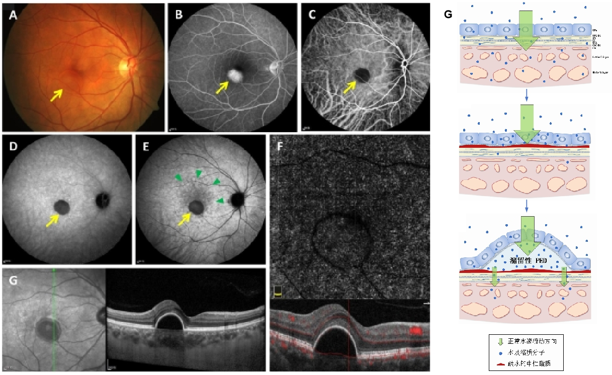
11、Su Y, Zhang X, Chen L, et al. Age-related retentional avascular
pigment epithelial detachment viewed with indocyanine green
angiography[ J]. Retina, 2022, 42(8): 1520-1528. DOI: 10.1097/
iae.0000000000003487.Su Y, Zhang X, Chen L, et al. Age-related retentional avascular
pigment epithelial detachment viewed with indocyanine green
angiography[ J]. Retina, 2022, 42(8): 1520-1528. DOI: 10.1097/
iae.0000000000003487.
12、Su Y, Wen F, Gan Y, et al. Natural course of age-related retentional
avascular pigment epithelial detachment: Support For The Lipid Barrier
Hypothesis[ J]. Retina, 2024, 44(11): 2001-2012. DOI: 10.1097/
iae.0000000000004210.Su Y, Wen F, Gan Y, et al. Natural course of age-related retentional
avascular pigment epithelial detachment: Support For The Lipid Barrier
Hypothesis[ J]. Retina, 2024, 44(11): 2001-2012. DOI: 10.1097/
iae.0000000000004210.
13、Li M, Zhang X, Ji Y, et al. Acute macular neuroretinopathy in
dengue fever: short-term prospectively followed up case series[ J].
JAMA Ophthalmol, 2015, 133(11): 1329-1333. DOI: 10.1001/
jamaophthalmol.2015.2687.Li M, Zhang X, Ji Y, et al. Acute macular neuroretinopathy in
dengue fever: short-term prospectively followed up case series[ J].
JAMA Ophthalmol, 2015, 133(11): 1329-1333. DOI: 10.1001/
jamaophthalmol.2015.2687.
14、Li M, Zhang X, Wen F, et al. Persistent placoid maculopathy: lichen�like lesions growing between retinal pigment epithelium and bruch's
membrane[ J]. Ophthalmol Retina, 2024, 8(3): 270-278. DOI:
10.1016/j.oret.2023.10.001.Li M, Zhang X, Wen F, et al. Persistent placoid maculopathy: lichen�like lesions growing between retinal pigment epithelium and bruch's
membrane[ J]. Ophthalmol Retina, 2024, 8(3): 270-278. DOI:
10.1016/j.oret.2023.10.001.