您的位置: 首页 > 2025年6月 第40卷 第6期 > 文字全文
2023年7月 第38卷 第7期11
目录

急性黄斑神经视网膜病变中视网膜前巨噬细胞样细胞和视网膜脉络膜血流变化

Characterization of epiretinalmacrophage-like cells and retinochoroidal blood flow in acute macular neuroretinopathy

来源期刊: 眼科学报 | 2025年6月 第40卷 第6期 454-463 发布时间:2025-6-28 收稿时间:2025/6/23 9:23:56 阅读量:18
作者:
关键词:
急性黄斑神经视网膜病变视网膜前巨噬细胞样细胞视网膜脉络膜血流
epiretinal macrophage-like cells retinochoroidal blood flow acute macular neuroretinopathy (AMN)
DOI:
10.12419/24121901
收稿时间:
2024-12-23 
修订日期:
2025-01-20 
接收日期:
2025-03-11 

目的:分析视网膜前巨噬细胞样细胞(epiretinal macrophage-like cellseMLC和视网膜脉络膜血流在急性黄斑神经视网膜病变(acute macular neuroretinopathyAMN)患眼的临床特征。方法回顾性分析202212—20235月在中山大学中山眼科中心就诊的病程2周内的AMN患者2137眼)及年龄匹配的健康对照组3338眼)的临床资料通过黄斑区内界膜上μmeface 光学相干断层扫描(optical coherence tomographyOCT)分层信息eMLC进行半自动分析提取和定量,同时测量OCT血管成像(optical coherence tomography angiographyOCTA6 mm² x 6 mm²的黄斑区域内视网膜脉络膜血流参数进行分析对比。结果 AMN黄斑区eMLC数量329.78±77.38)个,密度为(9.16±2.15/mm2,均较对照组(202.63±41.72、(5.63±1.16/mm2增加P0.001)。AMN组浅层和深层视网膜血流密度分别为33.00±6.49)(32.59±7.41%,均较对照组(36.18±5.63)(37.08±5.65%减少(P0.05。视网膜全层中心凹无血管区(foveal avascular zoneFAZ面积、视网膜全层血流密度、脉络膜毛细血管和脉络膜大中血管血流密度在两组间比较差异无统计学意义(P0.05)。浅层及深层视网膜血流密度和FAZ面积对eMLC密度无影响(P0.05)。13AMN患者(231个月时的随访资料显示:AMN组末次随访时eMLC数量248.70±59.88)个、密度为6.91±1.66)个/mm2初次就诊时(307.87±82.98和(8.55±2.30)个/mm2减少(P0.001,但仍高于同期对照组(176.58±27.89和(4.91±0.77/mm2P0.001)。视网膜和脉络膜血流参数较初次就诊时比较差异无统计学意义(P0.05)。结论AMN患眼中eMLC异常增多和聚集,同时存在轻度的视网膜血流密度下降,但无脉络膜血流参数变化,且eMLC变化与AMN病程相关但与视网膜血流变化无关,提示eMLC所代表的炎症可能独立参与了AMN的发生

Objective: To investigate the clinical characteristics of epiretinal macrophage-like cells (eMLC) and retinochoroidal blood flow in eyes affected by acute macular neuroretinopathy (AMN). Methods: This retrospective case series study included 21 (37 eyes) patients diagnosed with AMN and 28 (33 eyes) healthy age-matched subjects. A 3 mm En-face optical coherence tomography (OCT) slab on the inner limiting membrane of the macular region was used to visualize and binarize the MLCs. The MLCs were binarized and quantified using a semiautomated method. Optical coherence tomography angiography (OCTA) was used to evaluate the perfusion status and obtain the structural data of macular within a 6 x 6 mm² macular region. Results: The number and density of macular eMLC in AMN eyes were significantly increased in comparison to control eyes: 329.78±77.38 vs. 202.63±41.72, (P0.001) and (9.16±2.15) cells/mm2 vs. (5.63±1.16) cells/mm2 (P0.001). In the macular region, both superficial and deep retinal vessel densities were significantly lower in the AMN eyes than in the control eyes: 33.00±6.49 vs. 36.18±5.63 (P=0.040); 32.59±7.41 vs. 37.08±5.65 (P=0.008). There were no significant differences in the vessel densities and foveal avascular zone (FAZ) area of full retina and choroidal vessel density between the two groups (P0.05). The eMLC density was not associated with the superficial and deep retinal FAZ area and vessel densities (all P0.05). At the one-month follow-up data of 13 patients (23 eyes), the number and density of macular eMLC were significantly lower in comparison to the initial visit: 248.70±59.88 vs. 307.87±82.98, P0.001 and (6.91±1.66) cells/mm2 vs. (8.55±2.30) cells/mm2 (P0.001). However, the number and density of macular eMLC are still noticeably higher than those of the control group during the same timeframe: 248.70±59.88 vs. 176.58±27.89 (P0.001) and (6.91±1.66) cells/mm2 vs. (4.91±0.77) cells/mm2 (P0.001). There was no significant difference in the vessel density of retina and choroidal during the follow-up (P0.05). Conclusions: The aggregation and activation of eMLC and a mild decrease in retinal blood flow density are observed in AMN, yet there is no corresponding shift in choroidal vessel densityThe changes of eMLC are linked to the course of AMN, but they are not related to retinal vessel density. The inflammatory response represented by eMLC might independently contribute to the pathogenesis and progression of AMN.

文章亮点

1. 关键发现

本文通过分析视网膜前巨噬细胞样细胞(epiretinal macrophage-like cells, eMLC)和视网膜脉络膜血流在急性黄斑神经视网膜病变(acute macular neuroretinopathy, AMN)患眼的临床特征,发现AMN患眼中eMLC异常增多和聚集,同时存在轻度的视网膜血流密度下降,但无脉络膜血流参数变化,且eMLC变化与AMN病程相关但与视网膜血流变化无关,提示eMLC所代表的炎症可能独立参与了AMN的发生。

2. 已知与发现

缺血和(或)炎症被认为是AMN潜在的病理机制, AMN缺血学说的血管闭塞的原因和位置一直存在争议。

3. 意义与改变

为AMN发生的病理生理机制和治疗方向提供了新的临床证据。

       急性黄斑神经视网膜病变(acute macular neuroretinopathy, AMN)是1975年由Bos和Deutman[1]首先描述的表现为黄斑中心凹的暗红色楔形视网膜内病灶,多见于中青年女性,56%~88%的患者双眼发病,10%的患者合并有棉绒斑[2- 3]。多模式影像有助于诊断AMN:红外光(infrared, IR)成像可于黄斑区见到弱反射的楔形或花瓣样病灶;光学相干断层扫描(optical coherence tomography, OCT)可以看到急性期病灶处光感受器全层反射增强。AMN的诱因多样,目前有报道与血管因素(高血压/低血压、过敏性休克、贫血等)、炎症、外伤及病毒感染(如:登革热、流感及疫苗接种等)等相关[4-6]。在突发公共卫生事件流行期间,据调查AMN发病率上升至8.79/10万[7]
       缺血和(或)炎症被认为是AMN潜在的病理机制,但对于AMN中两者是单独或共同还是某一方起主导作用尚无定论。OCT血管成像(optical coherence tomography angiography, OCTA)可通过检测红细胞的运动和使用多次OCT-B扫描实现对视网膜和脉络膜缺血的无创评估。有研究者推测,急性期AMN在OCT上的光感受器层强反射是深层视网膜血流障碍所导致[8];也有研究者认为,脉络膜毛细血管层为光感受器细胞提供营养和氧气,脉络膜毛细血管层的灌注不良可能参与了AMN的发生过程[9]。活体的炎症细胞影像观察近年来也取得突破,en-face OCT可实现对人眼体内的视网膜前巨噬细胞样细胞(epiretinal macrophage-like cells, eMLC)的定量分析而被用于眼底炎症程度的评估[10]。既往研究观察到eMLC在糖尿病性视网膜病变、视网膜静脉阻塞、白塞病性葡萄膜炎等视网膜血管性疾病中的聚集与活化[11-14]。但尚无研究对AMN患眼eMLC进行研究,本研究旨在利用OCT/OCTA观察AMN发病过程中eMLC和视网膜脉络膜血流改变,并探讨炎症和缺血与AMN的关系。

1 对象与方法

1.1 研究对象

       本研究通过了中山大学中山眼科中心伦理委员会审查标准(伦理批件号:2022KYPJ096),符合赫尔辛基宣言原则,回顾性纳入了2022年12月—2023年5月在中山大学中山眼科中心诊断为AMN的患者。AMN急性期诊断标准[15]:①突发视力下降伴或不伴眼前黑影遮挡,病程在2周内;②IR成像于黄斑区见局部弱反射病灶,对应眼底彩照可有放射状排列的微红褐色楔形或花瓣样病灶轮廓;③病灶处OCT对应光感受器全层反射增强。

1.2 AMN组纳入与排除标准

       纳入标准:1)符合AMN急性期诊断标准;2)于本院初次就诊及复查有行OCTA检查,且成像质量≥7分。排除标准:1)合并其他白点综合征;2)合并其他视网膜脉络膜血管性疾病;3)既往接受过眼底视网膜光凝等治疗;4)既往曾有眼部外伤或其他眼部手术史;5)合并与AMN诱因无关的全身其他系统疾病。

1.3 对照组纳入与排除标准

       纳入标准:1)于本院眼部体检已排除眼底疾病;2)于本院行初诊及复查有OCTA检查资料,且成像质量≥7分。排除标准:1)既往接受过眼底视网膜光凝等治疗;2)既往有眼部外伤或眼部手术史;3)合并全身其他系统疾病。

1.4 资料收集 

       收集入组成员初次就诊时的临床资料以及多模式影像检查结果:年龄、性别、既往史、家族史、用药史、裂隙灯检查、最佳矫正视力(best-corrected visual acuity,BCVA、IR(Spectralis HRA+OCT;海德堡工程,海德堡)、OCT、OCTA(图湃,TowardPi BM400K BMizar)原始影像资料。BCVA采用国际标准对数视力,在统计时转换为最小分辨角对数(logarithm of the minimum angle of resolution, logMAR)视力后进行分析。

1.5 研究方法

       1.5.1 eMLC的提取与分析
       收集AMN组及对照组的OCTA检查结果:以黄斑中心凹为中心,收集黄斑区6.0 mm× 6.0 mm的OCTA。按照文献所述方法提取eMLC [16-18]:获取黄斑区内界膜表面3 μm平面图像及内界膜以下27 μm神经纤维层图像;使用ImageJ中半自动二值化过程分离并定量eMLC,其主要过程包括:1)降噪以去除背景不规则噪点和血管伪影;2)增强信号以提高eMLC识别能力;3)二值化提取细胞形状。最后,使用ImageJ软件细胞计数统计二值化图像的eMLC个数及密度。
       1.5.2 血流分析
       以黄斑中心凹为中心,收集黄斑区6.0  mm× 6.0 mm的OCTA后,使用内置系统划分为以黄斑为中心的3 x 3网格,从而得出更小的九个方位区域:颞上方、上方、鼻上方、颞侧、中央、鼻侧、颞下方、下方和鼻下方,统计中央区域的浅层、深层及全层中心凹无血管区(foveal avascular zone, FAZ)面积和视网膜血流密度、脉络膜毛细血管血流密度、脉络膜大中血管血流密度、脉络膜基质体积指数(choroidal stromal index, CSI)、脉络膜血管体积指数(choroidal vascular index, CVI) 数据,其中FAZ面积和血流密度分别以平方毫米(mm2)和百分比(%)表示,其中浅层视网膜从内界膜到内丛状层以下9 μm;深层视网膜从内丛状层以下6 μm到外丛状层以下9 μm;全层视网膜从内界膜到外丛状层以下6 μm。

1.6 统计学分析

       数据分析采用 SPSS 27.0 软件。对符合正态分布的连续变量采用均数±标准差表示,非正态变量以中位数 (范围)表示。分类变量以频次报告。采用 Shapiro-Wilk 检验分析数据正态性,对于符合正态分布的数据采用独立样本t检验进行分析;采用Pearson χ2检验对AMN组及对照组的性别分布进行检验,为了控制患者之间的眼间相关性,采用广义估计方程(generalized estimating equations,GEE)分析不同组间的eMLC 、视网膜脉络膜血流密度等参数;采用配对样本 检验分析单眼发病的5例AMN患者患眼和对侧眼数据以及有随访资料的AMN患者初次就诊与末次随访时的数据;采用多重线性回归分析检验各变量与eMLC密度的独立相关性。 < 0.05被认为具有统计学意义。

2 结 果

2.1 入组患眼就诊时临床特征

       本研究共纳入AMN患者21例(37眼)作为AMN组,其中男5例、女16例,年龄(32.38±9.15)岁;并纳入对照组33例(38眼),其中男13例、女20例,年龄(34.00±10.74)岁。组间年龄和性别比较差异均无统计学意义(均P>0.05)。所有患者起病时均在突发公共卫生事件期间,其抗体检测阳性。AMN组患者最佳矫正视力0.45±0.46,对照组最佳矫正视力0.06±0.20,两组存在显著统计差异(P<0.001)。AMN患眼中有6只于视盘周围有小灶棉绒斑。两组患者的详细临床数据详见表1。

表 1 AMN 组与对照组临床资料
Table 1 Clinical characteristics of AMN group and control group


项目

AMN组

对照组

统计量值

P

入组人数(单位:人)

21

33


入组眼数(单位:只)

37

38 


年龄/

32.38±9.15

34.00±10.74

t=-0.571

0.571 

性别(男/女)/

5/16

13/20

χ2=1.403

0.236#

初次就诊时BCVA(logMAR

0.45±0.46

0.06±0.20

Exp(B)=1.478

<0.001

棉绒斑/

6

/


/

eMLC数量/

329.78±77.38

202.63±41.72

Exp(B)=1.665E+55

<0.001

eMLC密度/(个/mm2

9.16±2.15

5.63±1.16

Exp(B)=34.192

<0.001

浅层FAZ面积/mm2

0.34±0.10

0.29±0.11

Exp(B)=1.055

0.049

深层FAZ面积/mm2

0.74±0.23

0.64±0.16

Exp(B)=1.102

0.045

全层视网膜FAZ面积/mm2

0.36±0.11

0.31±0.12

Exp(B)=1.052

0.090 

浅层视网膜血流密度/%

33.00±6.49

36.18±5.63

Exp(B)=0.041

0.040 

深层视网膜血流密度/%

32.59±7.41

37.08±5.65

Exp(B)=0.011

0.008 

全层视网膜血流密度/%

30.68±8.74

34.76±7.96

Exp(B)=0.017

0.062

脉络膜毛细血管血流密度/%

44.81±3.25

46.08±2.12

Exp(B)=0.281

0.086 

脉络膜大中血管血流密度/%

43.19±3.46

42.71±4.45

Exp(B)=1.614

0.634 

CVI/%

57.89±5.35

60.66±5.77

Exp(B)=0.063

0.056 

CSI/%

42.11±5.35

39.34±5.77

Exp(B)=0.063

0.056 

FAZ:黄斑中心凹无血管区;CVI:脉络膜血管体积密度;CSI:脉络膜基质体密度;#:采用Pearson χ2检验。
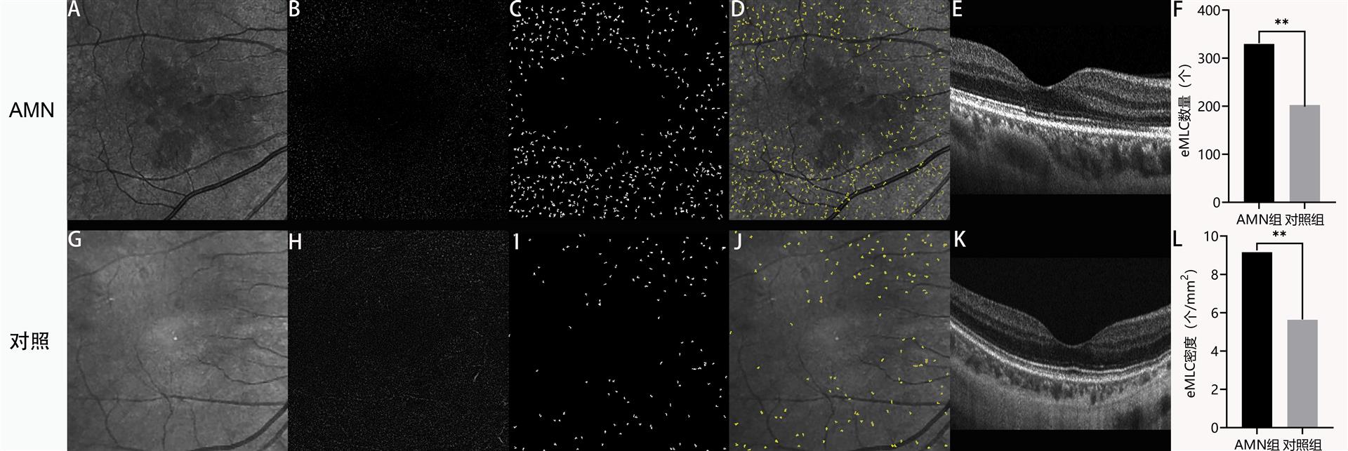
       本研究统计了eMLC在AMN组和对照组的数量及密度(表1):AMN组eMLC数量为(329.78±77.38)个,较对照组(202.63±41.72)个增加(P<0.001);AMN组eMLC密度为(9.16±2.15)个/mm2,较对照组(5.63±1.16)个/mm2升高(P<0.001),是对照组的1.6倍。图1为AMN眼与对照眼的eMLC分布,AMN眼中eMLC数量明显增加,且在AMN病灶周围聚集。

图1 AMN眼与对照眼eMLC对比
Figure 1 Comparison of eMLC between the AMN eye and the control eye

20250626150542_7454_thumb.png
(A) AMN眼IR图示黄斑区清晰的弱反射AMN病灶; AMN眼OCT内界膜表面3 μm平面图像eMLC数量(B)较对照眼(H)增加;AMN眼(C)和对照眼(I)经提取后的eMLC图像;AMN眼IR图示eMLC在病灶周围数量明显增加(D);对照眼eMLC分布( J);AMN眼的OCT图像示黄斑区外层中高反射病灶及中心凹下椭圆体带中断(E)。对照眼IR图(G)及OCT(K)视网膜结构未见明显异常;(F)初次就诊时AMN组eMLC数量较对照组增加(P<0.001);(L)初次就诊时AMN组eMLC密度较对照组增加;组间比较,**P<0.001。
(A) IR imaging in the AMN eye reveals a well-defined hypo-reflective lesion in the macular area. The en face OCT at the 3μm slab above the inner limiting membrane (ILM) in the AMN eye (B) shows an increased eMLC count compared to the control eye(H). Images of subtracted eMLC in the AMN eye (C) and the control eye (I). The number of eMLC increased significantly around the lesion in the AMN eye (D). The distribution of eMLC in the control eye ( J). The OCT shows hyperreflective lesions in the outer layer of the macula and disruption of the ellipsoid zone under the fovea in the AMN eye (E). In the control eye, no significant abnormalities were observed in the IR image (G) or the OCT (K). The number of eMLC in the AMN group was significantly higher than the control group (P<0.001) at the initial visit (F). The density of eMLC in the AMN group was significantly higher than the control group (P<0.001) at the initial visit (L).
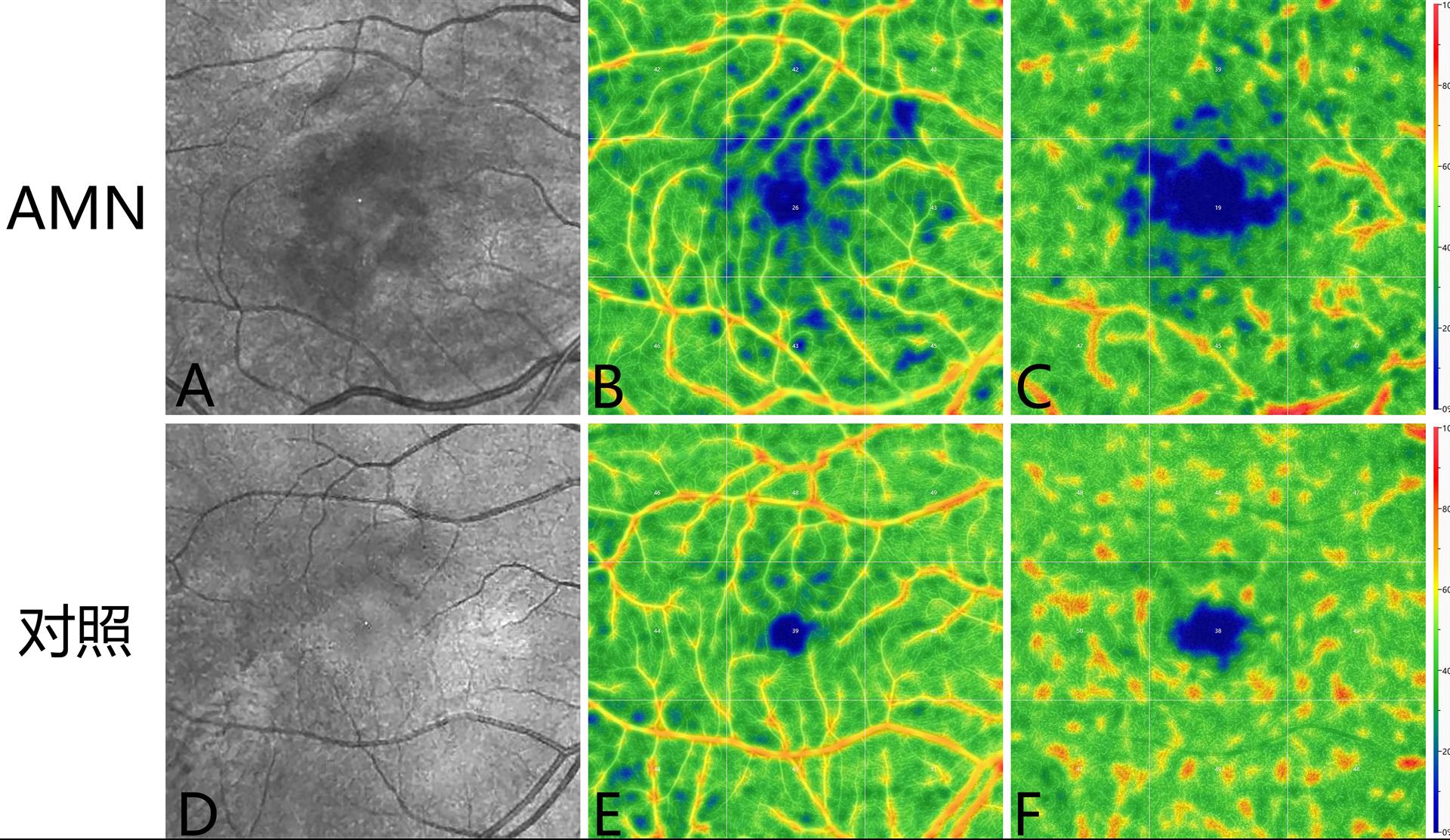
       本研究对比了AMN组及对照组视网膜和脉络膜血流密度情况(表1,图2):AMN组浅层及深层视网膜血流密度分别为(33.00±6.49)(32.59±7.41%),较对照组(36.18±5.63)(37.08±5.65)%减少(均< 0.05);AMN组全层视网膜血流密度、脉络膜毛细血管及大中血管血流密度分别为(30.68±8.74)(44.81±3.25)(43.19±3.46)%,与对照组(34.76±7.96)(46.08±2.12)(42.71±4.45)%比较差异无统计学意义(均P> 0.05);AMN组CVI及CSI分别为(57.89±5.35)(42.11±5.35)%,与对照组(60.66±5.77)(39.34±5.77%)比较差异无统计学意义(均> 0.05)。

图2 AMN眼与对照眼视网膜血流密度对比
Figure 2 Comparison of retina vessel density between the AMN eye and the normal eye

20250626150750_6772_thumb.png
(A)AMN眼IR图示黄斑区清晰的弱反射AMN病灶;(D)对照眼IR图;AMN眼浅层视网膜血流密度(B)较对照眼(E)下降;AMN眼深层视网膜血流密度(C)较对照眼(F)下降。
(A) IR imaging in the AMN eye reveals a well-defined hypo-reflective lesion in the macular area. D. IR imaging of the control eye. The superficial retinal blood flow density was lower in the AMN eye (B) than in the control eye (E). The deep retinal blood flow density was lower in the AMN eye (C) than in the control eye (F).
       本研究对比了中心凹无血管区面积在两组之间的差异(表1):AMN组浅层及深层FAZ面积分别为(0.34±0.10)(0.74±0.23)mm2,较对照组(0.29±0.11)(0.64±0.16)mm2扩大(均< 0.05);AMN组全层视网膜FAZ面积为(0.36±0.11)mm2,与对照组FAZ面积(0.31±0.12)mm2比较差异无统计学意义(= 0.090)。
多重线性回归模型对有统计学差异的浅层及深层视网膜血流密度和FAZ面积与eMLC密度分析(表2)显示,纳入模型的4个自变量对eMLC密度的影响均无统计学意义(均P>0.05)。

表 2 AMN 患眼血流与 eMLC 密度的多重线性回归分析
Table2 Multiple linear regression analysis of eMLC and retinochoroidal blood flow in eyes with AMN

变量

β

95%置信区间

标准化系数β

P

浅层FAZ面积

3.43

-4.70, 11.55

0.16

0.04

深层FAZ面积

2.25

-1.75, 6.78

0.27

0.24

浅层视网膜血流密度

0.01

-0.16, 0.18

0.04

0.89

深层视网膜血流密度

0.11

-0.07, 0.29

0.37

0.22

2.2 eMLC及视网膜脉络膜血流在随访期间的变化

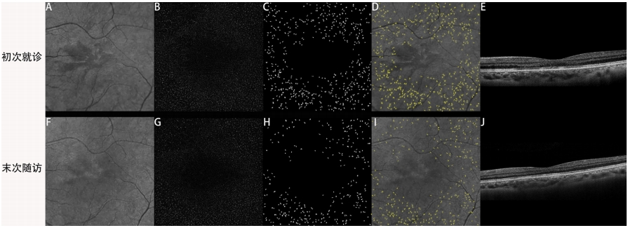
       本研究对初诊和1个月左右末次随访时均有OCTA资料的13例(23眼)AMN患者以及12例(12眼)对照组的随访资料进行了统计分析(见表3、4,图3、4):初次就诊时AMN组eMLC数量为(307.87±82.98)个,密度为(8.55±2.30)个/mm2,均较对照组(178.17±23.92)个、(4.95±0.66)个/mm2增加(均P<0.001)。末次随访时AMN组eMLC数量为(248.70±59.88)个,密度为(6.91±1.66)个/mm2,仍高于对照组(176.58±27.89)个、(4.91±0.77)个/mm2(均P<0.001)。

图3 AMN眼初次就诊和末次随访时eMLC对比
Figure 3 Comparison of eMLC in the AMN eye at the initial visit and the last follow-up

20250626153700_9799.png
IR图示AMN眼末次随访时(F)与初次就诊时(A)相比黄斑区的弱反射病灶消退; OCT内界膜表面3μm平面图像示末次随访时(G)eMLC数量较初次就诊时(B)减少;末次随访(H)及初次就诊(C)时经提取后的eMLC图像; 末次随访时病灶周围eMLC数量(I)较初次就诊时(D)明显减少;OCT示末次随访时病灶处椭圆体带(J)较初次就诊时(E)恢复。
The IR lesion had faded in the macular area of the AMN eye at the last follow-up (F) compared to the initial visit (A). The en face OCT at the 3μm slab above the inner limiting membrane (ILM) in the AMN eye shows a decreased eMLC count at the last follow-up (G) compared to the initial visit (B). Images of subtracted cells in the AMN eye at the last follow-up (H) compared to the initial visit (C). A significant decrease in eMLC count was found around the lesion in the AMN eye at the last follow-up (I) compared to the initial visit (D). The ellipsoid zone had normalized at the last follow-up ( J) compared to the initial visit (E).

图5 eMLC数量及密度变化统计图
Figure 5 The data distribution and comparison of the macular MLC densities and counts

20250626153744_4981.png
(A)初次就诊和末次随访时AMN组eMLC数量均较对照组增加(均P < 0.001),AMN组eMLC数量在初次就诊时与末次随访时相比增加(P < 0.001);(B)初次就诊时和末次随访时AMN组eMLC密度均较对照组增加(均P < 0.001),AMN组eMLC密度在初次就诊时与末次随访时相比加( < 0.001);(C)AMN组eMLC密度在初次就诊时与末次随访时相比升高(P < 0.001),对照组eMLC密度在初次就诊时较末次随访时差异无统计学意义(P > 0.05);组间比较,**P < 0.01。
A. The number of eMLC in the AMN group was significantly higher than the control group at the initial visit and the last follow-up (P<0.001). The number of eMLC in the AMN group was significantly higher at the initial visit compared to the last follow-up (P < 0.001). B. The density of eMLC in the AMN group was significantly higher than the control group (< 0.001) at the initial visit and the last follow-up. The density of eMLC in the AMN group was significantly higher at the initial visit compared to the last follow-up (< 0.001). C. The density of eMLC in the AMN group was significantly higher at the initial visit compared to the last follow-up (< 0.001). There was no significant difference in eMLC densities in the control group at the initial visit and the last follow-up (P > 0.05).

表 3 AMN 组与对照组初次就诊与末次随诊 eMLC 比较
Table 3 Comparison of eMLC in the AMN group at the initial visit and the last follow-up

项目

AMN组

对照组

Exp(B)

P

入组人数(单位:人)

13

12


入组眼数(单位:只)

23

12


初次就诊时eMLC数量/个

307.87±82.98

178.17±23.92

1.475E+56

<0.001*

初次就诊时eMLC密度/(个/mm2

8.55±2.30

4.95±0.66

36.328

<0.001*

末次随访时eMLC数量/个

248.70±59.88

176.58±27.89

1.798E+32

<0.001*

末次随访时eMLC密度/(个/mm2

6.91±1.66

4.91±0.77

7.870

<0.001*

       AMN患眼末次随访时eMLC数量为(248.70±59.88)个,密度为(6.91±1.66)个/mm2,较初次就诊时(307.87±82.98)个、(8.55±2.30)个/mm2降低(均P<0.001);末次随访时浅层、深层及全层FAZ面积分别为(0.34±0.14)(0.74±0.28)(0.39±0.19)mm2与初次就诊时(0.32±0.09)(0.72±0.20)(0.38±0.21)比较差异无统计学意义(均P>0.05);末次随访时浅层、深层及全层视网膜血流密度分别为(35.52±6.85)(34.65±6.19)(33.57±8.68)%,与初次就诊时(33.96±6.85)(34.17±7.15)(31.48±9.48)%比较差异无统计学意义(均P>0.05);末次随访时脉络膜毛细血管及脉络膜大中血管血流密度分别为(45.00±3.28)(43.26±4.71)%,与初次就诊时(44.91±3.73)(42.87±4.22)%比较差异无统计学意义(均P>0.05);末次随访时CVI及CSI分别为(58.65±6.09)(41.35±6.09)%,较初次就诊时(59.04±5.75)(40.96±5.75)%比较差异无统计学意义(均P>0.05)。

表 4 13 例(23 眼)AMN 患者的随访资料
Table 4 Follow-up data of 13 patients (23eyes) with AMN

项目

初次就诊

末次随访

t

P

eMLC数量/个

307.87±82.98

248.70±59.88

4.129

<0.001

eMLC密度/(个/mm2

8.55±2.30

6.91±1.66

4.129

<0.001

浅层FAZ面积/mm2

0.32±0.09

0.34±0.14

-1.057

0.302

深层FAZ面积/mm2

0.72±0.20

0.74±0.28

-0.359

0.723

视网膜FAZ面积/mm2

0.38±0.21

0.39±0.19

-0.272

0.788

浅层视网膜血流密度/%

33.96±6.85

35.52±6.85

-1.298

0.208

深层视网膜血流密度/%

34.17±7.15

34.65±6.19

-0.335

0.741

全层视网膜血流密度/%

31.48±9.48

33.57±8.68

-1.317

0.201

脉络膜毛细血管血流密度/%

44.91±3.73

45.00±3.28

-0.182

0.875

脉络膜大中血管血流密度/%

42.87±4.22

43.26±4.71

-0.574

0.572

CVI/%

59.04±5.75

58.65±6.09

-0.306

0.763

CSI/%

40.96±5.75

41.35±6.09

-0.306

0.763

3  讨论

       本研究首次观察了eMLC及视网膜脉络膜血流在AMN患眼的临床特征:AMN急性期eMLC数量及密度较对照组增加,浅层及深层视网膜血流密度下降,但黄斑区eMLC密度的变化与视网膜血流参数变化无关联,全层视网膜总FAZ面积、全层视网膜血流密度和脉络膜血流参数无明显变化;随着AMN的缓解eMLC数量及密度随之减少,但仍高于对照组,随访期间视网膜脉络膜血流参数无明显变化。eMLC的增加、浅层及深层视网膜血流密度的降低表明炎症和缺血参与了AMN的发生和发展过程,其中eMLC随AMN病程发生变化且独立于视网膜血流改变。
       AMN诱因多样,提示其中可能有共通机制。光感受器层是AMN 主要累及部位,该层靠间接滋养并没有直接的血管成分,因此AMN缺血学说[19]的血管闭塞的原因和位置一直存在争议[20]:多项研究发现,AMN患眼黄斑区深层视网膜灌注不足,且在IR所示病灶对应区域的深层视网膜发现血流空洞信号[9, 21-23];但该推测与黄斑区外层视网膜光感受器的实际解剖学血供来源不吻合,即深层视网膜缺血不应该累及光感受器层;光感受器全层血供并不来自视网膜中央血管系统,即便在视网膜中央动脉阻塞的案例和研究中,也并未发现或报道过光感受器层类似AMN的改变。有研究在脉络膜毛细血管层发现血流空洞信号,脉络膜毛细血管密度降低[24-25],认为可能是AMN在视网膜脉络膜缺氧的基础上,累及光感受器末端深层视网膜[4];还有一种假设认为原发性的深层视网膜灌注不良引起光感受器的损伤,导致脉络膜毛细血管灌注不足[9]。本研究中也证实AMN患眼浅层及深层视网膜血流轻度下降,但视网膜血流变化与AMN病程无关,观察到的视网膜血流密度下降更可能是AMN发生后的继发性改变,另外由于AMN病灶的IR弱反射影响了同样使用IR作为光源的OCTA对视网膜脉络膜血流的定量分析,也可使病灶所在部位的血流密度出现假性下降。光感受器层和RPE层的氧气和营养来自脉络膜毛细血管层,本研究未发现AMN中有脉络膜血流改变,在其他累及脉络膜血供的疾病中,比如急性后极部多灶性鳞状色素上皮病变,由于脉络膜小叶缺血后光感受器和RPE层同时受累,在恢复期RPE会出现对应的色素脱失或萎缩改变,而既往AMN的长期随访报道并未见到有RPE受累。因此缺血可能不是AMN发生的主要原因。
       既往视网膜血管性疾病的eMLC研究中,eMLC作为血管病变后继发的炎性改变,与视网膜血管渗漏或视网膜缺血程度以及黄斑水肿等并发症密切相关[11-14]。AMN患眼eMLC密度的增加(是对照组的1.6倍)提示了eMLC的异常聚集,且与AMN病程相关,同时不受视网膜血流改变影响,提示AMN中eMLC所代表的炎症并非由视网膜血流改变所继发,炎症可能独立参与了AMN的发生、发展。eMLC是位于视网膜表面的一类具有自我更新能力的免疫细胞群,主要包括小胶质细胞、玻璃体细胞及髓系单核巨噬细胞等[26]。近来动物实验表明eMLC在稳态下主要由玻璃体基底部透明细胞、血管周围的单核巨噬细胞及小胶质细胞组成,而在炎症状态下主要由内界膜表面单核细胞及血浆募集而来的髓系单核巨噬细胞组成[27-30]。本组AMN患者均与突发公共卫生事件的感染相关,但单眼发病的AMN患者其患眼较对侧眼eMLC数量和密度存在差异,尽管视网膜血流密度差异无统计学意义,但仍较对侧眼偏低;对侧眼数据较对照组差异无统计学意义,说明本研究中eMLC和视网膜血流密度等改变与AMN相关,而并非直接由突发公共卫生事件感染引起。
       本研究存在若干局限性:首先,本研究是一项回顾性研究,仅选择eMLC作为炎症的影像学指标,但缺乏其他全身或眼内的炎症因子检测佐证炎症在AMN发病中的作用;其次,AMN患者随访时间点有限,仅部分有1个月后的随访资料;最后该组研究仅有突发公共卫生事件相关的AMN,未纳入其他诱因导致的AMN患者。
       综上所述,AMN患眼中eMLC所代表的影像学炎症指标升高,同时存在轻度的视网膜血流密度降低,但无脉络膜血流参数变化,eMLC随AMN病程变化但与视网膜血流变化无关,提示eMLC所代表的炎症可能独立参与了AMN的发生。

声明

       在论文撰写过程中未使用生成式人工智能。论文撰写中的所有内容仅有作者独立完成,并对出版物的真实性和准确性承担全部责任。

开放获取声明

       本文适用于知识共享许可协议(Creative Commons),允许第三方用户按照“署名(BY)-非商业性使用(NC)-禁止演绎(ND)(CC BY-NC-ND)”的方式共享,即允许第三方对本刊发表的文章进行复制、发行、展览、表演、放映、广播或通过信息网络向公众传播,但在这些过程中必须保留作者署名、仅限于非商业性目的、不得进行演绎创作。


1、Bos PJ, Deutman AF. Acute macular neuroretinopathy[J]. Am J Ophthalmol, 1975, 80(4): 573-584. DOI: 10.1016/0002-9394(75)90387-6.Bos PJ, Deutman AF. Acute macular neuroretinopathy[J]. Am J Ophthalmol, 1975, 80(4): 573-584. DOI: 10.1016/0002-9394(75)90387-6.
2、Feng H, Zhao M, Mo J, et al. The characteristics of acute macular neuroretinopathy following COVID-19 infection[J]. BMC Ophthalmol, 2024, 24(1): 19. DOI: 10.1186/s12886-024-03283-2. Feng H, Zhao M, Mo J, et al. The characteristics of acute macular neuroretinopathy following COVID-19 infection[J]. BMC Ophthalmol, 2024, 24(1): 19. DOI: 10.1186/s12886-024-03283-2.
3、Song X, Yu Y, Zhou H, et al. Acute macular neuroretinopathy associated with COVID-19 pandemic: a real-world observation study[J]. Asia Pac J Ophthalmol, 2024, 13(5): 100103. DOI: 10.1016/j.apjo.2024.100103.Song X, Yu Y, Zhou H, et al. Acute macular neuroretinopathy associated with COVID-19 pandemic: a real-world observation study[J]. Asia Pac J Ophthalmol, 2024, 13(5): 100103. DOI: 10.1016/j.apjo.2024.100103.
4、Munk MR, Jampol LM, Cunha Souza E, et al. New associations of classic acute macular neuroretinopathy[J]. Br J Ophthalmol, 2016, 100(3): 389-394. DOI: 10.1136/bjophthalmol-2015-306845. Munk MR, Jampol LM, Cunha Souza E, et al. New associations of classic acute macular neuroretinopathy[J]. Br J Ophthalmol, 2016, 100(3): 389-394. DOI: 10.1136/bjophthalmol-2015-306845.
5、Ashfaq I, Vrahimi M, Waugh S, et al. Acute macular neuroretinopathy associated with acute influenza virus infection[J]. Ocul Immunol Inflamm, 2021, 29(2): 333-339. DOI: 10.1080/09273948.2019.1681470.Ashfaq I, Vrahimi M, Waugh S, et al. Acute macular neuroretinopathy associated with acute influenza virus infection[J]. Ocul Immunol Inflamm, 2021, 29(2): 333-339. DOI: 10.1080/09273948.2019.1681470.
6、Ikema S, Miura G, Shimizu D, et al. Long-term follow-up of a young male who developed acute macular neuroretinopathy following COVID-19 vaccination[J]. Clin Case Rep, 2023, 11(11): e8181. DOI: 10.1002/ccr3.8181. Ikema S, Miura G, Shimizu D, et al. Long-term follow-up of a young male who developed acute macular neuroretinopathy following COVID-19 vaccination[J]. Clin Case Rep, 2023, 11(11): e8181. DOI: 10.1002/ccr3.8181.
7、Azar%20G%2C%20Bonnin%20S%2C%20Vasseur%20V%2C%20et%20al.%20Did%20the%20COVID-19%20pandemic%20increase%20the%20incidence%20of%20acute%20macular%20neuroretinopathy%3F%5BJ%5D.%20J%20Clin%20Med%2C%202021%2C%2010(21)%3A%205038.%20DOI%3A%2010.3390%2Fjcm10215038.Azar%20G%2C%20Bonnin%20S%2C%20Vasseur%20V%2C%20et%20al.%20Did%20the%20COVID-19%20pandemic%20increase%20the%20incidence%20of%20acute%20macular%20neuroretinopathy%3F%5BJ%5D.%20J%20Clin%20Med%2C%202021%2C%2010(21)%3A%205038.%20DOI%3A%2010.3390%2Fjcm10215038.
8、Bhavsar KV, Lin S, Rahimy E, et al. Acute macular neuroretinopathy: a comprehensive review of the literature[J]. Surv Ophthalmol, 2016, 61(5): 538-565. DOI: 10.1016/j.survophthal.2016.03.003.Bhavsar KV, Lin S, Rahimy E, et al. Acute macular neuroretinopathy: a comprehensive review of the literature[J]. Surv Ophthalmol, 2016, 61(5): 538-565. DOI: 10.1016/j.survophthal.2016.03.003.
9、 Casalino G, Arrigo A, Romano F, et al. Acute macular neuroretinopathy: pathogenetic insights from optical coherence tomography angiography[J]. Br J Ophthalmol, 2019, 103(3): 410-414. DOI: 10.1136/bjophthalmol-2018-312197. Casalino G, Arrigo A, Romano F, et al. Acute macular neuroretinopathy: pathogenetic insights from optical coherence tomography angiography[J]. Br J Ophthalmol, 2019, 103(3): 410-414. DOI: 10.1136/bjophthalmol-2018-312197.
10、 Hammer DX, Agrawal A, Villanueva R, et al. Label-free adaptive optics imaging of human retinal macrophage distribution and dynamics[J]. Proc Natl Acad Sci USA, 2020, 117(48): 30661-30669. DOI: 10.1073/pnas.2010943117. Hammer DX, Agrawal A, Villanueva R, et al. Label-free adaptive optics imaging of human retinal macrophage distribution and dynamics[J]. Proc Natl Acad Sci USA, 2020, 117(48): 30661-30669. DOI: 10.1073/pnas.2010943117.
11、Zeng Y, Wen F, Mi L, et al. Changes in macrophage-like cells characterized by en face optical coherence tomography after retinal stroke[J]. Front Immunol, 2022, 13: 987836. DOI: 10.3389/fimmu.2022.987836.Zeng Y, Wen F, Mi L, et al. Changes in macrophage-like cells characterized by en face optical coherence tomography after retinal stroke[J]. Front Immunol, 2022, 13: 987836. DOI: 10.3389/fimmu.2022.987836.
12、Zeng Y, Zhang X, Mi L, et al. Characterization of macrophage-like cells in retinal vein occlusion using en face optical coherence tomography[J]. Front Immunol, 2022, 13: 855466. DOI: 10.3389/fimmu.2022.855466. Zeng Y, Zhang X, Mi L, et al. Characterization of macrophage-like cells in retinal vein occlusion using en face optical coherence tomography[J]. Front Immunol, 2022, 13: 855466. DOI: 10.3389/fimmu.2022.855466.
13、 Zeng Y, Wen F, Zhuang X, et al. Epiretinal macrophage-like cells on optical coherence tomography: Potential Inflammatory Imaging Biomarker of Severity in Diabetic Retinopathy[J]. Retina, 2024, 44(8): 1314-1322. DOI: 10.1097/iae.0000000000004100. Zeng Y, Wen F, Zhuang X, et al. Epiretinal macrophage-like cells on optical coherence tomography: Potential Inflammatory Imaging Biomarker of Severity in Diabetic Retinopathy[J]. Retina, 2024, 44(8): 1314-1322. DOI: 10.1097/iae.0000000000004100.
14、Zeng%20Y%2C%20Zhang%20X%2C%20Mi%20L%2C%20et%20al.%20Macrophage-like%20cells%20characterized%20by%20en%20face%20optical%20coherence%20tomography%20was%20associated%20with%20fluorescein%20vascular%20leakage%20in%20beh%C3%A7et%E2%80%99s%20uveitis%5BJ%5D.%20Ocul%20Immunol%20Inflamm%2C%202023%2C%2031(5)%3A%20999-1005.%20DOI%3A%2010.1080%2F09273948.2022.2080719.Zeng%20Y%2C%20Zhang%20X%2C%20Mi%20L%2C%20et%20al.%20Macrophage-like%20cells%20characterized%20by%20en%20face%20optical%20coherence%20tomography%20was%20associated%20with%20fluorescein%20vascular%20leakage%20in%20beh%C3%A7et%E2%80%99s%20uveitis%5BJ%5D.%20Ocul%20Immunol%20Inflamm%2C%202023%2C%2031(5)%3A%20999-1005.%20DOI%3A%2010.1080%2F09273948.2022.2080719.
15、Li M, Zhang X, Ji Y, Ye B, Wen F. Acute Macular Neuroretinopathy in Dengue Fever: Short-term Prospectively Followed Up Case Series. JAMA Ophthalmol. 2015 Nov;133(11):1329-33. doi: 10.1001/jamaophthalmol.2015.2687.Li M, Zhang X, Ji Y, Ye B, Wen F. Acute Macular Neuroretinopathy in Dengue Fever: Short-term Prospectively Followed Up Case Series. JAMA Ophthalmol. 2015 Nov;133(11):1329-33. doi: 10.1001/jamaophthalmol.2015.2687.
16、Castanos MV, Zhou DB, Linderman RE, et al. Imaging of macrophage-like cells in living human retina using clinical OCT[J]. Invest Ophthalmol Vis Sci, 2020, 61(6): 48. DOI: 10.1167/iovs.61.6.48. Castanos MV, Zhou DB, Linderman RE, et al. Imaging of macrophage-like cells in living human retina using clinical OCT[J]. Invest Ophthalmol Vis Sci, 2020, 61(6): 48. DOI: 10.1167/iovs.61.6.48.
17、Ong JX, Nesper PL, Fawzi AA, et al. Macrophage-like cell density is increased in proliferative diabetic retinopathy characterized by optical coherence tomography angiography[J]. Invest Ophthalmol Vis Sci, 2021, 62(10): 2. DOI: 10.1167/iovs.62.10.2. Ong JX, Nesper PL, Fawzi AA, et al. Macrophage-like cell density is increased in proliferative diabetic retinopathy characterized by optical coherence tomography angiography[J]. Invest Ophthalmol Vis Sci, 2021, 62(10): 2. DOI: 10.1167/iovs.62.10.2.
18、Schindelin J, Arganda-Carreras I, Frise E, et al. Fiji: an open-source platform for biological-image analysis. Nat Methods, 2012, 9(7): 676-682. DOI: 10.1038/nmeth.2019.Schindelin J, Arganda-Carreras I, Frise E, et al. Fiji: an open-source platform for biological-image analysis. Nat Methods, 2012, 9(7): 676-682. DOI: 10.1038/nmeth.2019.
19、 Iovino C, Au A, Ramtohul P, et al. Coincident PAMM and AMN and insights into a common pathophysiology[J]. Am J Ophthalmol, 2022, 236: 136-146. DOI: 10.1016/j.ajo.2021.07.004. Iovino C, Au A, Ramtohul P, et al. Coincident PAMM and AMN and insights into a common pathophysiology[J]. Am J Ophthalmol, 2022, 236: 136-146. DOI: 10.1016/j.ajo.2021.07.004.
20、Bellot L, Laurent C, Arcade PE, et al. Acute macular neuroretinopathy: En face OCT description, case series[J]. J Fr Ophtalmol, 2022, 45(2): 159-165. DOI: 10.1016/j.jfo.2021.09.013.Bellot L, Laurent C, Arcade PE, et al. Acute macular neuroretinopathy: En face OCT description, case series[J]. J Fr Ophtalmol, 2022, 45(2): 159-165. DOI: 10.1016/j.jfo.2021.09.013.
21、Fawzi AA, Pappuru RR, Sarraf D, et al. Acute macular neuroretinopathy: long-term insights revealed by multimodal imaging[J]. Retina, 2012, 32(8): 1500-1513. DOI: 10.1097/iae.0b013e318263d0c3.Fawzi AA, Pappuru RR, Sarraf D, et al. Acute macular neuroretinopathy: long-term insights revealed by multimodal imaging[J]. Retina, 2012, 32(8): 1500-1513. DOI: 10.1097/iae.0b013e318263d0c3.
22、Giacuzzo C, Eandi CM, Kawasaki A. Bilateral acute macular neuroretinopathy following COVID-19 infection[J]. Acta Ophthalmol, 2022, 100(2): e611-e612. DOI: 10.1111/aos.14913. Giacuzzo C, Eandi CM, Kawasaki A. Bilateral acute macular neuroretinopathy following COVID-19 infection[J]. Acta Ophthalmol, 2022, 100(2): e611-e612. DOI: 10.1111/aos.14913.
23、Capuano V, Forte P, Sacconi R, et al. Acute macular neuroretinopathy as the first stage of SARS-CoV-2 infection[J]. Eur J Ophthalmol, 2023, 33(3): NP105-NP111. DOI: 10.1177/11206721221090697. Capuano V, Forte P, Sacconi R, et al. Acute macular neuroretinopathy as the first stage of SARS-CoV-2 infection[J]. Eur J Ophthalmol, 2023, 33(3): NP105-NP111. DOI: 10.1177/11206721221090697.
24、Lee SY, Cheng JL, Gehrs KM, et al. Choroidal features of acute macular neuroretinopathy via optical coherence tomography angiography and correlation with serial multimodal imaging[J]. JAMA Ophthalmol, 2017, 135(11): 1177-1183. DOI: 10.1001/jamaophthalmol.2017.3790. Lee SY, Cheng JL, Gehrs KM, et al. Choroidal features of acute macular neuroretinopathy via optical coherence tomography angiography and correlation with serial multimodal imaging[J]. JAMA Ophthalmol, 2017, 135(11): 1177-1183. DOI: 10.1001/jamaophthalmol.2017.3790.
25、von der Burchard C, Gruben A, Roider J. Optical coherence tomography angiography suggests choriocapillaris perfusion deficit as etiology of acute macular neuroretinopathy[J]. Graefe’s Arch Clin Exp Ophthalmol, 2024, 262(8): 2471-2479. DOI: 10.1007/s00417-024-06436-7. von der Burchard C, Gruben A, Roider J. Optical coherence tomography angiography suggests choriocapillaris perfusion deficit as etiology of acute macular neuroretinopathy[J]. Graefe’s Arch Clin Exp Ophthalmol, 2024, 262(8): 2471-2479. DOI: 10.1007/s00417-024-06436-7.
26、Mat Nor MN, Guo CX, Green CR, et al. Hyper-reflective dots in optical coherence tomography imaging and inflammation markers in diabetic retinopathy[J]. J Anat, 2023, 243(4): 697-705. DOI: 10.1111/joa.13889.Mat Nor MN, Guo CX, Green CR, et al. Hyper-reflective dots in optical coherence tomography imaging and inflammation markers in diabetic retinopathy[J]. J Anat, 2023, 243(4): 697-705. DOI: 10.1111/joa.13889.
27、 Rajesh A, Droho S, Lavine JA. Macrophages in close proximity to the vitreoretinal interface are potential biomarkers of inflammation during retinal vascular disease[J]. J Neuroinflammation, 2022, 19(1): 203. DOI: 10.1186/s12974-022-02562-3. Rajesh A, Droho S, Lavine JA. Macrophages in close proximity to the vitreoretinal interface are potential biomarkers of inflammation during retinal vascular disease[J]. J Neuroinflammation, 2022, 19(1): 203. DOI: 10.1186/s12974-022-02562-3.
28、Taghavi Y, Hassanshahi G, Kounis NG, et al. Monocyte chemoattractant protein-1 (MCP-1/CCL2) in diabetic retinopathy: latest evidence and clinical considerations[J]. J Cell Commun Signal, 2019, 13(4): 451-462. DOI: 10.1007/s12079-018-00500-8. Taghavi Y, Hassanshahi G, Kounis NG, et al. Monocyte chemoattractant protein-1 (MCP-1/CCL2) in diabetic retinopathy: latest evidence and clinical considerations[J]. J Cell Commun Signal, 2019, 13(4): 451-462. DOI: 10.1007/s12079-018-00500-8.
29、Pichi F, Neri P, Aljeneibi S, et al. In vivo visualization of macrophage-like cells in patients with uveitis by use of En face swept source optical coherence tomography[J]. Ocul Immunol Inflamm, 2024, 32(8): 1532-1538. DOI: 10.1080/09273948.2023.2254369.Pichi F, Neri P, Aljeneibi S, et al. In vivo visualization of macrophage-like cells in patients with uveitis by use of En face swept source optical coherence tomography[J]. Ocul Immunol Inflamm, 2024, 32(8): 1532-1538. DOI: 10.1080/09273948.2023.2254369.
30、Carre%C3%B1o%20E%2C%20Hernanz%20I%2C%20Collado%20B%2C%20et%20al.%20Description%20of%20macrophage-like%20cells%20in%20active%20ocular%20toxoplasmosis%5BJ%5D.%20Ocul%20Immunol%20Inflamm%2C%202024%2C%2032(8)%3A%201893-1896.%20DOI%3A%2010.1080%2F09273948.2023.2263073.%20Carre%C3%B1o%20E%2C%20Hernanz%20I%2C%20Collado%20B%2C%20et%20al.%20Description%20of%20macrophage-like%20cells%20in%20active%20ocular%20toxoplasmosis%5BJ%5D.%20Ocul%20Immunol%20Inflamm%2C%202024%2C%2032(8)%3A%201893-1896.%20DOI%3A%2010.1080%2F09273948.2023.2263073.%20
1、Bos PJ, Deutman AF. Acute macular neuroretinopathy[J]. Am J Ophthalmol, 1975, 80(4): 573-584. DOI: 10.1016/0002-9394(75)90387-6. Bos PJ, Deutman AF. Acute macular neuroretinopathy[J]. Am J Ophthalmol, 1975, 80(4): 573-584. DOI: 10.1016/0002-9394(75)90387-6.
2、Feng H, Zhao M, Mo J, et al. The characteristics of acute macular neuroretinopathy following COVID-19 infection[J]. BMC Ophthalmol, 2024, 24(1): 19. DOI: 10.1186/s12886-024-03283-2.Feng H, Zhao M, Mo J, et al. The characteristics of acute macular neuroretinopathy following COVID-19 infection[J]. BMC Ophthalmol, 2024, 24(1): 19. DOI: 10.1186/s12886-024-03283-2.
3、Song X, Yu Y, Zhou H, et al. Acute macular neuroretinopathy associated with COVID-19 pandemic: a real-world observation study[J]. Asia Pac J Ophthalmol, 2024, 13(5): 100103. DOI: 10.1016/j.apjo.2024.100103. Song X, Yu Y, Zhou H, et al. Acute macular neuroretinopathy associated with COVID-19 pandemic: a real-world observation study[J]. Asia Pac J Ophthalmol, 2024, 13(5): 100103. DOI: 10.1016/j.apjo.2024.100103.
4、Munk MR, Jampol LM, Cunha Souza E, et al. New associations of classic acute macular neuroretinopathy[J]. Br J Ophthalmol, 2016, 100(3): 389-394. DOI: 10.1136/bjophthalmol-2015-306845. Munk MR, Jampol LM, Cunha Souza E, et al. New associations of classic acute macular neuroretinopathy[J]. Br J Ophthalmol, 2016, 100(3): 389-394. DOI: 10.1136/bjophthalmol-2015-306845.
5、Ashfaq I, Vrahimi M, Waugh S, et al. Acute macular neuroretinopathy associated with acute influenza virus infection[J]. Ocul Immunol Inflamm, 2021, 29(2): 333-339. DOI: 10.1080/09273948.2019.1681470. Ashfaq I, Vrahimi M, Waugh S, et al. Acute macular neuroretinopathy associated with acute influenza virus infection[J]. Ocul Immunol Inflamm, 2021, 29(2): 333-339. DOI: 10.1080/09273948.2019.1681470.
6、Ikema S, Miura G, Shimizu D, et al. Long-term follow-up of a young male who developed acute macular neuroretinopathy following COVID-19 vaccination[J]. Clin Case Rep, 2023, 11(11): e8181. DOI: 10.1002/ccr3.8181. Ikema S, Miura G, Shimizu D, et al. Long-term follow-up of a young male who developed acute macular neuroretinopathy following COVID-19 vaccination[J]. Clin Case Rep, 2023, 11(11): e8181. DOI: 10.1002/ccr3.8181.
7、Azar%20G%2C%20Bonnin%20S%2C%20Vasseur%20V%2C%20et%20al.%20Did%20the%20COVID-19%20pandemic%20increase%20the%20incidence%20of%20acute%20macular%20neuroretinopathy%3F%5BJ%5D.%20J%20Clin%20Med%2C%202021%2C%2010(21)%3A%205038.%20DOI%3A%2010.3390%2Fjcm10215038.%20Azar%20G%2C%20Bonnin%20S%2C%20Vasseur%20V%2C%20et%20al.%20Did%20the%20COVID-19%20pandemic%20increase%20the%20incidence%20of%20acute%20macular%20neuroretinopathy%3F%5BJ%5D.%20J%20Clin%20Med%2C%202021%2C%2010(21)%3A%205038.%20DOI%3A%2010.3390%2Fjcm10215038.%20
8、Bhavsar KV, Lin S, Rahimy E, et al. Acute macular neuroretinopathy: a comprehensive review of the literature[J]. Surv Ophthalmol, 2016, 61(5): 538-565. DOI: 10.1016/j.survophthal.2016.03.003. Bhavsar KV, Lin S, Rahimy E, et al. Acute macular neuroretinopathy: a comprehensive review of the literature[J]. Surv Ophthalmol, 2016, 61(5): 538-565. DOI: 10.1016/j.survophthal.2016.03.003.
9、Casalino G, Arrigo A, Romano F, et al. Acute macular neuroretinopathy: pathogenetic insights from optical coherence tomography angiography[J]. Br J Ophthalmol, 2019, 103(3): 410-414. DOI: 10.1136/bjophthalmol-2018-312197. Casalino G, Arrigo A, Romano F, et al. Acute macular neuroretinopathy: pathogenetic insights from optical coherence tomography angiography[J]. Br J Ophthalmol, 2019, 103(3): 410-414. DOI: 10.1136/bjophthalmol-2018-312197.
10、Hammer DX, Agrawal A, Villanueva R, et al. Label-free adaptive optics imaging of human retinal macrophage distribution and dynamics[J]. Proc Natl Acad Sci USA, 2020, 117(48): 30661-30669. DOI: 10.1073/pnas.2010943117. Hammer DX, Agrawal A, Villanueva R, et al. Label-free adaptive optics imaging of human retinal macrophage distribution and dynamics[J]. Proc Natl Acad Sci USA, 2020, 117(48): 30661-30669. DOI: 10.1073/pnas.2010943117.
11、 Zeng Y, Wen F, Mi L, et al. Changes in macrophage-like cells characterized by en face optical coherence tomography after retinal stroke[J]. Front Immunol, 2022, 13: 987836. DOI: 10.3389/fimmu.2022.987836. Zeng Y, Wen F, Mi L, et al. Changes in macrophage-like cells characterized by en face optical coherence tomography after retinal stroke[J]. Front Immunol, 2022, 13: 987836. DOI: 10.3389/fimmu.2022.987836.
12、Zeng Y, Zhang X, Mi L, et al. Characterization of macrophage-like cells in retinal vein occlusion using en face optical coherence tomography[J]. Front Immunol, 2022, 13: 855466. DOI: 10.3389/fimmu.2022.855466.Zeng Y, Zhang X, Mi L, et al. Characterization of macrophage-like cells in retinal vein occlusion using en face optical coherence tomography[J]. Front Immunol, 2022, 13: 855466. DOI: 10.3389/fimmu.2022.855466.
13、Zeng Y, Wen F, Zhuang X, et al. Epiretinal macrophage-like cells on optical coherence tomography: Potential Inflammatory Imaging Biomarker of Severity in Diabetic Retinopathy[J]. Retina, 2024, 44(8): 1314-1322. DOI: 10.1097/iae.0000000000004100. Zeng Y, Wen F, Zhuang X, et al. Epiretinal macrophage-like cells on optical coherence tomography: Potential Inflammatory Imaging Biomarker of Severity in Diabetic Retinopathy[J]. Retina, 2024, 44(8): 1314-1322. DOI: 10.1097/iae.0000000000004100.
14、Zeng%20Y%2C%20Zhang%20X%2C%20Mi%20L%2C%20et%20al.%20Macrophage-like%20cells%20characterized%20by%20en%20face%20optical%20coherence%20tomography%20was%20associated%20with%20fluorescein%20vascular%20leakage%20in%20beh%C3%A7et%E2%80%99s%20uveitis%5BJ%5D.%20Ocul%20Immunol%20Inflamm%2C%202023%2C%2031(5)%3A%20999-1005.%20DOI%3A%2010.1080%2F09273948.2022.2080719.Zeng%20Y%2C%20Zhang%20X%2C%20Mi%20L%2C%20et%20al.%20Macrophage-like%20cells%20characterized%20by%20en%20face%20optical%20coherence%20tomography%20was%20associated%20with%20fluorescein%20vascular%20leakage%20in%20beh%C3%A7et%E2%80%99s%20uveitis%5BJ%5D.%20Ocul%20Immunol%20Inflamm%2C%202023%2C%2031(5)%3A%20999-1005.%20DOI%3A%2010.1080%2F09273948.2022.2080719.
15、Li M, Zhang X, Ji Y, Ye B, Wen F. Acute Macular Neuroretinopathy in Dengue Fever: Short-term Prospectively Followed Up Case Series. JAMA Ophthalmol. 2015 Nov;133(11):1329-33. doi: 10.1001/jamaophthalmol.2015.2687.Li M, Zhang X, Ji Y, Ye B, Wen F. Acute Macular Neuroretinopathy in Dengue Fever: Short-term Prospectively Followed Up Case Series. JAMA Ophthalmol. 2015 Nov;133(11):1329-33. doi: 10.1001/jamaophthalmol.2015.2687.
16、Castanos MV, Zhou DB, Linderman RE, et al. Imaging of macrophage-like cells in living human retina using clinical OCT[J]. Invest Ophthalmol Vis Sci, 2020, 61(6): 48. DOI: 10.1167/iovs.61.6.48. Castanos MV, Zhou DB, Linderman RE, et al. Imaging of macrophage-like cells in living human retina using clinical OCT[J]. Invest Ophthalmol Vis Sci, 2020, 61(6): 48. DOI: 10.1167/iovs.61.6.48.
17、Ong JX, Nesper PL, Fawzi AA, et al. Macrophage-like cell density is increased in proliferative diabetic retinopathy characterized by optical coherence tomography angiography[J]. Invest Ophthalmol Vis Sci, 2021, 62(10): 2. DOI: 10.1167/iovs.62.10.2. Ong JX, Nesper PL, Fawzi AA, et al. Macrophage-like cell density is increased in proliferative diabetic retinopathy characterized by optical coherence tomography angiography[J]. Invest Ophthalmol Vis Sci, 2021, 62(10): 2. DOI: 10.1167/iovs.62.10.2.
18、Schindelin J, Arganda-Carreras I, Frise E, et al. Fiji: an open-source platform for biological-image analysis. Nat Methods, 2012, 9(7): 676-682. DOI: 10.1038/nmeth.2019.Schindelin J, Arganda-Carreras I, Frise E, et al. Fiji: an open-source platform for biological-image analysis. Nat Methods, 2012, 9(7): 676-682. DOI: 10.1038/nmeth.2019.
19、Iovino C, Au A, Ramtohul P, et al. Coincident PAMM and AMN and insights into a common pathophysiology[J]. Am J Ophthalmol, 2022, 236: 136-146. DOI: 10.1016/j.ajo.2021.07.004.Iovino C, Au A, Ramtohul P, et al. Coincident PAMM and AMN and insights into a common pathophysiology[J]. Am J Ophthalmol, 2022, 236: 136-146. DOI: 10.1016/j.ajo.2021.07.004.
20、Bellot L, Laurent C, Arcade PE, et al. Acute macular neuroretinopathy: En face OCT description, case series[J]. J Fr Ophtalmol, 2022, 45(2): 159-165. DOI: 10.1016/j.jfo.2021.09.013. Bellot L, Laurent C, Arcade PE, et al. Acute macular neuroretinopathy: En face OCT description, case series[J]. J Fr Ophtalmol, 2022, 45(2): 159-165. DOI: 10.1016/j.jfo.2021.09.013.
21、 Fawzi AA, Pappuru RR, Sarraf D, et al. Acute macular neuroretinopathy: long-term insights revealed by multimodal imaging[J]. Retina, 2012, 32(8): 1500-1513. DOI: 10.1097/iae.0b013e318263d0c3. Fawzi AA, Pappuru RR, Sarraf D, et al. Acute macular neuroretinopathy: long-term insights revealed by multimodal imaging[J]. Retina, 2012, 32(8): 1500-1513. DOI: 10.1097/iae.0b013e318263d0c3.
上一篇
下一篇
其他期刊
  • 眼科学报

    主管:中华人民共和国教育部
    主办:中山大学
    承办:中山大学中山眼科中心
    主编:林浩添
    主管:中华人民共和国教育部
    主办:中山大学
    浏览
  • Eye Science

    主管:中华人民共和国教育部
    主办:中山大学
    承办:中山大学中山眼科中心
    主编:林浩添
    主管:中华人民共和国教育部
    主办:中山大学
    浏览
目录

点击右上角菜单,浏览器打开下载

我知道了