您的位置: 首页 > 2024年6月 第1卷 第2期 > 文字全文
2023年7月 第38卷 第7期11
目录

Ultrapulse carbon dioxide laser excision as a novel treatment for large divided nevus of the eyelid

Ultrapulse carbon dioxide laser excision as a novel treatment for large divided nevus of the eyelid

来源期刊: Eye Science | 2024年6月 第1卷 第2期 115-127 发布时间:2024-06-28 收稿时间:2024/7/26 8:42:13 阅读量:1177
作者:
关键词:
ultrapulse carbon dioxide laser excision divided nevus of the eyelid surgical excision laser ablation
DOI:
10.12419/es24060102
Received date:
2024-05-01 
Accepted date:
2024-06-28 
Published online:
2024-06-28 
Aims: Divided nevus of the eyelid is a congenital pigmented nevus that impacts eyelid function and aesthetics. While surgical excision and laser ablation are current treatment options, they have limitations when dealing with large lesions. This study aims to investigate the efficacy and safety of carbon dioxide (CO2) laser excision treatment for divided nevus of the eyelid. Methods: This retrospective study included 10 patients (5 males, 5 females) with a mean age of 23.7 years (9-54 years). All underwent CO2 laser excision and were followed up for 12 months. Treatment outcomes were assessed through clearance and recurrence rates, evaluated using digital photography. Postoperative complications were closely monitored throughout the 12-month follow-up period. Patient satisfaction was assessed using a comprehensive questionnaire. Results:All patients presented with unilateral divided nevus of the eyelid, with lesion diameters ranging from 25 to 50 mm and heights ranging from 0.3 to 6 mm (mean: 3.93 mm). Patients received between 1 and 5 laser treatment sessions. At the 12-month follow-up, a 100% clearance rate was achieved, with no recurrence observed in any patient. All patients maintained a continuous eyelid margin with acceptable irregularity. Complications were minimal, with partial eyelash loss in 8 patients, hyperpigmentation in 2 patients, and mild upper eyelid trichiasis in 1 patient. No severe complications, such as ectropion, eyelid margin notching, corneal erosion, or significant scar hypertrophy, were reported. All patients expressed being "very satisfied" with the functional and cosmetic outcomes in a questionnaire. Conclusions: CO2 laser excision offers a simple, precise, and effective treatment approach for divided nevus of the eyelid. This innovative technique simplifies the treatment process, achieves excellent cosmetic outcomes, and eliminates the need for skin grafting, making it a promising option for the management of large divided nevus.
Aims: Divided nevus of the eyelid is a congenital pigmented nevus that impacts eyelid function and aesthetics. While surgical excision and laser ablation are current treatment options, they have limitations when dealing with large lesions. This study aims to investigate the efficacy and safety of carbon dioxide (CO2) laser excision treatment for divided nevus of the eyelid. Methods: This retrospective study included 10 patients (5 males, 5 females) with a mean age of 23.7 years (9-54 years). All underwent CO2 laser excision and were followed up for 12 months. Treatment outcomes were assessed through clearance and recurrence rates, evaluated using digital photography. Postoperative complications were closely monitored throughout the 12-month follow-up period. Patient satisfaction was assessed using a comprehensive questionnaire. Results: All patients presented with unilateral divided nevus of the eyelid, with lesion diameters ranging from 25 to 50 mm and heights ranging from 0.3 to 6 mm (mean: 3.93 mm). Patients received between 1 and 5 laser treatment sessions. At the 12-month follow-up, a 100% clearance rate was achieved, with no recurrence observed in any patient. All patients maintained a continuous eyelid margin with acceptable irregularity. Complications were minimal, with partial eyelash loss in 8 patients, hyperpigmentation in 2 patients, and mild upper eyelid trichiasis in 1 patient. No severe complications, such as ectropion, eyelid margin notching, corneal erosion, or significant scar hypertrophy, were reported. All patients expressed being "very satisfied" with the functional and cosmetic outcomes in a questionnaire. Conclusions: CO2 laser excision offers a simple, precise, and effective treatment approach for divided nevus of the eyelid. This innovative technique simplifies the treatment process, achieves excellent cosmetic outcomes, and eliminates the need for skin grafting, making it a promising option for the management of large divided nevus.

HIGHLIGHTS

Divided nevus of the eyelid is a congenital pigmented nevus that impacts eyelid function and aesthetics. Traditional carbon dioxide (CO₂) laser ablation is limited to small lesions, while surgical excision with flap transfer is the current treatment for large lesions. However, surgical interventions can result in visible color differences in skin, as well as complications such as ectropion and scar hyperplasia.

This study introduces ultrapulse CO₂ laser excision, a novel treatment method that enables complete excision of large divided nevi (25-50 mm) from their base, expanding the treatment indication of traditional laser ablation. The 12-month follow-up demonstrated 100% clearance, no recurrence, minimal complications, and high patient satisfaction. This technique simplifiesthe treatment procedure and accelerates patient recovery time.

Ultrapulse CO₂ laser excision is a promising treatment for large divided eyelid nevus, offering a simpler, more effective approach with fewer complications and faster recovery. The results of this study may encourage further research and clinical adoption of this technique as an alternative treatment choice for large divided nevi, potentially improving patient outcomes and satisfaction.

INTRODUCTION

Divided nevus of the eyelid is a congenital pigmented nevus that occurs on the adjacent regions of the upper and lower eyelids.[1-2] When the eyes are closed, the nevus joins to form a complete circular or elliptical black lesion. It is a benign tumor of the melanocyte system, also known as kissing nevus or panda nevus.[3-4] Congenital nevi occur in around 1% of all newborns, with the vast majority being less than 15 mm in size. Due to its location in the periorbital area, it often causes abnormalities in the contour of the eyelid margin, impacting both function and aesthetics.[5-6]

Currently, the primary treatment for divided nevus is surgical intervention for large lesions, with laser ablation used for small lesions.[7-10] A review of the literature shows that current surgical treatments include direct excision, skin grafting, local flap transfer, and distant pedicled flap transfer.[6-7, 11-13] However, skin flap after surgical excision is a complex procedure that can result in visible differences in texture and color compared to the surrounding skin.[6] Moreover, when the extent of resection is large, complications such as ectropion and scar hyperplasia can occur.[8, 14-15] For the carbon dioxide (CO₂) laser technique, only two studies have investigated the CO2 laser in divided nevus treatment, and the size of the lesion was all within 35 mm.[16-17] Both studies used traditional carbon dioxide laser, and due to technical limitations, their laser procedures could not be used to treat large divided nevus.

To address this limitation, we have developed an innovative laser treatment method, ultrapulse CO2 laser excision, which differs from traditional layer-by-layer laser ablation. This method can completely excise the lesion from its base and may be suitable for treating large divided nevus. This technique aims to achieve treatment results comparable to surgical excision but with fewer complications and faster recovery.

In the present study, we conducted a retrospective analysis of 10 cases of divided nevus to evaluate the efficacy and safety of ultrapulse CO2 laser excision over a 12-month follow-up period, particularly for large lesions.

MATERIALS AND METHODS

Study Design and Participants

We performed a retrospective study at the Zhongshan Ophthalmic Center, analyzing 10 patients with divided nevus of the eyelid who underwent treatment between January 2016 and January 2023. Patients were identified by conducting a retrospective review of their medical records. Patients were included in the study if they had a confirmed clinical diagnosis of divided nevus of the eyelid, no history of photosensitivity, bleeding, or coagulation disorders, and had completed a minimum of 12 months of follow-up. Exclusion criteria encompassed pre-existing scarring or infection at the treatment site, lidocaine or alcaine allergy, presence of malignant lesions, and systemic immunological diseases. The primary reasons for seeking treatment were cosmetic concerns or discomfort. Before the commencement of ultrapulse CO₂ laser therapy, each patient provided informed consent for the procedure and photographic documentation. Post-treatment follow-up assessments were conducted at 1, 3, 6, and 12 months.

The study protocol was approved by the Institutional Review Board of the Zhongshan Ophthalmic Center, Sun Yat-sen University, China (2023KYPJ286), and adhered to the tenets of the Declaration of Helsinki.

Data Collection

Demographic information, including age, gender, medical history, medication use, and family history of divided nevus of the eyelid, was collected for each patient.

Baseline characteristics of the lesions were also recorded, such as anatomical location, size, height, and involvement of critical eyelid structures (such as eyelid margin, medial canthus, and lateral canthus). The number of laser treatment sessions was documented. Lesion size was determined by measuring the length and width at its longest point using a caliper while keeping the skin taut. Lesions were categorized based on their diameter: small (< 15 mm), medium (15 - 200 mm), and large (> 200 mm).[18] The maximum lesion height was measured using the mirror device described by Nagamatsu et al.,[19] which involves placing a device with a 45-degree angled mirror adjacent to the lesion base and reading the max height scale on the tilted mirror surface.

Laser Excision Technique

Equipment and preoperative preparation
All procedures were performed using an ultrapulse CO₂ laser system with a wavelength of 10,600 nm (Ultrapulse® , Lumenis Inc., Santa Clara, CA, USA), which was equipped with a TotalFX™ handpiece. Before commencing the excision, the treatment area was anesthetized using a local infiltration of lidocaine 1% with epinephrine diluted to 1:100,000. In cases where the lesion was close to or involved the eyelid margin, additional anesthesia was provided by instilling alcaine eye drops to ensure adequate ocular surface anesthesia.

Laser excision procedure
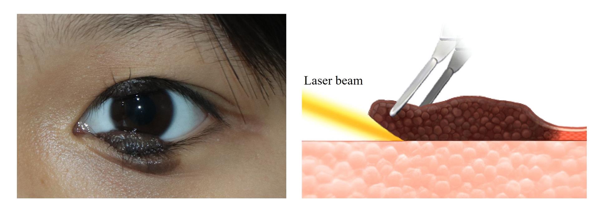
The ultrapulse CO₂ laser parameters were customized according to the specific characteristics of each lesion. The energy levels were adjusted between 100-200 mJ per pulse. The laser power was set within a range of 1.0-2.0 W, with a pulse duration of approximately 10 ms. The spot size was varied from 0.2 to 1 mm, depending on the lesion's size and location. The excision process commenced with a careful delineation of the treatment area. The laser was applied perpendicular to the skin surface, tracing the lesion's edge to create a well-defined base. The excision was performed along the marked base, with an additional 1 mm margin beyond the lesion's boundary to ensure complete removal and minimize the risk of recurrence.The depth of laser excision was set at 2 mm below the base of the divided nevus to ensure complete removal of the lesion. In cases where the lesion involved a depth greater than 2 mm, staged treatment was performed. (Figure 1)

Figure 1 Laser excision technique for divided nevus of the eyelid
The right panel offers a schematic illustration, highlighting the precise removal process of the divided nevus from the underlying dermal layers. 
The left panel provides an actual divided nevus photograph. It demonstrates the advantages of laser excision over traditional surgical methods, particularly in terms of achieving a smoother post-operative surface and thus reducing the risk of scarring.

The interval between treatment sessions was set at around one month, considering the time require for skin healing, which typically occurs over a 28-day period.

To prevent corneal thermal injury during the laser treatment, patients were instructed to keep their eyes closed, and in cases where the patient needed to keep their eyes open, a large metal blade was placed between the eyelid and the eyeball to protect the ocular surface. Further pathology examination were conducted to confirm pathological type.

After the procedure, The wound should be kept clean and dry. Tobramycin ointment and deproteinized calf blood extract eye gel were applied twice a day for 1-2 weeks.

Outcome Assessments

The study also recorded the number of treatment sessions required for each patient to achieve complete clearance of the divided nevus.

The efficacy of the ultrapulse CO₂ laser excision was primarily evaluated based on two key metrics: the clearance and recurrence rates, and the incidence of complications. To maintain objectivity, two independent plastic surgeons, who were not involved in the treatment, reviewed a series of photographs before and after the procedure for evaluation. In the event of a disagreement between the two reviewing ophthalmologists, a senior ophthalmologist was consulted to provide a final assessment.

Clearance rate evaluation
To quantify the degree of lesion clearance, a four-classification system was employed. The categories were defined as follows: less than 50% cleared, 50%- 75% cleared, 76%-95% cleared, and greater than 95% cleared. The proportion of the clearance was calculated by comparing the treated area to the total surface area of the lesion.

Recurrence rate assessment
Recurrence was defined as the reappearance of divided nevus within the previously treated area after the last treatment session. The development of new lesions outside the treated zones was not considered a recurrence.

Complication evaluation
Postoperative complications were closely monitored for 12 months. Adverse events were reviewed with postoperative photographs, including but not limited to, eyelash loss, eyelid trichiasis, ectropion, lagophthalmos, hypertrophic scarring, and pigmentary changes.

Patients' satisfaction survey
Patients’ satisfaction survey was conducted by a questionnaire to evaluate the subjective outcomes undergoing CO₂ laser excision for divided nevus (Supplementary File). The questionnaire consisted of five questions assessing various aspects, including the therapeutic effect, postoperative wound recovery, ocular function, cosmetic appearance, and comfort during the procedure. Each question was rated on a three-point scale: very satisfied (20 points), slightly satisfied (15 points), and dissatisfied (10 points). The total score ranged from 50 to 100 points, with scores ≥ 80 indicating very satisfied, 60 - 79 slightly satisfied, and < 60 dissatisfied.

Statistical Analysis

Statistical analysis was performed using SPSS version 23.0 software. Categorical variables were expressed as percentages. Statistical significance was set at a P-value < 0.05.

RESULTS

Efficacy outcomes

Our study included a total of 10 patients with divided nevus of the eyelid, consisting of 5 males and 5 females. The age range of the participants was 9 - 54 years, with a mean age of 23.7 years. All cases presented with unilateral involvement, and the eyelid margin was affected in all patients. Two cases had additional involvement, with one affecting the inner canthus and the other involving the outer canthus. The lesion diameters ranged from 25 to 50 mm and the size of the divided nevi were moderate (1.5 - 200 mm) in all cases, with lesion heights ranging from 0.3 to 6 mm (mean: 3.93 mm). All patients were verified as benign based on pathological examination.

Each patient received at least one laser excision session, with the number of treatments ranging from 1 to 5 sessions until complete clearance of the lesion was achieved. Among the ten individuals treated, the number of treatment sessions varied as follows: one patient required one session, two patients needed two sessions, three patients underwent three sessions, and four patients required more than three sessions to achieve the desired outcome, with one of these receiving five treatment sessions.

The lesions were completely cleared in all 10 patients, achieving a clearance rate of 100%. No recurrence was observed in any of the patients during the 12-month follow-up period. The demographic data and outcomes are detailed in Table 1. The resection size of the lesion for each treatment session and the follow-up time for each patient are provided in Supplementary Table 1.

Table 1. Patient demographics and treatment outcomes.


Patient NO.

Age (years)

Sex

Rt/Lt

Upper Size (mm)

Lower Size (mm)

Lesion diameters (mm)

Height (mm)

No. of sessions

Involvement

Clearance

Recurrence

Complications

Patient Satisfaction

1

22

M

R

25 x 20

13 x 15

38

2

3

Eyelid margin

100%

n/a

Partial absent eyelashes, hyperpigmentation

Very satisfied

2

9

M

L

15 x 6

15 x 3

30

3

1

Eyelid margin

100%

n/a

Partial absent eyelashes

Very satisfied

3

24

F

R

30 x 25

5 x 3

35

5

5

Eyelid margin

100%

n/a

Partial absent eyelashes, hyperpigmentation

Very satisfied

4

22

M

L

20 x 8

5 x 3

25

4

2

Eyelid margin

100%

n/a

Eyelid trichiasis

Very satisfied

5

11

M

R

12 x 13

17 x 26

39

0.3

4

Eyelid margin

100%

n/a

Partial absent eyelashes 

Very satisfied

6

16

F

L

15 x 9

20 x 18

35

5

2

Eyelid margin

100%

n/a

n/a

Very satisfied

7

17

M

R

15 x 22

5 x 3

25

2

3

Eyelid margin Medial canthus

100%

n/a

Partial absent eyelashes

Very satisfied

8

30

F

L

6 x 22

10 x 3

25

6

2

Eyelid margin, Lateral canthus

100%

n/a

Partial absent eyelashes

Very satisfied

9

54

F

L

20 x 10

18 x 22

38

6

4

Eyelid margin

100%

n/a

Partial absent eyelashes

Very satisfied

10

32

F

R

30 x 15

20 x 15

50

6

4

Eyelid margin

100%

n/a

Partial absent eyelashes

Very satisfied

M: Male; F: Female; R: Right; L: Left

Post-treatment complications

The eyelid margin remained continuous in all 10 patients, with acceptable irregularity forming a continuous semi-circular scar along the eyelid margin (diameter ≤ 2 mm). A reduction in eyelashes in the treatment area was observed in 8 patients. Two patients had hyperpigmentation, and one patient experienced mild upper eyelid trichiasis. No significant complications, such as ectropion, eyelid margin notching, corneal erosion, or marked scar hypertrophy, were observed.

Patient Satisfaction

Each patient scored above 80 points on the 5 questions on the questionnaire, indicating a rating of "very satisfied". The "very satisfied" rate reached 100% among the study participants.

Case Illustrations

The following examples illustrate the outcomes of ultrapulse CO₂ laser excision for divided nevus of the eyelid (Figure 2-3).

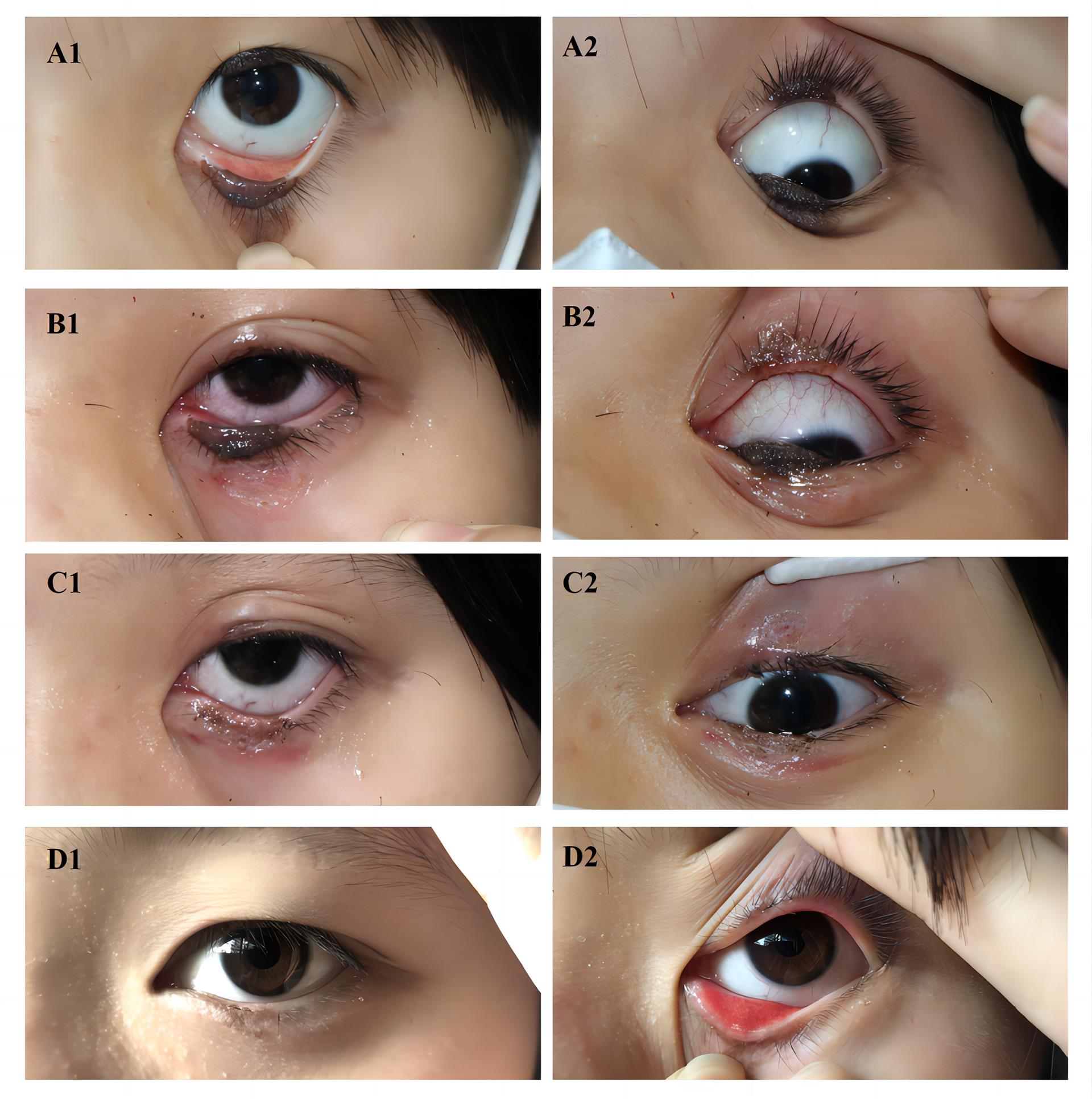
Case 1: A 16-year-old female patient was diagnosed with divided nevus of the eyelid on the left eye, presenting with a lesion diameter of 35 mm when the eye was closed. The upper eyelid lesion measured 15 mm × 9 mm, while the lower eyelid lesion measured 20 mm × 18 mm, with a maximum lesion height of 5 mm. The patient underwent a total of two staged laser treatments. Postoperatively, divided nevi were completely removed, and there was no evidence of recurrence or complications. (Figure 2)


Figure 2 Staged laser treatment of divided nevus of the eyelid in a 16-year-old female patient
(A1-A2) Preoperative appearance of the lesion with a diameter of 35 mm when the eye was closed, upper eyelid lesion measuring 15 x 9 mm, lower eyelid lesion measuring 20 x 18 mm, and a maximum lesion height of 5 mm. (B1-B2) After the first laser treatment, over 50% of the tumor was excised. (C1-C2) Following the second laser treatment, the last 50% tumor was completely eradicated. (D1-D2) After 12 months of follow-up, there was no evidence of recurrence or complications.

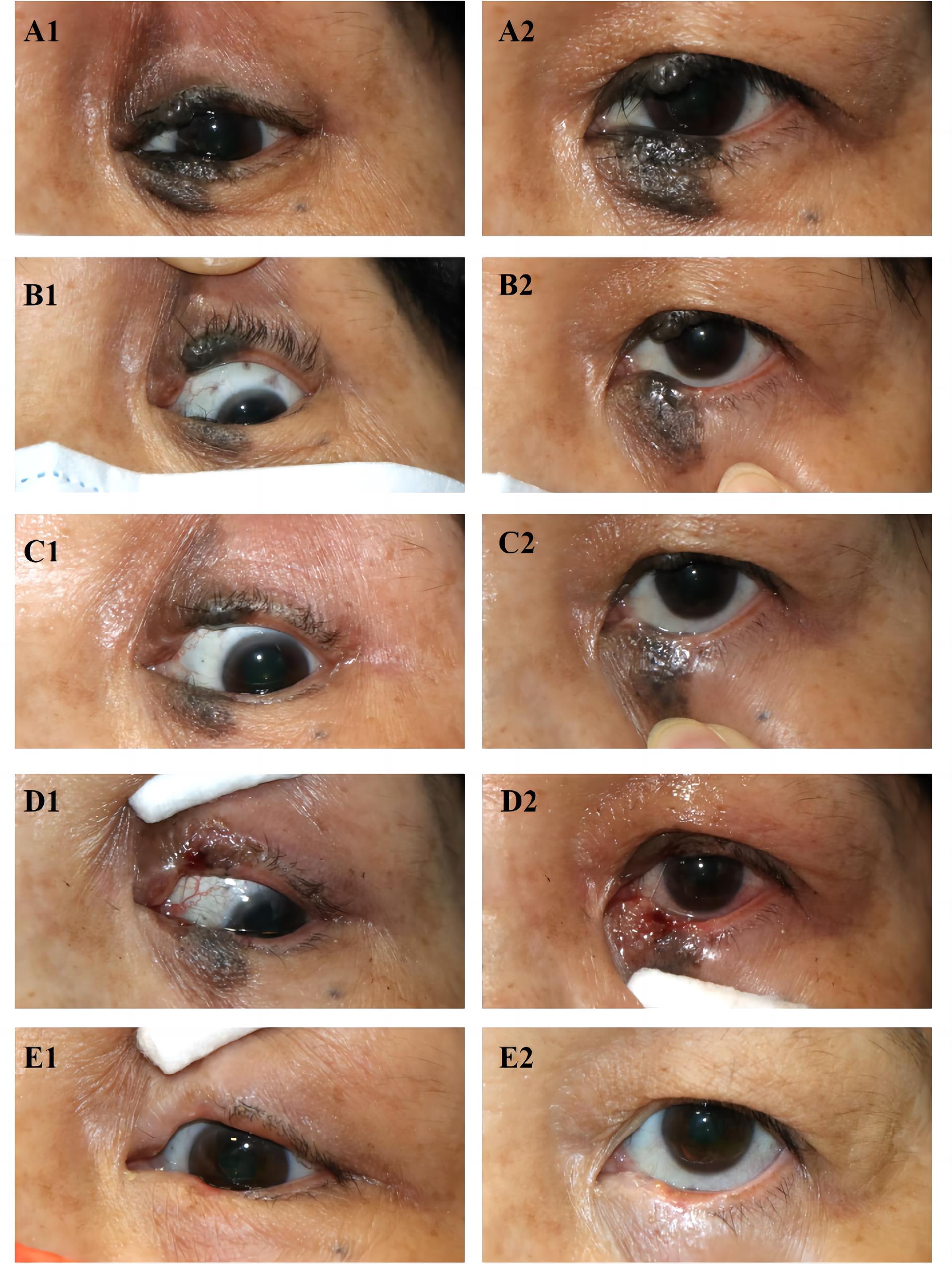
Case 2: A 54-year-old female patient was diagnosed with divided nevus of the eyelid on the left eye, presenting with a lesion diameter of 38 mm when the eye was closed. The upper eyelid lesion measured 20 mm × 10 mm, while the lower eyelid lesion measured 18 mm × 22 mm, with a maximum lesion height of 6 mm. The patient underwent a total of four staged laser treatments. After the surgery, the divided nevi were completely removed, resulting in a slight and acceptable irregularity in the eyelid margin and a partial reduction of eyelashes. (Figure 3)

Figure 3 Staged laser treatment of divided nevus of the eyelid in a 54-year-old female patient
(A1-A2) Preoperative appearance of the lesion with a diameter of 38 mm when the eye was closed, upper eyelid lesion measuring 20 mm × 10 mm, lower eyelid lesion measuring 18 mm × 22 mm, and a maximum lesion height of 6 mm. (B1-B2) After the first laser treatment, over 30% of the tumor was excised. (C1-C2) Following the second laser treatment, more than 50% of the tumor was removed. (D1-D2) After the third laser treatment, over 80% of the tumor was excised. (E1-E2) After the fourth laser treatment, the tumor was completely eradicated, with the lid margin removed and an acceptable minimal irregularity of the eyelash-bearing area and partial absence of eyelashes observed.

DISCUSSION

Our study provides a comprehensive evaluation of ultrapulse CO₂ laser excision for the treatment of divided nevus of the eyelid. The technique has proven to be highly effective, achieving a 100% clearance rate with no recurrence in all patients within 12 months. Postoperative complications were minimal, with partial sparse to absent eyelashes being the most common finding, two cases with hyperpigmentation, and only one case of mild upper eyelid trichiasis. Moreover, patients’ satisfaction with the functional and cosmetic outcomes was high.

For small lesions (diameter less than 25 mm), laser excision of divided nevus has a less complications compared to conventional laser ablation. In our study of 10 patients, complications were minor, such as partial sparse to absent eyelashes, while Cho et al.[16] reported a 13% incidence of eyelid margin discontinuity following laser ablation. One possible explanation is that the laser excision technique employs a direct incision from the base of the lesion, resulting in a smoother excision surface and causing less damage to the surrounding tissues and eyelid margin, thereby reducing the risk of complications.

The advantage of laser excision lies in its ability to treat large lesions, particularly those exceeding 25 mm in diameter, which were previously treated primarily through surgical excision. Our study demonstrates that the therapeutic efficacy of ultrapulse CO₂ laser excision is comparable to that of traditional surgical treatment. This technique effectively minimizes intraoperative bleeding, shortens the duration of the procedure, and decreases postoperative recovery time. Moreover, it has fewer complications than surgical excision which can lead to severe lower eyelid ectropion (15% reported by Zloto et al.[20]) and insufficient eyelid closure (one case reported by Hu et al.[21]). Also, skin grafts after surgery in Asian patients can result in significant hyperpigmentation, compromising the cosmetic outcome.[21] Thus, laser excision provides a more precise and less invasive alternative treatment for medium to large-sized divided nevus.

Ultrapulse CO2 laser excision technique is also specifically beneficial for removing lesions adjacent to the lacrimal punctum. Surgical excision of lesions in this area can be complex, and lacrimal duct reconstruction may be required, with a higher risk of postoperative epiphora (excessive tearing). While the clear vision and precise excision provided by the CO2 laser allow for maximal preservation of the lacrimal punctum function, helping to avoid damage even when the lesion is in the inner canthus. In our study, one case involved a lesion in the inner canthus, adjacent to the lacrimal region. After laser excision, the patient's punctum remained intact, and no secondary epiphora was observed.

There are some treatment considerations. When treating divided nevus involving critical eyelid structures, excessive tissue removal may cause severe functional impairments. To avoid this, a staged treatment approach, particularly around the lid margin, can be employed to prevent excessive tissue loss and subsequent scarring. Additionally, when treating lesions in the eyelid margin area, it is crucial to protect the cornea from laser energy damage by placing a large metal shield between the eye globe and the eyelid to prevent inadvertent corneal exposure to the laser beam. For the timing of surgery, divided nevus of the upper eyelid can cause mechanical ptosis due to their weight and excess tissue, potentially leading to irreversible visual developmental disorders.[18-22] If surgical treatment is planned, it should be performed as early as possible to prevent visual deficit and uncorrectable amblyopia.[23-24] Finally, it is crucial to note that even minor residual lesions pose a potential risk of recurrence or malignant transformation, underscoring the necessity for prolonged monitoring.[21-25]

The limitations of this study include a relatively small sample size and its single-center nature. Moreover, CO2 laser excision may require multiple sessions for larger divided nevi, potentially increasing time and financial costs for patients. Future research should involve larger, multi-center studies to further validate the efficacy and safety of ultrapulse CO₂ laser excision for the treatment of divided nevus of the eyelid.

CONCLUSIONS

In conclusion, CO₂ laser excision is a simple, precise, and effective treatment modality. It can help simplify the treatment process, achieve excellent cosmetic results, and avoid the need for skin grafting. This technique represents a promising approach for the management of large divided nevus of the eyelid.

Correction notice

None of the authors has any conflicts of interest to disclose.

Acknowledgement

We acknowledge the support of the State Key Laboratory of Ophthalmology, Zhongshan Ophthalmic Center, Sun Yat-sen University. We thank our colleagues for their contributions to this study.

Author Contributions

(Ⅰ) Conception and design: ZM and XL
(Ⅱ) Administrative support: XL
(Ⅲ) Provision of study materials or patients: DW, YQ and ZL
(Ⅳ) Collection and assembly of data: DW, WG, XL
(Ⅴ) Data analysis and interpretation: WG and XL
(Ⅵ) Manuscript writing: All authors
(Ⅶ) Final approval of manuscript: All authors

Fundings

None

Conflict of Interests

None of the authors has any conflicts of interest to disclose. All authors have declared in the completed the ICMJE uniform disclosure form.

Patient consent for publication

None

Ethical Statement

All experimental protocols were approved by the ethics committee of Zhongshan Ophthalmic Center (2023KYPJ286 Guangzhou, China).

Provenance and Peer Review

This article was a standard submission to our journal. The article has undergone peer review with our anonymous review system.

Data Sharing Statement

None

Open Access Statement

This is an Open Access article distributed in accordance with the Creative Commons Attribution-NonCommercial-NoDerivs 4.0 International License (CC BY-NC-ND 4.0), which permits the non-commercial replication and distribution of the article with the strict proviso that no changes or edits are made and the original work is properly cited (including links to both the formal publication through the relevant DOI and the license). See: https://creativecommons.org/ licenses/by-nc-nd/4.0/.

Supplementary table 1 Sizes of Lesions Removed in Each Treatment Session

Patient NO.

Follow up times (m)

No. of sessions

Lesion size removed at each session (mm)

1st

2nd

3th

4th

5th

1

12.2

3

10 x 20 (Upper)

7 x 13 (Lower)

10 x 20 (Upper)

8 x 13 (Lower)

5 x 20 (Upper)


-

-

2

13.2

1

15 x 6 (Upper)

15 x 3 (Lower)

-

-

-

-

3

12.5

5

10 x 15 (Upper)

5 x 3 (Lower)

10 x 10 (Upper)


10 x 10 (Upper)


10 x 10 (Upper)


10 x 10 (Upper)


4

12.0

2

10 x 8 (Upper)

5 x 3 (Lower)

10 x 8 (Upper)

-


-

5

12.1

4

12 x 6 (Upper)

15 x 13 (Lower)

12 x 7 (Upper)

15 x 13 (Lower)


2 x 13 (Lower)


2 x 13 (Lower)

-

6

11.7

2

9 x 5 (Upper)

20 x 10 (Lower)

10 x 9 (Upper)

20 x 8 (Lower)

-

-

-

7

11.9

3

15 x 7 (Upper)

5 x 3 (Lower)

15 x 7 (Upper)


15×8 (Upper)


-

-

8

12.0

2

6 x 10 (Upper)

10 x 3 (Lower)

6 x 12 (Upper)


-

-

-

9

12.8

4

10 x 10 (Upper)

9 x 11 (Lower)

10 x 5 (Upper)

9 x 11 (Lower)

10 x 5 (Upper)

9 x 11 (Lower)


9 x 11 (Lower)

-

10

11.8

4

10 x 10 (Upper)

15 x 10 (Lower)

14 x 10 (Upper)

15 x 10 (Lower)

16 x 10 (Upper)


10 x 5 (Upper)


-


1、Desai SC, Walen S, Holds JB, et al. Divided nevus of the eyelid: review of embryology, pathology and treatment. AmJOtolaryngol. 2013, 34(3): 223-229. DOI: 10.1016/ j.amjoto.2013.01.004.Desai SC, Walen S, Holds JB, et al. Divided nevus of the eyelid: review of embryology, pathology and treatment. AmJOtolaryngol. 2013, 34(3): 223-229. DOI: 10.1016/ j.amjoto.2013.01.004.
2、Papadopoulos O. Divided nevus of the eyelid. PlastReconstrSurg. 1991, 88(2): 331-333. DOI: 10.1097/00006534-199108000-00028.Papadopoulos O. Divided nevus of the eyelid. PlastReconstrSurg. 1991, 88(2): 331-333. DOI: 10.1097/00006534-199108000-00028.
3、Yap LH, Earley MJ. The panda naevus: management of synchronous upper- and lower-eyelid pigmented naevi. BrJPlastSurg. 2001, 54(2): 102-105. DOI: 10.1054/ bjps.2000.3514.Yap LH, Earley MJ. The panda naevus: management of synchronous upper- and lower-eyelid pigmented naevi. BrJPlastSurg. 2001, 54(2): 102-105. DOI: 10.1054/ bjps.2000.3514.
4、Ghosh YK, Abu-Ain M, Ahluwalia HS. One stage management of a giant kissing naevus. Orbit. 2010, 29(3): 174-175. DOI: 10.3109/01676830903488811.Ghosh YK, Abu-Ain M, Ahluwalia HS. One stage management of a giant kissing naevus. Orbit. 2010, 29(3): 174-175. DOI: 10.3109/01676830903488811.
5、Marghoob AA, Borrego JP, Halpern AC. Congenital melanocytic nevi: treatment modalities and management options. SeminCutanMedSurg. 2007, 26(4): 231-240. DOI: 10.1016/j.sder.2008.03.007.Marghoob AA, Borrego JP, Halpern AC. Congenital melanocytic nevi: treatment modalities and management options. SeminCutanMedSurg. 2007, 26(4): 231-240. DOI: 10.1016/j.sder.2008.03.007.
6、Ashfaq I, Kyprianou I, Ahluwalia H. A large kissing (divided) naevus presenting with complete mechanical ptosis and lower lid ectropion. JPlastReconstrAesthetSurg. 2009, 62(5): e87-e88. DOI: 10.1016/j.bjps.2008.08.029.Ashfaq I, Kyprianou I, Ahluwalia H. A large kissing (divided) naevus presenting with complete mechanical ptosis and lower lid ectropion. JPlastReconstrAesthetSurg. 2009, 62(5): e87-e88. DOI: 10.1016/j.bjps.2008.08.029.
7、Yildirim N, Sahin A, Erol N. The surgical treatment of congenital divided nevus of the eyelid: a case report and review of the literature. IntOphthalmol. 2009, 29(1): 45-47. DOI: 10.1007/s10792-007-9163-5.Yildirim N, Sahin A, Erol N. The surgical treatment of congenital divided nevus of the eyelid: a case report and review of the literature. IntOphthalmol. 2009, 29(1): 45-47. DOI: 10.1007/s10792-007-9163-5.
8、Camargo CP, Saliba M, Saad EA, et al. Treatments of palpebral congenital melanocytic nevus: a systematic review. Acta CirBras. 2023, 38: e384823. DOI: 10.1590/ acb384823.Camargo CP, Saliba M, Saad EA, et al. Treatments of palpebral congenital melanocytic nevus: a systematic review. Acta CirBras. 2023, 38: e384823. DOI: 10.1590/ acb384823.
9、Liu J, Sun J, Wang Z, et al. Treatment of divided eyelid nevus with orbicularis oculi myocutaneous flap: report of 17 cases. AnnPlastSurg. 2020, 85(6): 626-630. DOI: 10.1097/ SAP.0000000000002507.Liu J, Sun J, Wang Z, et al. Treatment of divided eyelid nevus with orbicularis oculi myocutaneous flap: report of 17 cases. AnnPlastSurg. 2020, 85(6): 626-630. DOI: 10.1097/ SAP.0000000000002507.
10、Li J, Wang Y, Li D, et al. Application of allogeneic sclera combined with tarso-conjunctival flap in total excision of divided eyelid nevus. JPlastReconstrAesthetSurg. 2022, 75(10): 3789-3794. DOI: 10.1016/j.bjps.2022.06.028.Li J, Wang Y, Li D, et al. Application of allogeneic sclera combined with tarso-conjunctival flap in total excision of divided eyelid nevus. JPlastReconstrAesthetSurg. 2022, 75(10): 3789-3794. DOI: 10.1016/j.bjps.2022.06.028.
11、 Papadopoulos O, Chrisostomidis C, Konofaos P, et al. Divided naevus of the eyelid, seven cases. JPlastReconstrAesthetSurg. 2007, 60(3): 260-265. DOI: 10.1016/j.bjps.2005.12.032. Papadopoulos O, Chrisostomidis C, Konofaos P, et al. Divided naevus of the eyelid, seven cases. JPlastReconstrAesthetSurg. 2007, 60(3): 260-265. DOI: 10.1016/j.bjps.2005.12.032.
12、Jacobs SM, Couch SM, Custer PL. Divided eyelid nevus: a lid-sparing, staged surgical approach. AmJOphthalmol. 2013, 156(4): 813-818. DOI: 10.1016/j.ajo.2013.05.032.Jacobs SM, Couch SM, Custer PL. Divided eyelid nevus: a lid-sparing, staged surgical approach. AmJOphthalmol. 2013, 156(4): 813-818. DOI: 10.1016/j.ajo.2013.05.032.
13、 Naik MN, Ali MJ, Kaliki S. Epithelial stripping for divided (kissing) nevus of the eyelid: aminimally invasive technique. DermatolSurg. 2020, 46(6): 842-844. DOI: 10.1097/DSS.0000000000001937. Naik MN, Ali MJ, Kaliki S. Epithelial stripping for divided (kissing) nevus of the eyelid: aminimally invasive technique. DermatolSurg. 2020, 46(6): 842-844. DOI: 10.1097/DSS.0000000000001937.
14、Klapper S, Patrinely J. Management of cosmetic eyelid surgery complications. SeminPlastSurg. 2007, 21(1): 80-93. DOI: 10.1055/s-2007-967753.Klapper S, Patrinely J. Management of cosmetic eyelid surgery complications. SeminPlastSurg. 2007, 21(1): 80-93. DOI: 10.1055/s-2007-967753.
15、Carniciu AL, Jovanovic N, Kahana A. Eyelid complications associated with surgery for periocular cutaneous malignancies. Facial PlastSurg. 2020, 36(2): 166-175. DOI: 10.1055/s-0040-1709515.Carniciu AL, Jovanovic N, Kahana A. Eyelid complications associated with surgery for periocular cutaneous malignancies. Facial PlastSurg. 2020, 36(2): 166-175. DOI: 10.1055/s-0040-1709515.
16、Cho HJ, Lee W, Jeon MK, et al. Staged mosaic punching excision of a kissing nevus on the eyelid. Aesthetic PlastSurg. 2019, 43(3): 652-657. DOI: 10.1007/s00266-019- 01362-0.Cho HJ, Lee W, Jeon MK, et al. Staged mosaic punching excision of a kissing nevus on the eyelid. Aesthetic PlastSurg. 2019, 43(3): 652-657. DOI: 10.1007/s00266-019- 01362-0.
17、Zeng Y. Divided nevus of the eyelid: successful treatment with CO2 laser. JDermatologTreat. 2014, 25(4): 358-359. DOI: 10.3109/09546634.2012.756970.Zeng Y. Divided nevus of the eyelid: successful treatment with CO2 laser. JDermatologTreat. 2014, 25(4): 358-359. DOI: 10.3109/09546634.2012.756970.
18、Suzuki A, Yotsuyanagi T, Yamashita K, et al. Reconstruction of the congenital divided nevus of the eyelids and proposal of new classification. PlastReconstrSurgGlobOpen. 2019, 7(6): e2283. DOI: 10.1097/GOX.0000000000002283.Suzuki A, Yotsuyanagi T, Yamashita K, et al. Reconstruction of the congenital divided nevus of the eyelids and proposal of new classification. PlastReconstrSurgGlobOpen. 2019, 7(6): e2283. DOI: 10.1097/GOX.0000000000002283.
19、Nagamatsu S, Sasaki A, Uchiki T, et al. An angled mirror device facilitates measurement of skin lesion height. PlastReconstrSurgGlobOpen. 2020, 8(11): e3233. DOI: 10.1097/GOX.0000000000003233.Nagamatsu S, Sasaki A, Uchiki T, et al. An angled mirror device facilitates measurement of skin lesion height. PlastReconstrSurgGlobOpen. 2020, 8(11): e3233. DOI: 10.1097/GOX.0000000000003233.
20、Zloto O, Landau Prat D, Katowitz JA, et al. The surgical management and outcomes of kissing nevi of the eyelids. Eye. 2023, 37(14): 3015-3019. DOI: 10.1038/s41433-023- 02463-6.Zloto O, Landau Prat D, Katowitz JA, et al. The surgical management and outcomes of kissing nevi of the eyelids. Eye. 2023, 37(14): 3015-3019. DOI: 10.1038/s41433-023- 02463-6.
21、Hu L, Jin Y, Tremp M, et al. Reconstruction of a large divided nevus of the eyelid. DermatolSurg. 2017, 43(12): 1483-1486. DOI: 10.1097/dss.0000000000000905.Hu L, Jin Y, Tremp M, et al. Reconstruction of a large divided nevus of the eyelid. DermatolSurg. 2017, 43(12): 1483-1486. DOI: 10.1097/dss.0000000000000905.
22、Zhu L, Qiao Q, Liu Z, et al. Treatment of divided eyelid nevus with island skin flap: report of ten cases and review of the literature. Ophthalmic PlastReconstrSurg. 2009, 25(6): 476-480. DOI: 10.1097/IOP.0b013e3181b81eb7.Zhu L, Qiao Q, Liu Z, et al. Treatment of divided eyelid nevus with island skin flap: report of ten cases and review of the literature. Ophthalmic PlastReconstrSurg. 2009, 25(6): 476-480. DOI: 10.1097/IOP.0b013e3181b81eb7.
23、Ritz M, Corrigan B. Congenital melanocytic nevi of the eyelids and periorbital region. PlastReconstrSurg. 2010, 125(5): 1568-1569. DOI: 10.1097/PRS.0b013e3181d512aa.Ritz M, Corrigan B. Congenital melanocytic nevi of the eyelids and periorbital region. PlastReconstrSurg. 2010, 125(5): 1568-1569. DOI: 10.1097/PRS.0b013e3181d512aa.
24、Zhang X, Tang W, Yi L, et al. Divided eyelid nevus: surgical repair discussion and case reports from northwestern China. PlastSurg. 2020, 28(4): 249-253. DOI: 10.1177/2292550320928559.Zhang X, Tang W, Yi L, et al. Divided eyelid nevus: surgical repair discussion and case reports from northwestern China. PlastSurg. 2020, 28(4): 249-253. DOI: 10.1177/2292550320928559.
25、%20Lema%C3%AEtre%20S%2C%20Gardrat%20S%2C%20Vincent-Salomon%20A%2C%20et%20al.%20Malignant%20%0Atransformation%20of%20a%20multi-operated%20divided%20nevus%20of%20the%20%0Aeyelids.%20OculOncolPathol.%202018%2C%204(2)%3A%20112-115.%20DOI%3A%20%0A10.1159%2F000479069.%20Lema%C3%AEtre%20S%2C%20Gardrat%20S%2C%20Vincent-Salomon%20A%2C%20et%20al.%20Malignant%20%0Atransformation%20of%20a%20multi-operated%20divided%20nevus%20of%20the%20%0Aeyelids.%20OculOncolPathol.%202018%2C%204(2)%3A%20112-115.%20DOI%3A%20%0A10.1159%2F000479069.
上一篇
下一篇
出版者信息
目录