您的位置: 首页 > 2024年12月 第1卷 第4期 > 文字全文
2023年7月 第38卷 第7期11
目录

Evaluating FFA and UWF SS-OCTA abnormalities in X-linked retinoschisis: a cross-sectional study of 46 eyes

Evaluating FFA and UWF SS-OCTA abnormalities in X-linked retinoschisis: a cross-sectional study of 46 eyes

来源期刊: Eye Science | 2024年12月 第1卷 第4期 398-411 发布时间:2024-12-20 收稿时间:2024/12/17 9:23:38 阅读量:97
作者:
关键词:
X-linked retinoschisis retinal vascular changes FFA ultrawide field swept source OCT peripheral schisis
DOI:
10.12419/es24080801
Received date:
2024-08-08 
Accepted date:
2024-11-28 
Published online:
2024-12-20 
Aims: This study describes vascular abnormalities in X-linked retinoschisis (XLRS) using fundus fluorescein angiography (FFA) and ultra-widefield swept-source optical coherence tomography angiography (UWF SS-OCTA) to better understand the disease's vascular features and impact. Methods: A retrospective cross-sectional study was conducted on 26 XLRS patients (46 eyes). A comprehensive ophthalmic examination was performed, including FFA and UWF SS-OCTA. FFA abnormalities were divided into peripheral schisis-associated and optic disc-associated types. Results: The mean age of patients was 11.3±6.5 years. Macular schisis appeared in 97.8% of eyes, peripheral schisis in 89.1%, and peripheral bullous schisis (PBS) in 67.39%. Major vascular changes identified by FFA included dendritic capillary dilation/leakage (91.3%), internal residual vessel leakage (78.3%), and capillary dropout/ischemia (71.7%). Minor changes included zonal retinal pigment epithelium (RPE) proliferation (6.5%), bridging vessels (4.4%), and capillary sheathing (4.4%). peripapillary choroidal neovascularization (PPCNV) was noted in 10.9% and situs inversus of optic disc in 13.0% of eyes. Additionally, situs inversusof optic disc and zonal RPE proliferation were novel findings. Major FFA changes correlated with broader PBS (= 0.045) (P < 0.001) (= 0.003). Clock hours of PBS were significant predictors for internal residual vessel leakage (OR = 0.30, P = 0.03). No significant correlation was found between gene mutation type and FFA abnormalities(= 1.000)(= 0.539). Conclusions: This study highlighted the significant prevalence (95.7%) of vascular abnormalities in XLRS and emphasized the importance of combining FFA with UWF SS-OCTA for comprehensive evaluation, enhancing the understanding of XLRS pathophysiology and aiding in targeted treatment approaches.
Aims: This study describes vascular abnormalities in X-linked retinoschisis (XLRS) using fundus fluorescein angiography (FFA) and ultra-widefield swept-source optical coherence tomography angiography (UWF SS-OCTA) to better understand the disease's vascular features and impact. Methods: A retrospective cross-sectional study was conducted on 26 XLRS patients (46 eyes). A comprehensive ophthalmic examination was performed, including FFA and UWF SS-OCTA. FFA abnormalities were divided into peripheral schisis-associated and optic disc-associated types. Results: The mean age of patients was 11.3±6.5 years. Macular schisis appeared in 97.8% of eyes, peripheral schisis in 89.1%, and peripheral bullous schisis (PBS) in 67.39%. Major vascular changes identified by FFA included dendritic capillary dilation/leakage (91.3%), internal residual vessel leakage (78.3%), and capillary dropout/ischemia (71.7%). Minor changes included zonal retinal pigment epithelium (RPE) proliferation (6.5%), bridging vessels (4.4%), and capillary sheathing (4.4%). peripapillary choroidal neovascularization (PPCNV) was noted in 10.9% and situs inversus of optic disc in 13.0% of eyes. Additionally, situs inversusof optic disc and zonal RPE proliferation were novel findings. Major FFA changes correlated with broader PBS (= 0.045) (P < 0.001) (= 0.003). Clock hours of PBS were significant predictors for internal residual vessel leakage (OR = 0.30, P = 0.03). No significant correlation was found between gene mutation type and FFA abnormalities(= 0.539). Conclusions: This study highlighted the significant prevalence (95.7%) of vascular abnormalities in XLRS and emphasized the importance of combining FFA with UWF SS-OCTA for comprehensive evaluation, enhancing the understanding of XLRS pathophysiology and aiding in targeted treatment approaches.

HIGHLIGHTS

• Vascular abnormalities were prevalent in 95.7% of XLRS cases with novel findings of optic disc situs inversus and zonal RPE proliferation.

• Combination of FFA and UWF SS-OCTA enabled comprehensive evaluation of XLRS, particularly its vascular features.

• Findings enhance understanding of XLRS pathophysiology and support targeted treatment development for improved patient outcomes.

INTRODUCTION

X-linked retinoschisis (XLRS) is an early-onset inherited retinal disorder predominantly affecting males. It is primarily caused by mutations in the RS1 gene and is characterized by intraretinal splitting in the macular and/or periphery.[1] The estimated prevalence ranges from 1:5,000 to 1:25,000.[2] Although the exact pathophysiology of XLRS has not been fully elucidated, initial theories involved Müller cell dysfunction.[3] However, identification of more than 200 different mutations in the retinoschisin (RS1) gene suggests that the pathology rooted in the adhesive protein properties within photoreceptors and bipolar cells.[4-6] Clinically, age of presentation in XLRS ranges from 3 months to school age with features that include strabismus, nystagmus, and decreased vision.[7-8] Complications include vitreous hemorrhage (VH) (reported prevalence; 4 to 40%), retinal detachment (RD) (0-22%) of rhegmatogenous, traction, or exudative origin, retinal vascular abnormalities with or without exudation (6-11%), macular holes, macular folds, and neovascular glaucoma ect.[4,9-12]

Advances in retinal imaging contribute significantly to both the diagnosis and understanding of the disease process. Optical coherence topography (OCT) accurately demonstrates the characteristic intraretinal splitting observed in XLRS-affected patients.[3,13]However, fundus fluorescein angiography (FFA), which traditionally reveals normal findings without apparent vascular leakage, has received less emphasis in XLRS diagnosis.[3,14] Interestingly, vascular changes were initially reported using FFA by Green and Jampolin 197915. Previous studies have documented various changes including vascular sheathing, dendritic vascular opacification, aneurysm formation in the schisis cavity, vascular tortuosity, capillary nonperfusion, and retinal neovascularization (RNV), not only within the schisis areas but also in nonschitic retina.[1,11,15-18]

This study aimed to provide a comprehensive description of vascular abnormalities in patients with XLRS by combining FFA with ultra-widefield sweptsource optical coherence tomography (UWF SSOCT) and SS-OCT angiography (OCTA), with the hope of elucidating their pathophysiology and potential impact. The objective of this research is to broaden the understanding of the vascular component in XLRS and facilitate the exploration of novel and earlier treatment approaches for XLRS.

MATERIALS AND METHODS

This was a retrospective cross-sectional study of the FFA and UWF SS-OCT(A) in patients with XLRS.26 subjectsdiagnosed with XLRS by clinical presentations and genetic testing were included from the retinal department at Zhongshan Ophthalmic Center between November 2023 and March 2024. Eyes with poor-quality imaging data or history of intraocular surgery were excluded. The research was conducted in compliance with the tenets of the Declaration of Helsinki and gained approval from the Institutional Review Board of Zhongshan Ophthalmic Center, Sun Yat-Sen University. Written informed consent was acquired from all participants, with additional parental consent for individuals under 18.

All participants underwent comprehensive ocular assessments. Best corrected visual acuity (BCVA) was determined using a Snellen chart and subsequently converted to the logarithm of the minimum angle of resolution (logMAR). Evaluations included slit-lamp examination, fundus photography using either a Clarus 500 (Carl Zeiss Meditec AG, Jena, Germany) or an Optos California FA (Gaush Meditech Ltd, Shanghai, China). FFA was conducted using either the Spectralis device (Heidelberg Engineering, Heidelberg, Germany) or Optos California FA. Additionally, UWF SS-OCT and SS-OCTA were performed using a VG200D (Intrasight, Henan, China) or BM400K BMizar system (Toward Pi Medical Technology Co., Ltd, Beijing, China). Image analysis was independently conducted by two ophthalmologists (L.Z and X.Z), with any discrepancies resolved through consultation with a senior ophthalmologist (X.D.).

The study recorded demographic data, genetic diagnosis, and details of clinical presentation, Changes observed in FFA, along with findings from fundus photography and UWF SS-OCT(A), were independently reviewed by two observers (Z.L and Z.X). The UWF SS-OCT(A) assessment involved evaluating macular and peripheral schisis, with particular attention to areas corresponding to characteristic features identified in FFA. Abnormalities detected through FFA were reclassified into two groups based on their locations. 1) Abnormalities located in and around the peripheral retinoschisis. These included dendritic capillary dilation/leakage, internal residual vessel leakage, capillary dropout/ischemia,[15,19]zonal proliferation of retinal pigment epithelium (RPE), bridging vessels, and the redefinition of previously studied ‘dendritic vascular opacification’[4,15,18] as ‘capillary sheathing’. 2) Abnormalities associated with optic disc:situs inversus of optic disc and retinal vessels, peripapillary choroidal neovascularization (PPCNV).

STATISTICAL ANALYSIS

The statistics was performed using the IBM SPSS (version 25.0, SPSS, Chicago, Illinois, USA). Continuous variables were reported as mean±standard deviation (Mean±SD), while the categorical variables were presented as number and percentage (n, %). The level of agreements between the two ophthalmologists were assessed using Cohen’s κ analysis.[20] Comparisons between groups were performed using the Student’s t-test for continuous variables and the Chi-square test or Fisher’s exact test for categorical variables. Linear regression was applied to model the relationship between continuous dependent variables and multiple independent variables, allowing for the assessment of the impact of each predictor. Logistic regression was utilized for categorical dependent variables, enabling the evaluation of the probability of occurrence of a particular outcome based on the independent variables. A significance level of< 0.05 was used for all statistical tests.

RESULTS

Demographic characteristics and clinical findings in patients with XLRS

In this study, 26 subjects (46 eyes) diagnosed with XLRS were included.Eyes with poor quality of FFA images due to vitreous opacity (1 eye) and those with a history of vitrectomy (5 eyes) were excluded. The mean age was 11.3±6.5 years (range: 5 to 33 years). The mean BCVA (logMAR) was 0.78±0.48, ranging from 0.15 to 2.60. Macular schisis was observed in 45 eyes(97.8%), while one eye (2.2%) exhibited macular hole (Supplementary material, Figure 1). Peripheral schisis was noted in 41eyes (89.1%), with peripheral bullous schisis (PBS) observed in 31 eyes(67.39%). Among these 31 eyes, involvement averaged 3.41±2.85 clock hours (range: 0.00 to 8.50).

FFA and OCTA findings in patients with XLRS

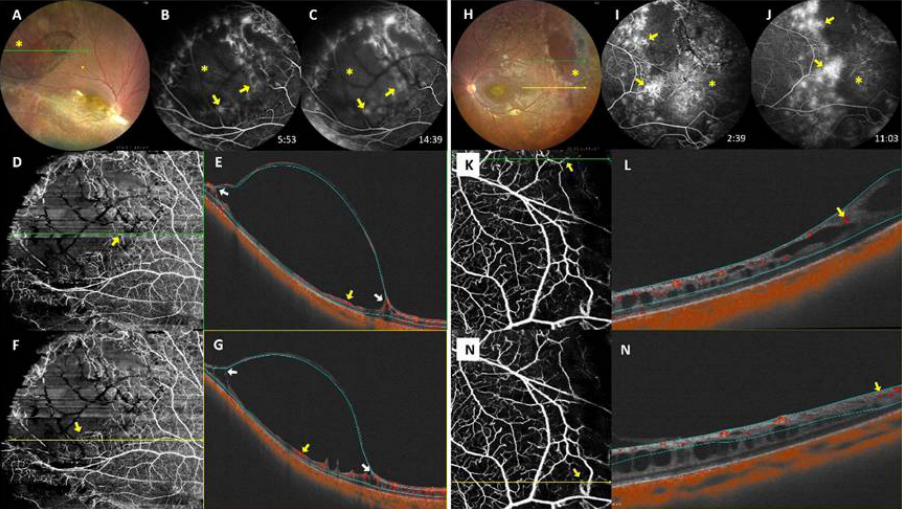
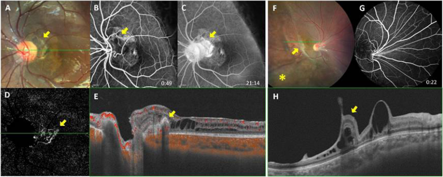
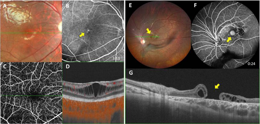
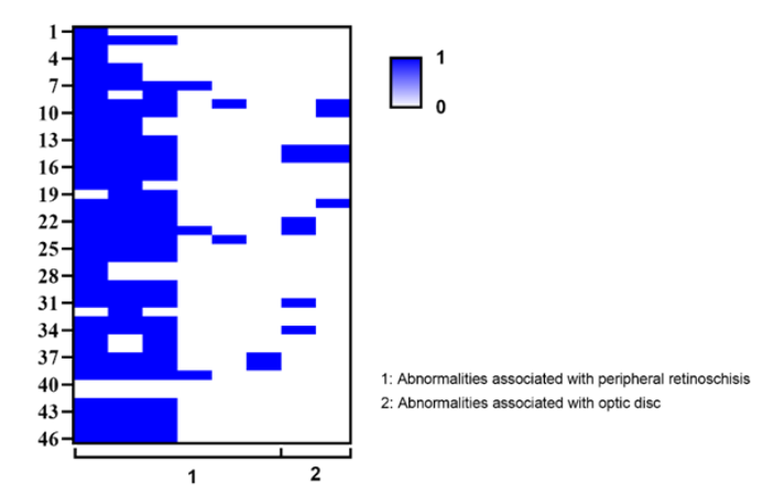
Overall, inter-observer agreement was 0.850 (Cohen’s kappa) .Our study identified six types of FFA changes in patients with XLRS. Dendritic capillary dilation/leakage was present in 42 eyes (91.3%) (Figure 1, A-N) (Figure2, O-R), internal residual vessel leakage in 36 eyes (78.3%) (Figure 2, A-D), and capillary dropout/ischemia in 33 eyes (71.7%) (Figure 1, B-C and I-J) (Figure 2, B). Other findings included zonal RPE proliferation in 3 eyes (6.5%) (Figure 3, F-H) across 3 patients, bridging vessels in 2 eyes(4.4%) (Figure 2, O-Q and S) across 2 patients, capillary sheathing in 2 eyes (4.4%) (Figure 2, I-N) across 1 patient. PPCNV were noted in 5 eyes (10.9%)(Figure 3, A-E) across 3 patients and situs inversus of the optic disc in 6 eyes (13.0%) (Figure 3, F-H) across4 patients. We categorized dendritic capillary dilation/leakage, internal residual vessel leakage, and capillary dropout/ischemia as major vascular changes, and zonal RPE proliferation, bridging vessels, and capillary sheathing as minor changes as well. Among these, marginal dendritic capillary dilation/leakage(Figure 1, A-G), typically observed at the inner border of the peripheral schisis, manifested as dendritiform abnormal capillary dilation and perivascular leakage evident in FFA. The corresponding retinoschisis observed in OCTA involved not only the outer nuclear layer (ONL) or outer retina but also the retinal nerve fiber layer (RNFL).Additionally, dendritic capillary dilation/leakage within the peripheral flat retinoschisis presented FFA leakage traceable back to inner retinal vessels(Figure 1, H-N). The spatial distribution of them across the retina is presented in Supplementary material, Figure 2.
Figure 1 Dendritic capillary dilation/leakage at inner border of bullous peripheral schisis (A-G)and in peripheral flat schisis (H-N)
(A) Large bullous peripheral schisis (yellow asterisk)insuperior temporal retina. (B-C) On FFA, marginal dendritic capillary dilation/leakage (yellow arrow) at the inner border of bullous peripheral schisis, and capillary dropout/ischemia in the central retina of bullous peripheral schisis (yellow asterisk). (D-G) Corresponding flow signals of the deep capillary plexus were visible on UWF SS-OCTA (yellow arrow).(E) (G)The corresponding retinoschisis observed on UWF SS-OCTA B-scan involved not only the outer nuclear layer (ONL) or outer retina but also the RNFL(white arrow).(H) Peripheral schisis (yellow asterisk)inthe temporal and superior retina. (I-J) On FFA, dendritic capillary dilation/leakage (yellow arrow) in the areas of temporal peripheral schisis, and capillary dropout/ischemia in the temporal peripheral retina (yellow asterisk). (K-N) Internal retinal vessel flow signals visible on UWF SS-OCTA (yellow arrow), with structural disorganization or atrophy of inner retinal layers at the peripheral flat schisis.
Figure 2 Other FFA findings associated with peripheral schisis
(A) Large bullous peripheral schisis (yellow asterisk) and residual internal residual vessel(yellow arrow) insuperior temporal retina. (B) On FFA, there were leakage from the residual internal vessel (yellow arrow) and capillary dropout/ischemia(yellow asterisk)in the bullous peripheral schisis.(C-D) Corresponding flow signals of inner retinal vessels visible on UWF SS-OCTA (yellow arrow). (E)An arcuate band of pigment abnormalitywasobserved in the posterior pole (yellow arrow), running parallel to the border of the inferotemporal peripheral bullous schisis (white arrow). (F) On FFA, this appeared as an arc-shaped background fluorescence masking (yellow arrow). (G-H) RPE proliferation was visible at the corresponding location on UWF SS-OCT B-scan (yellow arrow). (I) Ring-shaped or arcuate clustered distribution of capillary sheathing in the temporal periphery (yellow arrow). ( J-K) Capillary sheathing appeared as dendritic-like capillary dilation with leakage on FFA (yellow arrow). (L-N) UWF SS-OCTA showed no retinoschisis in the corresponding area, with high reflectivity material around the vessels (yellow arrow). (O) Peripheral schisis in the temporal retina (yellow asterisk), with temporal inferior retinoschisis involving the posterior pole. (P) Late-phase FFA demonstratingnumerous vertically oriented bridging vessels (yellow arrow) at the border of peripheral schisis and dendritic-like capillary dilation with leakage (white arrow) between the leaflets of the schisis cavity.(Q) (R) UWF SS-OCTA shows corresponding flow signals in the deep capillary plexus (white arrow) and internal retinal vessel flow signals (yellow arrow), with structural disorganization or atrophy of inner retinal layers at the border of peripheral flat schisis. (Q) (S) Internal retinal vessel flow signals corresponding to the bridging vessels were visible on UWF SS-OCTA (yellow arrow), with structural disorganization or atrophy of residual inner retinal layers at the border of peripheral schisis.
Figure 3 FFA findings associated with optic disc
(A) Irregular yellow-white lesion with pigmentation (yellow arrow) on the temporal side of the optic disc, along with situs inversus of optic disc. (B-C) On FFA, fluorescence staining of PPCNV (postanti-VEGF) lesion showed no significant leakage(yellow arrow). (D) Irregular PPCNV lesion on SS-OCTA Enface (yellow arrow). (E) Subtle vessel flow signals of the PPCNV on SS-OCTA B-scan (yellow arrow). (F) Bullous peripheral schisis (yellow asterisk) intemporal and inferior retina and retinal fold (yellow arrow) at the border of bullous peripheral schisis, with situs inversus of optic disc. (G) Situs inversus of optic disc on FFA. (H) Retinal fold at the border of peripheral retinoschisis (yellow arrow), with macular schisis and atrophic changes in the residual retina in the area of bullous schisis.

Correlation analysis between FFA changes and peripheral schisis

We conducted a correlation analysis between three major and three minor FFA abnormalities and age, BCVA, and the presence and severity of peripheral schisis, particularly PBS. Eyes with internal residual vessel leakage or capillary dropout/ischemia tended to have worse BCVA (P=0.039, P=0.055, respectively). The presence of PBS correlated positively with internal residual vessel leakage or capillary dropout/ischemia (P = 0.014, P = 0.023, respectively). In terms of severity, all major FFA changes (including dendritic capillary dilation/leakage, internal residual vessel leakage and capillary dropout/ischemia) correlated with broader PBS (P = 0.045, < 0.001, P = 0.003, respectively; refer to Table 1).

Table 1. Clinical Characteristics of Eyes with abnormal FFA Findings


Dendritic capillary dilation/leakage

P value

Internal residual vessel leakage

P 

value

Capillary dropout/

ischemia

P value

Zonal RPE proliferation

P value

Bridging vessels

P value

Capillary sheathing

P 

value

Situs inversus

P value

PPCNV

P 

value


With

 (n=42)

Without (n=4)

With (n=36)

Without (n=10)

With (n=33)

Without (n=13)

With 

(n=3)

Without (n=43)

With (n=2)

Without (n=44)

With (n=2)

Without (n=44)

With (n=6)

Without (n=40)

With (n=5)

Without (n=41)

Age y

11.3±6.5

11.5±8.5

0.962-

11.8±7.2

11.3±6.5

0.374-

10.6±6.0

13.2±7.7

0.225-

17.7±14.2

10.9±5.8

0.497-

12.0±1.4

11.3±6.7

0.887-

17.0±0.0

11.1±6.6

0.216-

7.5±3.0

11.9±6.8

0.124-

9.6±1.51

11.6±6.9

0.533-

BCVA (LogMAR)


0.81±0.49

0.50±0.16

0.227-

0.86±0.49

0.81±0.49

0.039-

0.87±0.49

0.57±0.41

0.055-

0.81±0.11

0.78±0.50

0.909-

0.76±0.09

0.78±0.49

0.948-

1.70±1.28

0.74±0.40

0.481-

0.81±0.39

0.78±0.50

0.880-

0.88±0.37

0.77±0.49

0.644-

With PBS


29(69.0%)

2(50.0%) 

0.827#

28(77.8%)

3(30.0%)

0.014#

26(78.8%)

5(38.5%)

0.023#

2(66.7%)

29(67.4%)

1.000#

2(100.0%)

29(65.9%)

1.000

0(0.0%)

31(70.5%)

0.101

5(83.3%)

26(65.0%)

0.670#

5(100.0%)

26(63.4%)

0.157

Without  PBS


13(31.0%)

2(50.0%) 

8(22.2%)

7(70.0%) 

7(21.2%)

8(61.5%) 

1(33.3%)

14(32.6%) 

0(0.0%)

15(34.1%) 

2(100.0%)

13(29.5%) 

1 (16.7%)

14(35.0%)

0(0.0%)

15(36.6%)

Clock hours
of
 PBS

7.4±3.9

3.3±2.2

0.045-

4.1±2.8

7.4±3.9

0.001-

4.2±2.7

1.5±2.2

0.003-

4.5±4.3

3.3±2.8

0.501-

6.0±0.7

3.3±2.9

0.026-

0.0±0.0

3.6±2.9

0.001-

4.3±2.7

3.3±2.9

0.402-

5.8±1.2

3.1±2.9

0.002-

- t test; †Pearson χ2; ‡ Fisher exact test; # continuity correction, α=0.05.

There was no difference found between the three minor FFA abnormalities and age, BCVA, or the presence of PBS (= 0.497, = 0.887, P = 0.216; P = 0.909, P = 0.948, P = 0.481; P = 1.000, P = 1.000, P = 0.101, respectively). Zonal RPE proliferation showed no difference with the clock hours of PBS (P = 0.501). However, bridging vessels tended to be present in eyes with broader PBS (P = 0.026), while capillary sheathing was exclusively observed in eyes without PBS ( P < 0.001). Situs inversus and PPCNV exhibited no statistical differences with respect to age (P =0.124, = 0.533), visual acuity (P = 0.880, P = 0.644), or the presence of peripheral PBS (P = 0.670, P = 0.157). There was no statistically significant contrast between situs inversus and clock hours of PBS involvement (P = 0.402), whereas there was a statistically significant disparity between PPCNV and clock hours of PBS involvement (P = 0.002) (Table 1).

In the multivariate analysis, we dropped, due to lack of statistical significance, PBS (P = 0.45), dendritic capillary dilation (P = 0.27), capillary dropout/ischemia (= 0.09), zonal RPE proliferation (P = 0.77), bridging vessels (P = 0.95), situs inversus(P = 0.68), and PPCNV(P = 0.47). In the final model (regression coefficient r: 1.75), capillary sheathing and clock hours of PBS were significantly associated with BCVA (LogMAR), with capillary sheathing negatively impacting BCVA (P < 0.001) and clock hours of PBS showing a positive association (P < 0.001). For internal residual vessel leakage, clock hours of PBS were significant predictors (OR = 0.30, P = 0.03), while age and PBS were not statistically significant (Table 2). No significant factors of the other FFA changes were found. Phenotypic and genotypic correlation analysis Pathogenic mutations in the RS1 gene were identified in 24 subjects, consisting of missense mutations in 19 patients, splicing mutations in 2, and truncating mutations in 3. FFA abnormalities associated with peripheral schisis (P = 1.000) or optic disc irregularities (P = 0.539) showed no correlation with gene mutation types.

Table 2. Associations (multivariate analysis) between the FFA and UWF SS-OCTA abnormalities and ocular and demographic parameters

BCVA (LogMAR)

Parameters

R

R2

R2

F

B

β

Capillary sheathing

0.411

0.169

0.150

8.936***

-1.200

-0.516

Clock hours of PBS

0.552

0.305

0.273

9.44***

0.141

0.384

Internal residual vessel leakage

Parameters

B

Standard error

Wald

P value

OR

95% Confidence
Interval


Age

-0.20

0.11

3.24

0.07

1.22

1.01, 1.60


PBS(1)

2.88

2.20

1.71

0.19

0.06

0.00, 3.44


Clock hours of PBS

-1.20

0.56

4.58

0.03

3.33

1.31, 13.03


Intercept

2.03

1.31

2.41

0.12

0.13

0.00, 1.32


Overall model suitability test

χ 2=15.80***Hosmer-Lemeshow test value=5.55Sig.=0.48


 *P<0.05;**P<0.01;***P<0.001; (1) Dummy variable.

DISCUSIION

Our study, distinguished by its comprehensive approach and the inclusion of a sizable sample, revealed a significant prevalence of vascular anomalies in XLRS, with 95.7% (44/46 eyes) of cases affected. Although previous examinations employing FFA in XLRS cases have been scarce, seminal work by Green and Jampol in 1979 was among the first to identify capillary nonperfusion and vascular leakage in a small case series.[15] A later series examining FFA findings in 22 XLRS eyes also found that 59% eyes demonstrated vascular leakage.[18] In recent Al-khersan’s study,[19]majority of the patients had capillary dropout/ ischemia and vascular leakage. In this current relative cohort with large sample, we led to further delineation of dendritic capillary dilation/leakage(91.3%),internal residual vessel leakage(78.3%) and capillary dropout/ischemia(71.7%) as the most frequent alterations. These changes appear to correlate with the presence of peripheral schisis, which is considered a risk factor for complications in XLRS.[16,21] Our study also identified two less common, but relatively novel alterations, including situs invertus of the optic disc and zonal RPE proliferation, parallel to the cavity margin. Their prevalence and potential clinical significance were also discussed. 

Our study presents several significant and intriguing observations. First, 91.3% eyes presented with dendritic capillary dilation/leakage, which was likely associated with the separation of retinal layers, particularly in areas with highly separated intraretinal layers. We observed that dendritic capillary dilation/leakage predominantly occurred in regions where the inner retinal layers were damaged in peripheral schisis, manifesting as retinoschisis with involvement of the RNFL or atrophy of the inner retinal structure. On the other hand, the capillary sheathing mentioned in this study is actually the dendritic vascular opacification reported in previous studies, for which we have provided a redefinition.[4,15]It manifests as white areas resembling dendritiform structures within the peripheral schisis areas. According to the findings from UWF SS-OCTA, this alteration commonly appears at the margins of closed or collapsed peripheral schisis, corresponding to the location of marginal dendritic capillary dilation/leakage.The distinct locations and characteristics of these vascular abnormalities provide insights into the dynamic nature of schisis cavities in XLRS. Furthermore, two other major abnormalities—internal residual vessel leakage and capillary dropout/ischemia—showed a clear association with PBS. These abnormalities, along with PBS,[16] may further compromise vision and represent poor prognostic indicators for XLRS. In addition, in the structure-vision correlation analysis, capillary sheathing appears to be a late-stage change related to vascular occlusion, which leads to severe visual impairment. Thus, internal residual vessel leakage, capillary dropout/ischemia, and capillary sheathing may be considered potential risk factors for XLRS.

Second, our study represents the first documented instance of situs inversus of the optic disc and zonal RPE proliferation. Situs inversus was identified in 6 eyes (13.0%) across 4 patients (15.4%). This condition is characterized by an atypical emergence of XLRS initially directed nasally, followed by an abrupt turn towards the temporal side, associated with dysversion of the optic nerve head.[22-23] Situs inversus of the optic disc is generally considered a congenital anomaly and is also noted in tilted disc syndrome.[24] In comparison, a previous study reported situs inversus in only 0.21% of 4324 Korean subjects at a glaucoma clinic and in 0.5% of 862 pairs of healthy twins from the UK Adult Twin Registry.[25] Additionally, zonal RPEproliferation was observed in 3 eyes (6.5%) belonging to 3 patients (11.5%). Interestingly, this proliferation occurred precisely parallel to the margins of extensively opened schisis cavities, which exhibited significant traction. Notably, 2 of these cases were in relatively older boys, aged 15 and 24 years respectively. This observation suggests that zonal RPE proliferation may be a response to chronic inflammation and subsequent proliferation, potentially arising from ongoing but subtle traction forces. Unfortunately, we were unable to meaningfully calculate association for situs inversus, as well as zonal RPE proliferation with XLRS because the number of subjects with these phenotypes was too low.Further observations in larger samples could provide valuable insights into the pathogenesis of XLRS.

Third, our study identified the presence of PPCNV in 5 eyes (10.9%) of 3 patients (11.5%). This prevalence aligns with our previous findings in a larger cohort indicating that choroidal neovascularization (CNV) is a relatively common but often overlooked complication in XLRS.[26] In our previous study, all CNVs were active and of type 2 (corresponding to the classic CNV identified in FFA). Additionally, PPCNV was identified as the most common location for CNV. Extensive PPCNV, particularly those involving the macula, can lead to severe vision loss and complications such as scar contraction, hemorrhage, or aggressive fibrovascular vessel growth. These findings underscore the importance of timely intervention for vision preservation.Forth, our investigation did not reveal any instances of RNV. Conducting a thorough assessment for angiogenic indicators, such as RNV in eyes with XLRS, is crucial to determine if chronic retinal ischemia is a contributing factor. Understanding whether RNV is a component in the disease progression and potentially the underlying cause of VH in XLRS is of significant interest.

However, it remained inconclusive whether they result from vascular developmental anomalies or were caused by retinoschisis. Further clinical studies with larger sample sizes, along with animal experiments, will help elucidate the pathophysiological mechanisms underlying these abnormalities.

Strengths and limitations

This is the first study characterizing the FFA changes in XLRS using OCT(A). In this study, UWF SS-OCT(A) was employed, allowing for a more comprehensive and accurate comparative analysis of peripheral vasculature features between FFA and OCTA.We recognize several limitations in our study. Firstly, the cross-sectional observational nature of our research inherently restricts its scope. To address this, a future study incorporating long-term observation would be valuable. Secondly, despite this series representing the largest cohort in existing literature to our knowledge, there remains a possibility that our study may lack sufficient power to detect all vascular anomalies in XLRS. Thirdly, the acquisition of FFA data was challenging due to the limited cooperation from young children, particularly in infants and toddlers. Therefore, further evaluation of FFA in early life stages is warranted to gain a more comprehensive understanding of XLRS in younger populations. Nonetheless, our current findings significantly contribute to the understanding of vascular anomalies in XLRS, potentially offering critical insights into the pathophysiology of the disease.

CONCLUSION

In summary, our study reveals a high prevalence of vascular abnormalities (95.7%) in patients with XLRS, highlighting the crucial role of combining FFA with UWF SS-OCTA in assessing these vascular abnormalities.We identify three major vascular changes associated with peripheral retinal schisis: dendritic capillary dilation/leakage, internal residual vessel leakage, and capillary dropout/ischemia, along with three minor changes including zonal proliferation of RPE, bridging vessels, and capillary sheathing. Additionally, we describe two novel alternations: situs inversus of the optic disc and zonal RPE proliferation. Although the precise etiology of these vascular anomalies remains unclear, FFA should be considered a crucial component in the diagnostic evaluation of patients with XLRS, particularly when combined with UWF SS-OCT(A), as it greatly enhances our understanding of the vascular changes in XLRS.

Correction notice

None

Acknowledgement

None

Author Contributions

(Ⅰ) Conception and design: Xiongze Zhang and Linyan Zhang 
(Ⅱ) Administrative support: Xiaoyan Ding
(Ⅲ) Provision of study materials or patients: Xiaoyan Ding, Miner Yuan and Xianghua Wu
(Ⅳ) Collection and assembly of data: Lingyan Zhang and Shuya Ke
(Ⅴ) Data analysis and interpretation: Linyan Zhang and Xiongze Zhang
(Ⅵ) Manuscript writing: All authors
(Ⅶ) Final approval of manuscript: All authors

Funding

This study was supported by the Construction Project of High-Level Hospitals in Guangdong Province (303020107, 303010303058); the National Natural Science Foundation of China (82271092, 82401282); Guangdong Basic and Applied Research Foundation (2023A1515010430); Guangzhou Municipal Science and Technology Key Project (2024A03J0171). The sponsor or funding organization had no role in the design or conduct of this research.

Conflict of Interests

None of the authors has any conflicts of interest to disclose. All authors have declared in the completed the ICMJE uniform disclosure form.

Patient consent for publication

None

Ethical Statement

The study was conducted in accordance with the tenets of the Declaration of Helsinki. The protocol was approved by the Ethics Committee of Zhongshan Ophthalmic Center (2022KYPJ009), Sun Yat-sen University. 

Provenance and Peer Review

This article was a standard submission to our journal. The article has undergone peer review with our anonymous review system.

Data Sharing Statement

None

Open Access Statement

This is an Open Access article distributed in accordance with the Creative Commons Attribution-Evaluating FFA and UWF SS-OCTA Abnormalities in X-Linked Retinoschisis: A Cross-Sectional Study of 46 Eyes Linyan Zhang, et al. 409NonCommercial-NoDerivs 4.0 International License (CC BY-NC-ND 4.0), which permits the non-commercial replication and distribution of the article with the strict proviso that no changes or edits are made and the original work is properly cited (including links to both the formal publication through the relevant DOI and the license).

Supplementary material

Figure 1 FFA findings associated with macular changes
(A) Loss of foveal reflex in the fundus photography. (B) Petaloid pattern of macular fluorescein defect on FFA. (C-D) Macular schisis and retinal vessels (including superficial vascular plexus, intermediate capillary plexus, and the deep capillary plexus) flow signals on SS-OCTA. (E) Macular hole (yellow arrow) with pigmentation around it, and inner structure of peripheral retinoschisis floating in the vitreous. (F) On FFA, fluorescein defect at the macular hole (yellow arrow), and temporal inferior band-like fluorescence masking. (G) Macular hole with macular atrophy on SS-OCT B-scan.
Figure 2 Spatial distribution of vascular abnormalities across the retina


1、George ND, Yates JR, Moore AT. X linked retinoschisis. Br J Ophthalmol. 1995,79(7):697-702. DOI:10.1136/ bjo.79.7.697.George ND, Yates JR, Moore AT. X linked retinoschisis. Br J Ophthalmol. 1995,79(7):697-702. DOI:10.1136/ bjo.79.7.697.
2、Vijayasarathy C, Sardar Pasha SPB, Sieving PA. Of men and mice: human X-linked retinoschisis and fidelity in mouse modeling[J]. Prog Retin Eye Res, 2022, 87: 100999. DOI: 10.1016/j.preteyeres.2021.100999.Vijayasarathy C, Sardar Pasha SPB, Sieving PA. Of men and mice: human X-linked retinoschisis and fidelity in mouse modeling[J]. Prog Retin Eye Res, 2022, 87: 100999. DOI: 10.1016/j.preteyeres.2021.100999.
3、Molday RS, Kellner U, Weber BHF. X-linked juvenile retinoschisis: clinical diagnosis, genetic analysis, and molecular mechanisms[J]. Prog Retin Eye Res, 2012, 31(3): 195-212. DOI: 10.1016/j.preteyeres.2011.12.002.Molday RS, Kellner U, Weber BHF. X-linked juvenile retinoschisis: clinical diagnosis, genetic analysis, and molecular mechanisms[J]. Prog Retin Eye Res, 2012, 31(3): 195-212. DOI: 10.1016/j.preteyeres.2011.12.002.
4、Wakabayashi T, Chang E, Nudleman E, et al. Typical and atypical clinical presentations of X-Linked retinoschisis: a case series and literature review[J]. Surv Ophthalmol, 2023, 68(3): 347-360. DOI: 10.1016/ j.survophthal.2023.01.008.Wakabayashi T, Chang E, Nudleman E, et al. Typical and atypical clinical presentations of X-Linked retinoschisis: a case series and literature review[J]. Surv Ophthalmol, 2023, 68(3): 347-360. DOI: 10.1016/ j.survophthal.2023.01.008.
5、Molday LL, Hicks D, Sauer CG, et al. Expression of X-linked retinoschisis protein RS1 in photoreceptor and bipolar cells[J]. Invest Ophthalmol Vis Sci, 2001, 42(3): 816-825.Molday LL, Hicks D, Sauer CG, et al. Expression of X-linked retinoschisis protein RS1 in photoreceptor and bipolar cells[J]. Invest Ophthalmol Vis Sci, 2001, 42(3): 816-825.
6、Weber BHF, Schrewe H, Molday LL, et al. Inactivation of the murine X-linked juvenile retinoschisis gene, Rs1h, suggests a role of retinoschisin in retinal cell layer organization and synaptic structure[J]. PNAS, 2002, 99(9): 6222-6227. DOI: 10.1073/pnas.092528599.Weber BHF, Schrewe H, Molday LL, et al. Inactivation of the murine X-linked juvenile retinoschisis gene, Rs1h, suggests a role of retinoschisin in retinal cell layer organization and synaptic structure[J]. PNAS, 2002, 99(9): 6222-6227. DOI: 10.1073/pnas.092528599.
7、Sikkink SK, Biswas S, Parry NRA, et al. X-linked retinoschisis: an update[J]. J Med Genet, 2007, 44(4): 225- 232. DOI: 10.1136/jmg.2006.047340.Sikkink SK, Biswas S, Parry NRA, et al. X-linked retinoschisis: an update[J]. J Med Genet, 2007, 44(4): 225- 232. DOI: 10.1136/jmg.2006.047340.
8、George ND, Yates JR, Bradshaw K, et al. Infantile presentation of X linked retinoschisis[J]. Br J Ophthalmol, 1995, 79(7): 653-657. DOI: 10.1136/bjo.79.7.653.George ND, Yates JR, Bradshaw K, et al. Infantile presentation of X linked retinoschisis[J]. Br J Ophthalmol, 1995, 79(7): 653-657. DOI: 10.1136/bjo.79.7.653.
9、Parra MM, Hartnett ME. Vitreous hemorrhage in X-linked retinoschisis[J]. Am J Ophthalmol Case Rep, 2022, 25: 101395. DOI: 10.1016/j.ajoc.2022.101395.Parra MM, Hartnett ME. Vitreous hemorrhage in X-linked retinoschisis[J]. Am J Ophthalmol Case Rep, 2022, 25: 101395. DOI: 10.1016/j.ajoc.2022.101395.
10、Lee Y, Oh BL. Retinal detachment in X-linked retinoschisis[J]. N Engl J Med, 2020, 382(12): 1149. DOI: 10.1056/NEJMicm1904890.Lee Y, Oh BL. Retinal detachment in X-linked retinoschisis[J]. N Engl J Med, 2020, 382(12): 1149. DOI: 10.1056/NEJMicm1904890.
11、Patel NA, Laura D, Tran KD, et al. Retinal vasproliferative tumor in a case of X-linked retinoschisis detachment[J]. Am J Ophthalmol Case Rep, 2018, 9: 48-50. DOI: 10.1016/j.ajoc.2018.01.002.Patel NA, Laura D, Tran KD, et al. Retinal vasproliferative tumor in a case of X-linked retinoschisis detachment[J]. Am J Ophthalmol Case Rep, 2018, 9: 48-50. DOI: 10.1016/j.ajoc.2018.01.002.
12、Fan KC, McAllister MA, Yannuzzi NA, et al. X-Linked retinoschisis and a Coats-Like response in the setting of retinopathy of prematurity. J Vitreoretin Dis. 2020, 4(6): 525-529. DOI: 10.1177/2474126420939734.Fan KC, McAllister MA, Yannuzzi NA, et al. X-Linked retinoschisis and a Coats-Like response in the setting of retinopathy of prematurity. J Vitreoretin Dis. 2020, 4(6): 525-529. DOI: 10.1177/2474126420939734.
13、Yu J, Ni Y, Keane PA, et al. Foveomacular schisis in juvenile X-linked retinoschisis: an optical coherence tomography study[J]. Am J Ophthalmol, 2010, 149(6): 973-978.e2. DOI: 10.1016/j.ajo.2010.01.031.Yu J, Ni Y, Keane PA, et al. Foveomacular schisis in juvenile X-linked retinoschisis: an optical coherence tomography study[J]. Am J Ophthalmol, 2010, 149(6): 973-978.e2. DOI: 10.1016/j.ajo.2010.01.031.
14、Tantri A, Vrabec TR, Cu-Unjieng A, et al. X-linked retinoschisis: a clinical and molecular genetic review[J]. Surv Ophthalmol, 2004, 49(2): 214-230. DOI: 10.1016/ j.survophthal.2003.12.007.Tantri A, Vrabec TR, Cu-Unjieng A, et al. X-linked retinoschisis: a clinical and molecular genetic review[J]. Surv Ophthalmol, 2004, 49(2): 214-230. DOI: 10.1016/ j.survophthal.2003.12.007.
15、Green JL Jr, Jampol LM. Vascular opacification and leakage in X-linked (juvenile) retinoschisis[J]. Br J Ophthalmol, 1979, 63(5): 368-373. DOI: 10.1136/ bjo.63.5.368.Green JL Jr, Jampol LM. Vascular opacification and leakage in X-linked (juvenile) retinoschisis[J]. Br J Ophthalmol, 1979, 63(5): 368-373. DOI: 10.1136/ bjo.63.5.368.
16、Fahim AT, Ali N, Blachley T, et al. Peripheral fundus findings in X-linked retinoschisis[J]. Br J Ophthalmol, 2017, 101(11): 1555-1559. DOI: 10.1136/ bjophthalmol-2016-310110.Fahim AT, Ali N, Blachley T, et al. Peripheral fundus findings in X-linked retinoschisis[J]. Br J Ophthalmol, 2017, 101(11): 1555-1559. DOI: 10.1136/ bjophthalmol-2016-310110.
17、George ND, Yates JR, Moore AT. Clinical features in affected males with X-linked retinoschisis[J]. Arch Ophthalmol, 1996, 114(3): 274-280. DOI: 10.1001/ archopht.1996.01100130270007.George ND, Yates JR, Moore AT. Clinical features in affected males with X-linked retinoschisis[J]. Arch Ophthalmol, 1996, 114(3): 274-280. DOI: 10.1001/ archopht.1996.01100130270007.
18、Rao P, Robinson J, Yonekawa Y, et al. Wide-field imaging of nonexudative and exudative congenital X-linked retinoschisis[J]. Retina, 2016, 36(6): 1093-1100. DOI: 10.1097/IAE.0000000000000897.Rao P, Robinson J, Yonekawa Y, et al. Wide-field imaging of nonexudative and exudative congenital X-linked retinoschisis[J]. Retina, 2016, 36(6): 1093-1100. DOI: 10.1097/IAE.0000000000000897.
19、Al-Khersan H, Sengillo J, Fan KC, et al. Widefield fluorescein angiography findings in pediatric patients with X-linked retinoschisis[J]. Ophthalmol Retina, 2023, 7(7): 639-643. DOI: 10.1016/j.oret.2023.02.005.Al-Khersan H, Sengillo J, Fan KC, et al. Widefield fluorescein angiography findings in pediatric patients with X-linked retinoschisis[J]. Ophthalmol Retina, 2023, 7(7): 639-643. DOI: 10.1016/j.oret.2023.02.005.
20、Faatz H, Rothaus K, Ziegler M, et al. Vascular analysis of type 1, 2, and 3 macular neovascularization in agerelated macular degeneration using swept-source optical coherence tomography angiography shows new insights into differences of pathologic vasculature and may lead to a more personalized understanding[J]. Biomedicines, 2022, 10(3): 694. DOI: 10.3390/biomedicines10030694.Faatz H, Rothaus K, Ziegler M, et al. Vascular analysis of type 1, 2, and 3 macular neovascularization in agerelated macular degeneration using swept-source optical coherence tomography angiography shows new insights into differences of pathologic vasculature and may lead to a more personalized understanding[J]. Biomedicines, 2022, 10(3): 694. DOI: 10.3390/biomedicines10030694.
21、Piquin G , Abdelmassih Y, Martin G , et al . SYMPTOM ATICEARLY- ONSETX - LINKED RETINOSCHISIS: Clinical Presentation and Outcomes. Retina. 2023, 43(2): 348-355. DOI:10.1097/IAE. 0000000000003667Piquin G , Abdelmassih Y, Martin G , et al . SYMPTOM ATICEARLY- ONSETX - LINKED RETINOSCHISIS: Clinical Presentation and Outcomes. Retina. 2023, 43(2): 348-355. DOI:10.1097/IAE. 0000000000003667
22、Brodsky MC. Congenital optic disk anomalies[J]. Surv Ophthalmol, 1994, 39(2): 89-112. DOI: 10.1016/0039- 6257(94)90155-4.Brodsky MC. Congenital optic disk anomalies[J]. Surv Ophthalmol, 1994, 39(2): 89-112. DOI: 10.1016/0039- 6257(94)90155-4.
23、Caccamise WC. Situs inversus of the optic disc with inferior conus and variable myopia: a case report[J]. Am J Ophthalmol, 1954, 38(6): 854-856. DOI: 10.1016/0002- 9394(54)90418-3.Caccamise WC. Situs inversus of the optic disc with inferior conus and variable myopia: a case report[J]. Am J Ophthalmol, 1954, 38(6): 854-856. DOI: 10.1016/0002- 9394(54)90418-3.
24、Cohen SY, Vignal-Clermont C, Trinh L, et al. Tilted disc syndrome (TDS): new hypotheses for posterior segment complications and their implications in other retinal diseases[J]. Prog Retin Eye Res, 2022, 88: 101020. DOI: 10.1016/j.preteyeres.2021.101020.Cohen SY, Vignal-Clermont C, Trinh L, et al. Tilted disc syndrome (TDS): new hypotheses for posterior segment complications and their implications in other retinal diseases[J]. Prog Retin Eye Res, 2022, 88: 101020. DOI: 10.1016/j.preteyeres.2021.101020.
25、Kang S, Jin S, Roh KH, et al. Peripapillary retinal nerve fiber layer and optic nerve head characteristics in eyes with situs inversus of the optic disc[J]. J Glaucoma, 2015, 24(4): 306-310. DOI: 10.1097/IJG.0b013e31829e1ba2.Kang S, Jin S, Roh KH, et al. Peripapillary retinal nerve fiber layer and optic nerve head characteristics in eyes with situs inversus of the optic disc[J]. J Glaucoma, 2015, 24(4): 306-310. DOI: 10.1097/IJG.0b013e31829e1ba2.
26、Zhang L, Liu X, Sun L, et al. Choroidal neovascularisation secondary toX-linked retinoschisis[J]. Br J Ophthalm ol,2024,108(11):1564-1570. DOI: 10.1136/bjo-2023- 324165.Zhang L, Liu X, Sun L, et al. Choroidal neovascularisation secondary toX-linked retinoschisis[J]. Br J Ophthalm ol,2024,108(11):1564-1570. DOI: 10.1136/bjo-2023- 324165.
1、This study was supported by the Construction Project of High-Level Hospitals in Guangdong Province (303020107, 303010303058); the National Natural Science Foundation of China (82271092, 82401282); Guangdong Basic and Applied Research Foundation (2023A1515010430); Guangzhou Municipal Science and Technology Key Project (2024A03J0171). The sponsor or funding organization had no role in the design or conduct of this research.()
上一篇
下一篇
出版者信息
目录