您的位置: 首页 > 2024年月 第卷 第Articles In Press期 > 文字全文
2023年7月 第38卷 第7期11
目录

Progression of visual impairment in a patient harboring OPA1 mutation: a case report and literature review

Progression of visual impairment in a patient harboring OPA1 mutation: a case report and literature review

来源期刊: Eye Science | 237-243 发布时间:2024-09-20 收稿时间:2024/11/7 16:02:52 阅读量:143
作者:
关键词:
Dominant optic atrophy OPA1 mutation hydroxychloroquine
DOI:
10.12419/es24081801
Received date:
2024-08-02 
Accepted date:
2024-09-05 
Published online:
2024-09-20 
Dominant optic atrophy (DOA) is an inherited optic neuropathy and more than 75% of DOA patients harbor pathogenic mutations in OPA1. We reported a 39-year-old female harboring c.2119G>T mutation of OPA1 and manifested progressive visual impairment after hydroxychloroquine (HCQ) therapy. The patient’s visual impairment remained stable for 10 years until she began to take HCQ 13 months ago. She complained about progressively decreased vision in both eyes. Bilateral pale temporal optic disc was similar with that of 11 years ago. Optical coherence tomography showed bilateral moderate retinal nerve fiber layer thinning other than the nasal quadrant and general thinning of the inner retina in the macular. Microcystic macular edema was noted in nasal macular in both eyes. Visual field testing showed paracentral scotoma and microperimetry showed decrease sensitivity in the macular in both eyes. After the patient stopped taking HCQ, her functional tests including visual acuity, field testing and microperimetry testing was stable compared with those of 2 years ago. However, progressive inner macular and RNFL thinning was shown by OCT. OPA1 c.2119 G>T found in this patient was a mutation that had been rarely reported in previous studies. The patient has been followed up for over 10 years and her visual acuity stayed stable for decades long until she took HCQ for 13 months. Her vision decline terminated after she stopped taking HCQ. Although HCQ toxicity is highly related to the duration and daily dose, HCQ may aggravate visual impairment in certain individuals harboring OPA1 mutation. Patients with DOA should avoid using neurotoxic HCQ and other medications that may interfere mitochondrial metabolism.

Dominant optic atrophy (DOA) is an inherited optic neuropathy and more than 75% of DOA patients harbor pathogenic mutations in OPA1. We reported a 39-year-old female harboring c.2119G>T mutation of OPA1 and manifested progressive visual impairment after hydroxychloroquine (HCQ) therapy. The patient’s visual impairment remained stable for 10 years until she began to take HCQ 13 months ago. She complained about progressively decreased vision in both eyes. Bilateral pale temporal optic disc was similar with that of 11 years ago. Optical coherence tomography showed bilateral moderate retinal nerve fiber layer thinning other than the nasal quadrant and general thinning of the inner retina in the macular. Microcystic macular edema was noted in nasal macular in both eyes. Visual field testing showed paracentral scotoma and microperimetry showed decrease sensitivity in the macular in both eyes. After the patient stopped taking HCQ, her functional tests including visual acuity, field testing and microperimetry testing was stable compared with those of 2 years ago. However, progressive inner macular and RNFL thinning was shown by OCT. OPA1 c.2119 G>T found in this patient was a mutation that had been rarely reported in previous studies. The patient has been followed up for over 10 years and her visual acuity stayed stable for decades long until she took HCQ for 13 months. Her vision decline terminated after she stopped taking HCQ. Although HCQ toxicity is highly related to the duration and daily dose, HCQ may aggravate visual impairment in certain individuals harboring OPA1 mutation. Patients with DOA should avoid using neurotoxic HCQ and other medications that may interfere mitochondrial metabolism.

HIGHLIGHTS

We reported a 39-year-old female with DOA, harboring c.2119G>T mutation of OPA1 and manifested progressive visual impairment after hydroxychloroquine (HCQ) therapy.

OPA1 c.2119 G>T found in this patient was a mutation that had been rarely reported in previous studies. The patient has been followed up for over 10 years and her visual acuity stayed stable for decades long until she took HCQ for 13 months. Her vision decline terminated after she stopped taking HCQ.

Patients with DOA should avoid using neurotoxic HCQ and other medications that may interfere mitochondrial metabolism.

INTRODUCTION

Dominant optic atrophy (DOA), also known as Kjer's Optic Atrophy, was first reported by the Danish ophthalmologist Dr. Poul Kjer in 1959. [1] DOA is an inherited optic neuropathy that primarily affects the retinal ganglion cells (RGCs) and their axons, typically starting during childhood and progressing slowly thereafter. Its prevalence is 1:30000 in most populations worldwide, but can reach up to 1:10 000 in Denmark. [2]

More than 75% of DOA patients harbor pathogenic mutations in OPA1 (3q28-q29). Other rarer DOA- associated loci include OPA2 (Xp11.4-p11.21), OPA3 (19q13.2-q13.3), OPA4 (18q12.2-q12.3), OPA5 (22q12.1-q13.1), OPA6 (8q21-q22), OPA7 (11q14.1-q21), OPA8 (16q21-q22). To date, 3 genes responsible for DOA have been identified, OPA1OPA3 and TMEM126A (OPA7), all encode ubiquitously expressed proteins that associated to the inner mitochondrial membrane. Therefore, DOA is basically an entity of mitochondrial disorders of nuclear origin.

Among all the known molecular mechanisms of OPA1, the c.2119G>T variant was very rare, [3] and was reported in only two DOA cases to the best of our knowledge. We herein reported a patient harboring c.2119G>T mutation of OPA1 and manifested progressive visual impairment after hydroxychloroquine (HCQ) therapy.

CASE

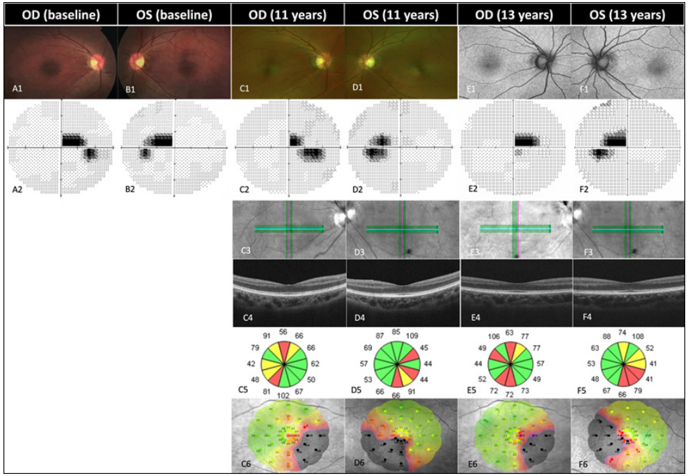
A 39-year-old female was evaluated for progressive bilateral visual decline over 1 year. The patient denied smoking, drinking alcohol, or exposure to toxins. Family history was negative. She had a decades-long history of stable, moderate visual impairment in both eyes. Her best corrected visual acuity (BCVA) was 0.4 (20/50) in both eyes in a routine eye check when she was 16-year- old. No further examinations were performed at that point. Twelve years later, at the age of 28, her BCVA remained 0.4 (20/50), no anterior segment abnormality was found, and pale temporal optic disc was noted in both eyes (Figure 1. A1 and B1). Visual field testing (Humphrey Visual Field Analyzer II; Carl Zeiss Meditec, Dublin, CA) showed central scotoma in both eyes with a mean threshold of -2.46 dB OD and -2.96 dB OS, respectively (Figure 1. A2 and B2). Brain and orbital magnetic resonance imaging (MRI) was normal and no enhancement of the optic nerve or chiasm was detected. Multifocal electroretinogram (mfERG, RETIscan, Roland consult, Brandenburg, Germany) recorded normal retinal electrical responses in all tested regions. A diagnosis of optic atrophy OU was made.

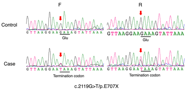
At the age of 38, the patient developed lupus erythematosus, and only hydroxychloroquine was prescribed. The usage of hydroxychloroquine lasted 13 months and the daily dose was 0.2 g. After that, the patient was treated at the age of 39 for a progressive decrease in binocular vision for nearly one year. BCVA was 0.2 (20/100) OU. Bilateral pale temporal optic disc was stable and similar with that of 11 years ago (Figure 1. C1 and D1). Optical coherence tomography (Cirrus OCT; Carl Zeiss Meditec, Dublin, CA, USA) showed bilateral moderate retinal nerve fiber layer (RNFL) thinning other than the nasal quadrant and general thinning of the inner retina in the macular (Figure 1. C5 and D5). Microcystic macular edema (MME) was noted in nasal macular in both eyes (Figure 1. C4 and D4). Visual field testing showed non-progressive central scotoma was detected with a mean threshold of -3.19 dB OD and -3.51 dB OS, respectively (Figure 1. C2 and D2). Microperimetry (MAIA, CenterVue, Padova, Italy) using full threshold 4-2 strategy found that the area nasally and inferiorly to the fovea had relatively low sensitivity with a mean threshold of 19.2 dB OD and 12.6 dB OS, respectively (Figure 1. C6 and D6). A known heterozygous nonsense mutation in OPA1, c.2119G>T/p.E707X, was identified using whole exome sequencing (WES) strategy, from which the diagnosis of DOA in both eyes was confirmed(Figure 2). Hydroxychloroquine was replaced by other immunosuppressant. The patient was closely followed in the following 2 years. Her functional tests including visual acuity, field testing and microperimetry testing was stable compared with those of 2 years ago. However, progressive inner macular and RNFL thinning was shown by OCT (Figure 1. E1, F1, E2, F2, E4, F4, E5, F5, E6, F6).

Figure 1 Longitudinal morphological and functional tests illustrate slow progression with a duration of 13 years.
(A1 and B1) Fundus photography at baseline, OD and OS, respectively; (A2 and B2) Visual field testing at baseline showing paracentral scotoma, OD and OS, respectively. Eleven years later, the patient complained about progressively decreased vision in both eyes after HCQ intake for 13 months. (C1 and D1) fundus photography, and (C2 and D2) visual field testing showed minimum progression; (C3 and D3)infrared image, OD and OS, respectively; (C4 and D4) OCT B-scan showed inner retinal thinning and microcystic macular edema (MME) in the inner nuclear layer (INL), OD and OS, respectively; (C5 and D5) Peripapillary retinal nerve fiber layer (RNFL) thickness evaluated by OCT, showing RNFL thinning in the superior, temporal and inferior quadrant, OD and OS, respectively; (C6 and D6) Evaluated by microperimetry, the mean sensitivity in the macular region was 19.2 dB in the right eye (C6) and 12.6 dB in the left eye (D6), respectively. Thirteen years since baseline, her vision decline terminated after she stopped taking HCQ. (E1 and F1) autofluorescence imaging; (E2 and F2) Visual field testing; (E3 and F3)infrared image, OD and OS, respectively; (E4 and F4) OCT B-scan showing progressive inner macular thinning and MME; (E5 and F5) RNFL thinning aggravates in the superior, temporal and inferior quadrant; (E6 and F6) The mean sensitivity evaluated by microperimetry was 19.0 dB in the right eye (E6) and 14.0 dB in the left eye (F6), respectively.

Figure 2 Sequences of the patient showing the c.2119G>T/p.E707X variation.

DISCUSSION

In most cases, DOA presents as an occult, slowly progressive bilateral optic neuropathy that usually cause moderate, irreversible visual impairment. DOA is typically diagnosed in school-aged children and visual acuity usually declines by one line per decade. But the severity of visual impairment and progression rate could be highly variable between affected family members. Rapid vision decline has been reported even years after vision stays stable. On the contrary, some patients who are diagnosed during routine fundus examination may experience no visual symptoms of a lifetime. [4]

The optic atrophycan be insidiousin early stage and become more distinct in the temporal optic disc as it develops gradually. Typical visual field manifestation is central or paracentral scotoma, usually without peripheral visual field defect. [5] Dischromatopsy, particularly a blue- yellow axis of color confusion, is strongly indicative of DOA. [2]

DOA primarily affects the retinal ganglion cells (RGCs) whose axons form the optic nerve. RGCs are neurons originating from an extension of the diencephalon, so DOA could also be considered as a disorder of the central nervous system that may manifest as DOA plus, accounting for some 20% of all DOA cases. DOA plus patients typically experience more severe visual deficits and may have extraocular symptoms, such as sensorineural hearing loss, progressive external ophthalmoplegia, myopathy, peripheral neuropathy, and ataxia. [6-8]

OPA1 is one of the major loci that are currently known for DOA, accounting for at least 75% of DOA cases in total. [9] OPA1 generates both the anchored long form L-OPA1 and the soluble short form S-OPA1 through proteolytic cleavage, which work synergistically to catalyze membrane fusion. The transmembrane domain of the anchored L-OPA1 molecule provides initial localization of OPA1 to the fusion site, initiating membrane remodeling and recruiting soluble S-OPA1 to rapidly polymerize into a flexible cylindrical scaffold that encapsulates the inner mitochondrial membrane, initiating the formation of moderately curved tubules in the inner membrane. This process generates bending stress, leading to the formation of unstable membranes that are prone to fusion. Further tubularization of the membranes enables the merging of the two bilayers. OPA1 assists in forming and stabilizing cristae junctions, which are narrow openings connecting the cristae to the intermembrane space. These junctions are crucial for controlling the diffusion of molecules, including cytochrome c, and maintaining the integrity of the respiratory chain. Most OPA1 mutations are substitutions or deletions, and in about 50% of cases, pathogenic mutations introduce a premature stop codon, resulting in the truncation of the open reading frame, which leads to mRNA decay and ultimately results in the loss of function of the mutant allele. This causes the fragmentation of the mitochondrial network and downregulation of membrane stability, subsequently leading to the release of cytochrome c and a reduction in ATP production. [10-12] As a result, higher energy consumption cells, such as RGC, are prone to degeneration and apoptosis. [13] The apoptosis of RGCs that forms the papillomacular bundle leads to central or paracentral scotoma in typical DOA cases.

Currently, over 500 OPA1 mutations have been found. [14] The mutation found in this patient, c.2119G>T, was a nonsense mutation and was previously reported by Chen et al [3] and Weisschuh et al. [14] Considering that there was a lack of descriptions on clinical manifestation and follow-up data in previous studies, this case was noteworthy for its detailed clinical data with a long- term follow-up. The fact that the visual function of this patient stayed stable for 22 years until oral hydroxychloroquine was prescribed raised a question that if the hydroxychloroquine was responsible for her vision decline. The ocular toxicity of hydroxychloroquine is manifested as cycloplegia, corneal deposition and retinopathy. Cycloplegia and corneal deposition is basically reversible, but hydroxychloroquine-related retinopathy is believed to be irreversible. The mechanism of HCQ damage to the retina is still unclear. An older antimalarial, chloroquine (CQ), was found to have a strong affinity with melanin-rich cells, and tend to form an irreversible conjugate with melanin in retinal cells,affecting cellular metabolism in the retina. [15] Marmor etal and Yusuf et al suggested that HCQ could affect the function of lysosome, decline the autophagy function of RPE and the stability of photoreceptor cell membrane, resulting in retinal toxicity. [16- 17]

HCQ toxicity is highly related to the duration and daily dose. The maximum recommended daily dose of HCQ by the American Academy of Ophthalmology is 5 mg/kg. At recommended doses, the risk of toxicity up to 5 years is under 1% and up to 10 years is under 2%, but it rises to almost 20% after 20 years. After 20 years, a patient without retinal toxicity has only a 4% risk of converting in the subsequent year. [18] Our patient had a significant decrease in visual acuity after taking HCQ at a dose of 4 mg/kg/day for 13 months, even within the recommended doses and duration. Although no significant changes in visual field testing and fundus photography before and after HCQ usage were found, MME was noted in nasal macular in both eyes, indicating RGC degeneration or apoptosis. It is generally believed that the damage of HCQ to the retina is mainly manifested in the outer retina, but Pasadhika et al. showed selective thinning of the macular inner retina in those without clinically apparent fundus changes. [19] There are also studies that show the effects of long-term HCQ are not limited to the outer retina. Not only RGC loss correlates with an increasing cumulative dose of HCQ, but also the INL, consisting mostly of Müller glia and the cell bodies of bipolar, horizontal, and amacrine cells, shows a significant difference between short- term and long-term treatment groups. [20] Therefore, we believe that the INL change observed by OCT may be an early lesion of hydroxyquinoline retinal toxicity. Although the patient used HCQ in a smaller dose and a shorter course of treatment, minor retinal damage was observed. Whether HCQ can induce extra retinal cell damage disease in DOA patients is still uncertain. Take DOA cases caused by OPA1 mutation for example, OPA1 protein was expressed not only in RGC and photoreceptor cells, but also in nerve fiber layer, inner plexiform layer and outer plexiform layer. Therefore, the abnormality of OPAl protein may also affect the signal transduction in the inner retina. [21] Khosa et al found abnormalities in mitochondrial ridges in muscle specimen from patients with HCQ-related myopathy, suggesting that oxidative stress induced by HCQ could alter mitochondrial integrity, and that the OPA1 protein might also have some impact on mitochondrial ridges. [22] Presumably, it was possible that DOA and HCQ intake may have a synergetic mechanism and cause progressive visual impairment in our case.

In summary, OPA1c.2119 G>T found in this patient was a mutation that had been rarely reported in previous studies. Our patient has been followed up for over 10 years and her visual acuity stayed stable for decades long until she took HCQ for 13 months. Her vision decline terminated after she stopped taking HCQ. HCQ may aggravate visual impairment in certain individuals harboring OPA1 mutation. Patients with DOA should avoid using neurotoxic HCQ and other medications that may interfere mitochondrial metabolism.

Correction notice

None

Acknowledgement

None

Author Contributions

(Ⅰ) Conception and design: Hui Yang
(Ⅱ) Administrative support: Hui Yang
(Ⅲ) Provision of study materials or patients: Yurong Zhang
(Ⅳ) Collection and assembly of data: Jie Sun
(Ⅴ) Data analysis and interpretation: Yurong Zhang
(Ⅵ) Manuscript writing: All authors
(Ⅶ) Final approval of manuscript: All authors

Fundings

This clinical research was supported by grants from the Guangdong Basic and Applied Basic Research Foundation (2023A1515011250).

Conflict of Interests

None of the authors has any conflicts of interest to disclose. All authors have declared in the completed the ICMJE uniform disclosure form.

Patient consent for publication

None

Ethical Statement

None

Provenance and Peer Review

This article was a standard submission to our journal. The article has undergone peer review with our anonymous review system.

Data Sharing Statement

None

Open Access Statement

This is an Open Access article distributed in accordance with the Creative Commons Attribution- NonCommercial-NoDerivs 4.0 International License (CC BY-NC-ND 4.0), which permits the non-commercial replication and distribution of the article with the strict proviso that no changes or edits are made and the original work is properly cited (including links to both the formal publicationthrough the relevant DOI and the license).

ABBREVIATIONS

BCVA: best corrected visual acuity.
CQ: chloroquine.
DOA: Dominant optic atrophy.
HCQ: hydroxychloroquine.
MAIA: Microperimetry.
MME: Microcystic macular edema.
MRI: magnetic resonance imaging.
RGCs: the retinal ganglion cells.
RNFL: retinal nerve fiber layer.
WES: whole exome sequencing.
1、Kjer P. Infantile optic atrophy with dominant mode of inheritance: a clinical and genetic study of 19 Danish families. Acta Ophthalmol Suppl. 1959, 164(Supp 54): 1-147.Kjer P. Infantile optic atrophy with dominant mode of inheritance: a clinical and genetic study of 19 Danish families. Acta Ophthalmol Suppl. 1959, 164(Supp 54): 1-147.
2、Lenaers G, Hamel C, Delettre C, et al. Dominant optic atrophy. Orphanet J Rare Dis. 2012, 7: 46. DOI: 10.1186/1750-1172-7-46.Lenaers G, Hamel C, Delettre C, et al. Dominant optic atrophy. Orphanet J Rare Dis. 2012, 7: 46. DOI: 10.1186/1750-1172-7-46.
3、Chen Y, Jia X, Wang P, et al. Mutation survey of the optic atrophy 1 gene in 193 Chinese families with suspected hereditary optic neuropathy. Mol Vis. 2013, 19: 292-302.Chen Y, Jia X, Wang P, et al. Mutation survey of the optic atrophy 1 gene in 193 Chinese families with suspected hereditary optic neuropathy. Mol Vis. 2013, 19: 292-302.
4、Skidd PM, Lessell S, Cestari DM. Autosomal dominant hereditary optic neuropathy (ADOA): a review of the genetics and clinical manifestations of ADOA and ADOA+. Semin Ophthalmol. 2013, 28(5-6): 422-426. DOI: 10.3109/08820538.2013.825296.Skidd PM, Lessell S, Cestari DM. Autosomal dominant hereditary optic neuropathy (ADOA): a review of the genetics and clinical manifestations of ADOA and ADOA+. Semin Ophthalmol. 2013, 28(5-6): 422-426. DOI: 10.3109/08820538.2013.825296.
5、Yu-Wai-Man P, Griffiths PG, Burke A, et al. The prevalence and natural history of dominant optic atrophy due to OPA1 mutations. Ophthalmology. 2010, 117(8): 1538-1546, 1546.e1. DOI: 10.1016/j.ophtha.2009.12.038.Yu-Wai-Man P, Griffiths PG, Burke A, et al. The prevalence and natural history of dominant optic atrophy due to OPA1 mutations. Ophthalmology. 2010, 117(8): 1538-1546, 1546.e1. DOI: 10.1016/j.ophtha.2009.12.038.
6、Aleo SJ, Del Dotto V, Fogazza M, et al. Drug repositioning as a therapeutic strategy for neurodegenerations associated with OPA1 mutations. Hum Mol Genet. 2021, 29(22): 3631-3645. DOI: 10.1093/hmg/ddaa244.Aleo SJ, Del Dotto V, Fogazza M, et al. Drug repositioning as a therapeutic strategy for neurodegenerations associated with OPA1 mutations. Hum Mol Genet. 2021, 29(22): 3631-3645. DOI: 10.1093/hmg/ddaa244.
7、Payne M, Yang Z, Katz BJ, et al. Dominant optic atrophy, sensorineural hearing loss, ptosis, and ophthalmoplegia: a syndrome caused by a missense mutation in OPA1. Am J Ophthalmol. 2004, 138(5): 749-755. DOI: 10.1016/ j.ajo.2004.06.011.Payne M, Yang Z, Katz BJ, et al. Dominant optic atrophy, sensorineural hearing loss, ptosis, and ophthalmoplegia: a syndrome caused by a missense mutation in OPA1. Am J Ophthalmol. 2004, 138(5): 749-755. DOI: 10.1016/ j.ajo.2004.06.011.
8、Yu-Wai-Man P, Griffiths PG, Gorman GS, et al. Multi- system neurological disease is common in patients with OPA1 mutations. Brain. 2010, 133(Pt 3): 771-786. DOI: 10.1093/brain/awq007.Yu-Wai-Man P, Griffiths PG, Gorman GS, et al. Multi- system neurological disease is common in patients with OPA1 mutations. Brain. 2010, 133(Pt 3): 771-786. DOI: 10.1093/brain/awq007.
9、Reynier P, Amati-Bonneau P, Verny C, et al. OPA3 gene mutations responsible for autosomal dominant optic atrophy and cataract. J Med Genet. 2004, 41(9): e110. DOI: 10.1136/jmg.2003.016576.Reynier P, Amati-Bonneau P, Verny C, et al. OPA3 gene mutations responsible for autosomal dominant optic atrophy and cataract. J Med Genet. 2004, 41(9): e110. DOI: 10.1136/jmg.2003.016576.
10、Del%20Dotto%20V%2C%20Fogazza%20M%2C%20Lenaers%20G%2C%20et%20al.%20OPA1%3A%20how%20much%20do%20we%20know%20to%20approach%20therapy%3F%20Pharmacol%20Res.%202018%2C%20131%3A%20199-210.%20DOI%3A%2010.1016%2Fj.phrs.2018.02.018.Del%20Dotto%20V%2C%20Fogazza%20M%2C%20Lenaers%20G%2C%20et%20al.%20OPA1%3A%20how%20much%20do%20we%20know%20to%20approach%20therapy%3F%20Pharmacol%20Res.%202018%2C%20131%3A%20199-210.%20DOI%3A%2010.1016%2Fj.phrs.2018.02.018.
11、von der Malsburg A, Sapp GM, Zuccaro KE, et al. Structural mechanism of mitochondrial membrane remodelling by human OPA1. Nature. 2023, 620(7976): 1101-1108. DOI: 10.1038/s41586-023-06441-6.von der Malsburg A, Sapp GM, Zuccaro KE, et al. Structural mechanism of mitochondrial membrane remodelling by human OPA1. Nature. 2023, 620(7976): 1101-1108. DOI: 10.1038/s41586-023-06441-6.
12、Nyenhuis SB, Wu X, Strub MP, et al. OPA1 helical structures give perspective to mitochondrial dysfunction. Nature. 2023, 620(7976): 1109-1116. DOI: 10.1038/ s41586-023-06462-1.Nyenhuis SB, Wu X, Strub MP, et al. OPA1 helical structures give perspective to mitochondrial dysfunction. Nature. 2023, 620(7976): 1109-1116. DOI: 10.1038/ s41586-023-06462-1.
13、Olichon A, Landes T, Arnauné-Pelloquin L, et al. Effects of OPA1 mutations on mitochondrial morphology and apoptosis: relevance to ADOA pathogenesis. J Cell Physiol. 2007, 211(2): 423-430. DOI: 10.1002/jcp.20950.Olichon A, Landes T, Arnauné-Pelloquin L, et al. Effects of OPA1 mutations on mitochondrial morphology and apoptosis: relevance to ADOA pathogenesis. J Cell Physiol. 2007, 211(2): 423-430. DOI: 10.1002/jcp.20950.
14、Weisschuh N, Schimpf-Linzenbold S, Mazzola P, et al. Mutation spectrum of the OPA1 gene in a large cohort of patients with suspected dominant optic atrophy: identification and classification of 48 novel variants. PLoS One. 2021, 16(7): e0253987. DOI: 10.1371/journal. pone.0253987.Weisschuh N, Schimpf-Linzenbold S, Mazzola P, et al. Mutation spectrum of the OPA1 gene in a large cohort of patients with suspected dominant optic atrophy: identification and classification of 48 novel variants. PLoS One. 2021, 16(7): e0253987. DOI: 10.1371/journal. pone.0253987.
15、Rosenthal AR, Kolb H, Bergsma D, et al. Chloroquine retinopathy in the rhesus monkey. Invest Ophthalmol Vis Sci. 1978, 17(12): 1158-1175.Rosenthal AR, Kolb H, Bergsma D, et al. Chloroquine retinopathy in the rhesus monkey. Invest Ophthalmol Vis Sci. 1978, 17(12): 1158-1175.
16、Latasiewicz M, Gourier H, Yusuf IH, et al. Hydroxychloroquine retinopathy: an emerging problem. Eye, 2017, 31(6): 972-976. DOI: 10.1038/eye.2016.297.Latasiewicz M, Gourier H, Yusuf IH, et al. Hydroxychloroquine retinopathy: an emerging problem. Eye, 2017, 31(6): 972-976. DOI: 10.1038/eye.2016.297.
17、Marmor MF. Comparison of screening procedures in hydroxychloroquine toxicity. Arch Ophthalmol, 2012, 130(4): 461-469. DOI: 10.1001/archophthalmol.2011.371.Marmor MF. Comparison of screening procedures in hydroxychloroquine toxicity. Arch Ophthalmol, 2012, 130(4): 461-469. DOI: 10.1001/archophthalmol.2011.371.
18、Marmor MF, Kellner U, Lai TYY, et al. Recommendations on screening for chloroquine and hydroxychloroquine retinopathy (2016 revision). Ophthalmology. 2016, 123(6): 1386-1394. DOI: 10.1016/j.ophtha.2016.01.058.Marmor MF, Kellner U, Lai TYY, et al. Recommendations on screening for chloroquine and hydroxychloroquine retinopathy (2016 revision). Ophthalmology. 2016, 123(6): 1386-1394. DOI: 10.1016/j.ophtha.2016.01.058.
19、Kim KE, Kim YH, Kim J, et al. Macular ganglion cell complex and peripapillary retinal nerve fiber layer thicknesses in hydroxychloroquine retinopathy. Am J Ophthalmol. 2023, 245: 70-80. DOI: 10.1016/ j.ajo.2022.07.028.Kim KE, Kim YH, Kim J, et al. Macular ganglion cell complex and peripapillary retinal nerve fiber layer thicknesses in hydroxychloroquine retinopathy. Am J Ophthalmol. 2023, 245: 70-80. DOI: 10.1016/ j.ajo.2022.07.028.
20、Gil P, Nunes A, Farinha C, et al. Structural and functional characterization of the retinal effects of hydroxychloroquine treatment in healthy eyes. Ophthalmologica. 2019, 241(4): 226-233. DOI: 10.1159/000495308.Gil P, Nunes A, Farinha C, et al. Structural and functional characterization of the retinal effects of hydroxychloroquine treatment in healthy eyes. Ophthalmologica. 2019, 241(4): 226-233. DOI: 10.1159/000495308.
21、Pesch UEA, Fries JE, Bette S, et al. OPA1, the disease gene for autosomal dominant optic atrophy, is specifically expressed in ganglion cells and intrinsic neurons of the retina. Invest Ophthalmol Vis Sci. 2004, 45(11): 4217- 4225. DOI: 10.1167/iovs.03-1261.Pesch UEA, Fries JE, Bette S, et al. OPA1, the disease gene for autosomal dominant optic atrophy, is specifically expressed in ganglion cells and intrinsic neurons of the retina. Invest Ophthalmol Vis Sci. 2004, 45(11): 4217- 4225. DOI: 10.1167/iovs.03-1261.
22、KhosaS, KhanlouN, Khosa GS, et al. Hydroxychloroquine-induced autophagic vacuolar myopathy with mitochondrial abnormalities. Neuropathology. 2018, 38(6): 646-652. DOI: 10.1111/neup.12520.KhosaS, KhanlouN, Khosa GS, et al. Hydroxychloroquine-induced autophagic vacuolar myopathy with mitochondrial abnormalities. Neuropathology. 2018, 38(6): 646-652. DOI: 10.1111/neup.12520.
1、This clinical research was supported by grants from the Guangdong Basic and Applied Basic Research Foundation (2023A1515011250).()
上一篇
下一篇
出版者信息
目录