综述

小梁网泵衰竭在青光眼发病中的作用机制的研究进展

Research progress on the mechanism of trabecular meshwork pump failure in the pathogenesis of glaucoma

:325-331
 
青光眼是一组以视盘萎缩凹陷、视野缺损以及视力下降为共同特征的视神经退行性疾病,也是世界首位不可逆性致盲眼病,导致患者生活质量降低、引起极大卫生经济负担。但其发病机制尚不明确,促进房水排出从而降低眼内压仍是目前减缓疾病进展的唯一治疗手段。房水排出的主要途径是经由小梁网进入Schlemm's管最后汇入巩膜外静脉,因此小梁网在调节房水排出以及平衡眼内压方面发挥重要作用。近年来,体内以及体外房水排出测量技术和小梁网成像技术不断突破,众多研究表明小梁网存在压力依赖的节律性搏动,在房水的脉冲式排出中起到关键作用,但在青光眼中这种搏动随疾病的进展减弱甚至消失。文章以小梁网的泵理论为核心,总结青光眼中房水排出的最新研究进展,并从恢复小梁网功能的角度出发探索可能有效的治疗策略,为青光眼的临床诊治提供新的思路。
Glaucoma, a group of optic nerve degenerative diseases, is characterized by papillary atrophy, visual field defects, and decreased vision. It is also the leading cause of irreversible blindness worldwide, significantly reducing patients’ the quality of life of patients and posing considerable health economic burdens. However, the pathogenesis of glaucoma remains unclear, and promoting aqueous humor outflow to reduce intraocular pressure is the only treatment option available to slow disease progression. The main pathway for aqueous humor outflow is through the trabecular meshwork into Schlemm's canal and finally into the episcleral veins, highlighting the crucial role of the trabecular meshwork in regulating aqueous humor outflow and maintaining intraocular pressure balance. In recent years, there have been notable breakthroughs in in vivo and in vitro aqueous humor outflow measurement techniques and trabecular meshwork imaging technologies.Many studies suggest that the trabecular meshwork exhibits pressure-dependent rhythmic pulsation, playing a crucial role in the pulse-like outflow of aqueous humor. Unfortunately, in glaucoma, this pulsation weakens or even disappears as the disease progresses. This article focuses on the trabecular meshwork's pump theory and summarizes the latest research progress in aqueous humor outflow in glaucoma, exploring potential effective therapeutic strategies aimed at restoring trabecular meshwork function. This provides new insights for the clinical diagnosis and treatment of glaucoma.
Review Article

Extrinsic regulation of optic nerve regeneration

Extrinsic regulation of optic nerve regeneration

:145-159
 
Retinal ganglion cells (RGCs) extend through the optic nerve, connecting with neurons in visually related nuclei. Similar to most mature neurons in the central nervous system, once damaged, RGCs are unable to regenerate their axons and swiftly progress to cell death. In addition to cell-intrinsic mechanisms, extrinsic factors within the extracellular environment, notably glial and inflammatory cells, exert a pivotal role in modulating RGC neurodegeneration and regeneration. Moreover, burgeoning evidence suggests that retinal interneurons, specifically amacrine cells, exert a substantial influence on RGC survival and axon regeneration. In this review, we consolidate the present understanding of extrinsic factors implicated in RGC survival and axon regeneration, and deliberate on potential therapeutic strategies aimed at fostering optic nerve regeneration and restoring vision.
Retinal ganglion cells (RGCs) extend through the optic nerve, connecting with neurons in visually related nuclei. Similar to most mature neurons in the central nervous system, once damaged, RGCs are unable to regenerate their axons and swiftly progress to cell death. In addition to cell-intrinsic mechanisms, extrinsic factors within the extracellular environment, notably glial and inflammatory cells, exert a pivotal role in modulating RGC neurodegeneration and regeneration. Moreover, burgeoning evidence suggests that retinal interneurons, specifically amacrine cells, exert a substantial influence on RGC survival and axon regeneration. In this review, we consolidate the present understanding of extrinsic factors implicated in RGC survival and axon regeneration, and deliberate on potential therapeutic strategies aimed at fostering optic nerve regeneration and restoring vision
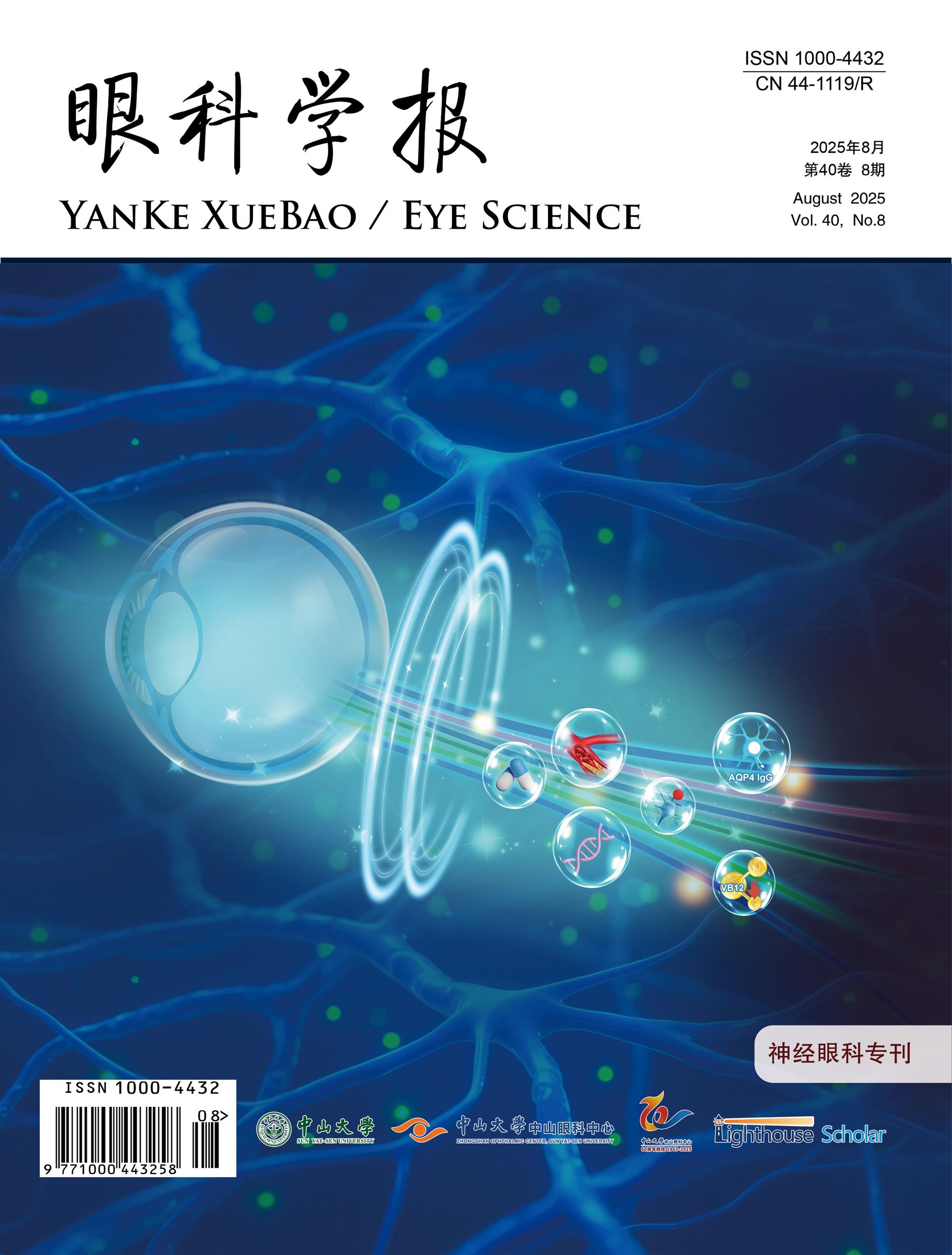
封面简介

小梁网泵衰竭在青光眼发病中的作用机制的研究发展

Research progress on the mechanism of trabecular meshwork pump failure in the pathogenesis of glaucoma

:-
 
青光眼是一组以视盘萎缩凹陷、视野缺损以及视力下降为共同特征的视神经退行性疾病,也是世界首位不可逆性致盲眼病,导致患者生活质量降低、引起极大卫生经济负担。但其发病机制尚不明确,促进房水排出从而降低眼内压力仍是目前减缓疾病进展的唯一治疗手段。房水排出的主要途径是经由小梁网进入Schlemm’ s管最后汇入巩膜外静脉,因此小梁网在调节房水排出以及平衡眼内压力方面发挥重要作用。近年以来体内以及体外房水排出测量技术和小梁网成像技术不断突破,众多研究表明小梁网存在压力依赖的节律性搏动,在房水的脉冲式排出中起到关键作用,但在青光眼中这种搏动随疾病的进展减弱甚至消失。文章将以小梁网的泵理论为核心,总结青光眼中房水排出的最新研究进展,并从恢复小梁网功能的角度出发探索可能有效的治疗策略,为青光眼的临床诊治提供新的思路。
出版者信息