您的位置: 首页 > 2024年12月 第1卷 第4期 > 文字全文
2023年7月 第38卷 第7期11
目录

Macular hemorrhage in high myopia

Macular hemorrhage in high myopia

来源期刊: Eye Science | 2024年12月 第1卷 第4期 422-442 发布时间:2024-11-28 收稿时间:2024/12/11 16:18:42 阅读量:176
作者:
关键词:
high myopia macular hemorrhage pathogenesis treatment
DOI:
10.12419/es24090201
Received date:
2024-09-02 
Accepted date:
2024-10-16 
Published online:
2024-11-28 
Macular hemorrhage (MH) is one of the most severe complications of high myopia, posing a significant threat to vision. MH can occur with or without choroidal neovascularization (CNV), with the CNV-associated form being the most prevalent. CNV-related MH may develop secondary to conditions such as pathological myopia, and punctate inner choroidopathy. Conversely, MH without CNV is often linked to factors like lacquer cracks, trauma, ocular surgery. While the exact mechanisms of CNV in high myopia are still not fully understood, anti-VEGF injections have been shown to be effective in improving visual function in patients with CNV. This review summarizes the clinical characteristics of various causes of MH and their respective treatments, providing valuable insights to help clinicians make informed diagnostic and therapeutic decisions.
Macular hemorrhage (MH) is one of the most severe complications of high myopia, posing a significant threat to vision. MH can occur with or without choroidal neovascularization (CNV), with the CNV-associated form being the most prevalent. CNV-related MH may develop secondary to conditions such as pathological myopia, and punctate inner choroidopathy. Conversely, MH without CNV is often linked to factors like lacquer cracks, trauma, ocular surgery. While the exact mechanisms of CNV in high myopia are still not fully understood, anti-VEGF injections have been shown to be effective in improving visual function in patients with CNV. This review summarizes the clinical characteristics of various causes of MH and their respective treatments, providing valuable insights to help clinicians make informed diagnostic and therapeutic decisions.

HIGHLIGHTS

• This review identifies the diverse etiologies of macular hemorrhage (MH) in high myopia (HM), distinguishing between CNV-related MH and non-CNV MH. CNV-associated MH is most frequently linked to pathological myopia and punctate inner choroidopathy (PIC), whereas non-CNV cases are often attributed to lacquer cracks, trauma, or surgery. Key discoveries include:
• Anti-VEGF therapy is the first-line treatment for CNV-related MH, demonstrating significant efficacy in improving visual outcomes.
• For non-CNV MH, conservative observation is generally recommended, highlighting the importance of tailored treatment strategies.
• Secondary CNV from PIC may benefit from adjunctive anti-inflammatory therapies, although more evidence is needed.
• This study employs a comprehensive review methodology to synthesize knowledge on the pathogenesis, clinical 
characteristics, and treatment options for MH in HM. Specific innovations include:
• Integration of findings on structural, hemodynamic, genetic, and systemic factors influencing CNV formation, providing a holistic understanding of MH pathogenesis.
• Highlighting the need for a diagnostic approach that differentiates CNV-associated MH from non-CNV MH to inform targeted therapies effectively.
• The review underscores critical gaps and potential advancements, suggesting several avenues for future work:
• Mechanistic Research: Investigating the molecular and genetic factors driving CNV and non-CNV MH development to inform preventative strategies.
• Diagnostic Improvements: Development of advanced imaging tools to improve early detection and enable precise differential diagnosis of MH types.
• Therapeutic Optimization: Refining anti-VEGF protocols, exploring combination therapies (e.g., anti-inflammatory treatments for PIC-related CNV), and evaluating long-term outcomes.
• Personalized Medicine: Advancing individualized treatment strategies based on the underlying etiology and patient-specific factors, ultimately improving care for high-myopia patients.

INTRODUCTION

High myopia (HM) is a significant vision-impairing disease, with a concerning increase in prevalence worldwide, particularly among younger generations.[1] Consequently, the complications associated with HM especially pathological myopia (PM) can have a profound impact on this population.[2] Among these complications, macular hemorrhage (MH) is one of the most severe, often resulting in a sudden and dramatic loss of vision. Although detailed prevalence data for MH in high myopia are currently lacking, it is estimated that the overall prevalence of MH in adults with HM in Asia is approximately 0.9%.[3] For myopic choroidal neovascularization (myopic CNV), the reported prevalence ranges from 0.9% to 2.7%.[4]MH refers to the accumulation of blood in the macular area due to the alternation of the retinal or choroidal circulation.Based on its position, MH can be classified into several categories which affect its natural course: preretinal (situated beneath the inner limiting membrane or hyaloid), intraretinal, subretinal, mixed (invading both the subretinal and subpigment epithelial layers), or encompassing multiple layers within the macular region.[5]In addition to the toxicity of blood to the retinal structure, the separation of photoreceptors from the pigment epithelium in cases of subretinal hemorrhage is also considered a mechanism of damage. 

In high myopia, MH is typically subretinal or mixed, with two major types identified: one involves isolated bleeding from the rupture of Bruch’s membrane (BM) without choroidal neovascularization (CNV), and the other involves bleeding associated with concurrent CNV. The latter can result from classical pathologic myopia or myopic CNV, punctate inner choroidopathy (PIC), choroiditis, and conditions concurrent with HM. While the presence of CNV may influence treatment choices, anti-VEGF therapy has been proven effective for CNV. However, for isolated MH without CNV, there are currently no effective treatment options available. 

Since various primary diseases can induce MH in high myopia, and these underlying conditions determine the treatment strategy and prognosis, it is essential to summarize the different characters of MH in high myopia. This allows clinicians to make more accurate diagnoses and choose appropriate treatment methods based on the distinct clinical features of MH from different etiologies.Therefore, the purpose of this review is to explore the varied etiology, clinical features, natural history, and treatment options for MH secondary to different causes. 

CNV SECONDARY TO PATHOLOGIC MYOPIA

The most common cause of MH in HM is myopic CNV. myopic CNV is considered the most frequent cause of CNV in young individuals in many countries, accounting for 62% of CNV cases in patients aged 50 years or younger. A recent systematic review summarized the epidemiology of CNV based on 39 reports and found that the prevalence of CNV in individuals with pathologic myopia ranged between 5.2% and 11.3%, with approximately 15% of cases being bilateral.[2]Furthermore, in individuals with myopic CNV (with disease duration ranging from less than 3 months to 21.5 years), a deterioration in best-corrected visual acuity (BCVA) was observed over time.[2]

Myopic CNV is more likely to occur in patients who are older, have more severe chorioretinal atrophy, thinner choroid thickness, and the presence of lacquer cracks (LC).[6-8] Factors associated with worse visual outcomes include older age (>40 years), subfoveal CNV location, larger baseline lesion size (>400 μm), and lower baseline BCVA.[2, 9] Conversely, younger age, smaller CNV, and better initial visual acuity are associated with a more favorable prognosis.[10] Patients with myopic CNV typically develop the condition in their mid-fifties, with a mean AL approximately 29 mm.[11] The development of CNV can significantly alter the natural course of myopia, leading to retinal hemorrhage, edema, and eventually a fibrotic scar, resulting in permanent loss of visual function. Studies have shown that the development of CNV, CNV-related macular atrophy, and the enlargement of CNV-related macular atrophy are strongly associated with loss of BCVA. Approximately 92.7% of myopic CNV cases progress to macular atrophy, resulting in significant visual acuity loss.[11]

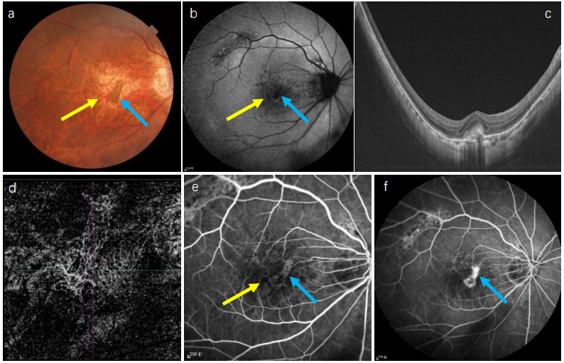
Typically, myopic CNV presents as a small, flat, grayish subretinal membrane, located between the neurosensory retina and the retinal pigment epithelium (RPE), often near the fovea, with or without associated hemorrhage.[12-13] On optical coherence tomography (OCT), these lesions appear as highly reflective signals with blurred upper boundaries of the RPE cells (Fig.1, typical of type 2 macular neovascularization) and are usually associated with minimal subretinal fluid.[14]The determination of activity on OCT mainly relies on four signs:[15] 1) intraretinal or subretinal fluid, 2) subretinal hyperreflective material, 3) disruption of the external limiting membrane, and 4) blurred lesion boundaries. Optical coherence tomography angiography (OCTA) is a valuable tool for detecting myopic CNV, showing a vascular network pattern in the outer retina and choriocapillaris slab, with high sensitivity (90.48%) and specificity (93.75%).[16] On OCTA, the assessment of activity is based on four indicators:[17] 1) a jellyfish-like or sea fan-shaped appearance of myopic macular neovascularization lesions, 2) numerous tiny capillary branches, 3) presence of vascular anastomoses or loops, and 4) a dark halo surrounding the lesion. The combined use of these methods can improve the sensitivity and specificity of activity assessment. However, fluorescein fundus angiography (FFA) remains the gold standard for diagnosis, showing early-phase well-defined hyperfluorescence of the lesion with late-phase fluorescent leakage. Indocyanine green angiography (ICGA) shows that ICG is minimally absorbed by the RPE and blood, allowing better differentiation of CNV from other pathologies, especially when hemorrhage is present.

Figure 1 Multimodal imaging of macular hemorrhage in pathological myopia-associated choroidal neovascularization (myopic CNV).
(a) Funduscopy image showing a small, flat gray lesion with macular hemorrhage at the edge of the lesion. (b) Autofluorescence (AF) image showing hypofluorescence in both the hemorrhage and CNV lesion. (c) Optical coherence tomography (OCT) image showing the CNV lesion with highly reflective signals and blurred boundaries. (d) Optical coherence tomography angiography (OCTA) image displaying a clear vascular network in myopic CNV. (e, f) Fluorescein angiography (FA) images showing early-phase well-defined hyperfluorescence of the lesion with late-phase fluorescent leakage and blocked fluorescence due to the hemorrhage. Yellow arrow: hemorrhage; Blue arrow: CNV lesion.

Understanding the natural history of myopic CNV is crucial for guiding treatment approaches. If left untreated, myopic CNV can cause scarring with expanding macular atrophy, leading to irreversible vision loss within a period as short as five years. In a 10-year follow-up study of 27 eyes with myopic CNV, the visual acuity of almost all eyes (approximately 96.3%) dropped to 20/200 or worse within 5 to 10 years after the onset of CNV, due to the development of chorioretinal atrophy around the regressed CNV.[18] The average time for complete bleeding absorption from myopic CNV was about 7.6 months (range, 1-15 months) without any intervention, and rebleeding occurred in 22.2% of eyes during followup.[18] These findings suggest that long-term visual acuity decline may be due to macular atrophy rather than the presence of CNV itself; however, the onset of CNV can definitely induce and accelerate chorioretinal atrophy. In another long-term follow-up study, chorioretinal atrophy developed around the regressed CNV in 26 eyes (96.3%) at 5 and 10 years after CNV onset.[18] After the absorption of MH, most patients exhibited intraretinal hyperreflective (IRH) signs on OCT. In a 24-month follow-up of a small sample with IRH signs after MH absorption in eyes with pathologic myopia, most signs remained stable. IRH may predict future myopic traction maculopathies.[19]

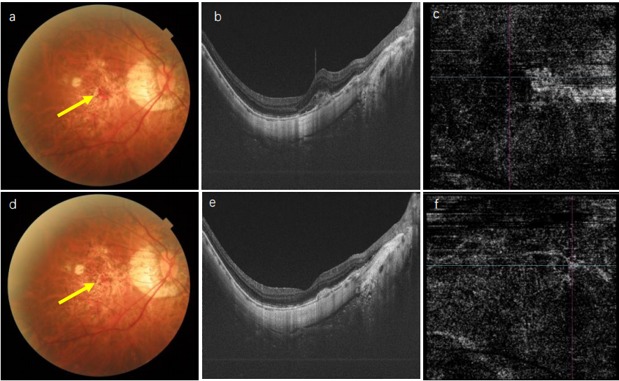
Few studies have reported favorable outcomes with observation alone; most studies describe a poor prognosis. Therefore, timely treatment of myopic CNV can lead to quick resolve of the CNV lesion and get better visual acuity outcomes. Previously, Laser photocoagulation, Photodynamic therapy[20] with verteporfin was applied to the treatment of myopic CNV, however, due to the high rate of recurrence and relative low benefit of visual acuity, and given the superior efficacy and long-term effect displayed by anti-vascular endothelial growth factor(anti-VEGF) therapy, Currently, anti-VEGF therapy is the standard of care and the recommended first-line treatment option for myopic CNV.[12, 21] It contributes to the reduction in CNV lesion size and central macular thickness improvement in myopic population (Fig.2). Long-term studies have demonstrated that early treatment of confirmed myopic CNV cases with an intravitreal anti-VEGF agent is effective in preventing late-stage complications.[21- 22]However, the optimal treatment regimen for anti-VEGF in myopic CNV remains debated. .Initially, a 3-loading-dose followed by pro re nata (PRN) approach, similar to that used in age-related macular degeneration (AMD), was suggested. , It was found that more than 70% myopic CNV eyes only required only three initial intravitreal injection of bevacizumab or ranibizumab, and average of 3.8 injection in 2 years follow up, about 65% had three or more lines visual gain.[23] Subsequently, studies began to compare different anti-VEGF regimens for myopic CNV, primarily focusing on 1+PRN versus 3+PRN. Wang et al. conducted a systematic review including three non-randomized controlled studies with a 1-year follow-up, comparing 1+PRN to 3+PRN in terms of BCVA and central retinal thickness (CRT). The 3+PRN group demonstrated better BCVA at 3 and 6 months (LogMAR MD = 0.11, 95% CI [0.01, 0.21]), but this difference diminished over time, with no significant differences at 1 year. There were also no significant differences in CRT between the two groups at 3, 6, or 12 months. Importantly, the 1+PRN group required, on average, 1.37 fewer injections than the 3+PRN group (P < 0.001).[24]

Figure 2 Anti-VEGF treatment for myopic CNV.
(a-c) Baseline imaging of myopic CNV, showing macular hemorrhage on fundus photography (a), a CNV lesion with highly reflective signals on OCT (b), and a vascular network on OCTA (c). (d-f ) Imaging one month after anti-VEGF injection, showing a reduction in macular hemorrhage (d), decreased CNV lesion size on OCT (e), and OCTA (f).

Li et al. conducted the SMILE study, a single-center randomized controlled trial comparing the efficacy of ranibizumab using 1+PRN versus 3+PRN in patients with active myopic CNV. After 12 months, there were no significant differences between the two groups in BCVA changes, anatomical parameters such as CRT, CNV thickness, CNV area, leakage area, or recurrence rates. The 1+PRN group required 1.54 fewer injections on average than the 3+PRN group over the 12-month period.[25] A retrospective study investigating the efficacy of conbercept for myopic CNV also compared the 1+PRN and 3+PRN regimens. At 12 months, there were no significant differences in BCVA or CRT between the two groups, but the 1+PRN group required fewer injections (2.14 ± 1.06 vs. 3.37 ± 0.76, P < 0.001).[26] Additionally, a network meta-analysis compared treatments for myopic CNV, including anti-VEGF, photodynamic therapy (PDT), intravitreal triamcinolone acetonide, and observation. The analysis found that anti-VEGF therapy resulted in the highest visual acuity gain, approximately 14.1 letters, and that the 1+PRN regimen was comparable to the 3+PRN regimen in terms of efficacy.[27] We also summarized all randomized controlled trials (RCTs) using anti-VEGF in myopicCNV,[25,28-34] which showed favorable results for improving visual acuity (Table 1).

Table 1 prospective and randomized clinical trials of anti-VEGF for the treatment of PM-CNV

Study

design

Sample size (eyes)

treatment

Anti-VEGF pattern

Mean Follow-up (y)

NO. of IV

Baseline VA


VA at last visit

RCT study [28]

182

Ranibizumab

(VA stabilization)

(1+PRN)

1

4.6 (2.4)

53.6 (12.6) letters

66.7 letters

184

Ranibizumab

(disease activity)

(1+PRN)

1

3.9 (2.5)

54.2 (13.0) letters

66.2 letters

91

vPDT

-

1

3.2 (2.3)

52.1 (12.7) letters

62.4 letters

prospective randomized

study [29]

24

Aflibercept

(3+PRN)

0.25

3

0.53 (0.10) LogMAR

0.38(0.11) LogMAR

24

Ranibizumab

(3+PRN)

0.25

3

0.55 (0.11) LogMAR

0.39(0.12 ) LogMAR

Prospective randomize  study[30]

16

Ranibizumab

(1+PRN)

0.5

2.81(1.17)

26.44 (12.58) letters

43.75 (9.92) letters

16

Bevacizumab

(1+PRN)

0.5

2.44(0.89)

29.50 (12.98 )letters

45.37 (9.95) letters

RCT study [31]

23

Ranibizumab

(1+PRN)

1.5

2.56 (1.61)

0.60 (0.29) LogMAR

0.40 (0.38) LogMAR


25

Bevacizumab

(1+PRN)

1.5

4.72(2.24)

0.60 (0.26) LogMAR

0.44 (0.32) LogMAR

RCT study [32]

50

Ranibizumab

(2+PRN)

2

2.5(0.9) 

0.21 (0.14) LogMAR

0.43 (0.24) LogMAR


47

Aflibercept

(2+PRN)

2

2.6(1.0) 

0.2 (0.14) LogMAR

0.41(0.2) LogMAR

RCT study [25]

26

Ranibizumab

(1+PRN)

1

2.04(1.22) 


54.1(15.5) letters

67.5(13.3) letters


24

Ranibizumab

(3+PRN)

1

3.58(0.72) 

48.7(13.1) letters

65.8(11.2) letters

RCT study [33]

90

Aflibercept

(1+PRN)

0.46

4.2

56.4 (9.8) letters

68.5 letters


Sham

 -

0.46

0

56.6 (9.8) letters

54.6 letters

RCT study [34]

40

Bevacizumab

(1+PRN)

1.53(0.40)

2.75(1.24)

0.52(0.34) LogMAR

0.53(0.33) LogMAR


38

Ranibizumab

(1+PRN)

1.65(0.43)

2.29(1.16)

0.62(0.36)LogMAR

0.50(0.35)LogMAR

CNV SECONDARY TO PUNCTATE INNER CHOROIDOPATHY

PIC is an uncommon idiopathic inflammatory chorioretinal disease that most commonly affects young, myopic females.[35] Myopia has been reported in 80% to 100% of PIC cases, with a mean refractive error ranging from -4.6 to -7.0 diopters, extending from -14 to +4 diopters, and a median age of 30 years (range, 15 to 55).[36-37] The etiology and pathogenesis of PIC remain unclear. Hypotheses suggest it is an autoimmune disease that manifests in individuals predisposed by polygenic susceptibility, triggered by environmental factors such as infection, immunization, or stress.[35]

Symptoms of PIC include photopsia, floaters, decreased night vision, blurred vision, and visual field loss. These symptoms are often acute and transient, without long-term visual consequences. Visual acuity at presentation varies and depends on the location of inflammatory lesions and their complications. Clinically, the fundus of PIC is characterized by multifocal, small (100-300 μm), yellow-gray lesions that are well-defined and confined to the posterior pole (Fig. 3), specifically at the inner choroid and outer retina.[38] Notably, the peripapillary and peripheral regions are spared from these lesions, and there is no sign of inflammation in the anterior chamber or vitreous. Over time, some lesions tend to resolve, while most result in permanent structural changes, especially the formation of atrophic chorioretinal scars at the previous sites of inflammation. These scars display a “punched-out” appearance and acquire pigmentation.[39] Although PIC typically presents unilaterally, more than 50% of cases eventually affect both eyes.[40] 

Figure 3 Multimodal imaging of macular hemorrhage in punctate inner choroidopathy (PIC). 
(a) Image showing multifocal, small, yellow-gray lesions that are well-defined and located around the macula, along with macular hemorrhage. (b) AF image showing hypofluorescence for both the PIC lesion and macular hemorrhage. (c) OCT image showing a small CNV lesion. (d) OCTA image displaying a clear vascular network of secondary CNV in PIC. Yellow arrow: hemorrhage; Purple arrow: PIC lesion.

MH in PIC is mainly due to secondary CNV. CNV is a common complication of PIC and tends to manifest as the disease progresses, presenting with MH similar to myopic CNV and resulting in vision loss. The risk of developing CNV in PIC is higher compared to other forms of posterior and panuveitis.[41] The rate of CNV development in PIC varies across studies, ranging from 17% to 75%.[36, 42] The presence of CNV in the fellow eye increases the risk of developing CNV, while previous oral corticosteroid treatment is associated with a reduced risk of developing CNV.[42] CNV secondary to PIC shares some features with inflammatory lesions, such as symptoms of vision loss and scotomata, and pathological manifestations including infiltrating lesions and heterogeneous material in the outer retina and subretinal space.

In FA, punctate hyperfluorescent choroidal lesions persist throughout the early and late venous phases, and CNV lesions are characterized by a zone of hypofluorescence surrounding a hyperfluorescent area in the early venous phase, associated with leakage in the late phase. ICGA shows lesions as hypofluorescent regions in both the early and late phases. OCT is effective for differentiating and monitoring the progression of inflammatory lesions and secondary CNV. Zhang et al. used OCT to divide PIC into five stages depending on disease activity.[43] In some cases, it is challenging to distinguish between active inflammatory lesions and CNV, as both exhibit disruption of the blood-retina barrier and infiltrative lesions.[44] Chen et al. found that RPE disruption and hyper-transmission are early signs of secondary CNV in PIC.[45] OCTA is a non-invasive technique for imaging the microvasculature of the retina and choroid, making it a useful alternative tool for detecting CNV in PIC and differentiating it from inflammatory lesions.[46] Distinguishing PIC from multifocal choroiditis with panuveitis (MCP) is challenging, as they share a similar disease spectrum. However, the absence of peripheral chorioretinal lesions and intraocular inflammation are key characteristics that differentiate PIC from MCP.[47] Additionally, PIC shows a preference for affecting young, myopic women. 

Due to the varying stages of PIC, limited evidence surrounding treatment strategies, and the scarcity of prospective clinical trials, the management of PIC remains a significant challenge. However, intervention is warranted when secondary CNV occurs, or when new or active inflammatory PIC lesions develop, especially those posing a threat to the fovea.[35] There is no standardized protocol for treating CNV resulting from PIC, and considerable debate persists regarding the appropriate management of CNV secondary to PIC. There are two major strategies for treating CNV secondary to PIC: corticosteroids and anti-VEGF therapy. Corticosteroids have both anti-inflammatory and anti-angiogenic effects. Several case reports and case series have demonstrated that oral corticosteroids not only stabilize or improve vision in PIC-associated CNV but also decrease the incidence and reduce the frequency of CNV recurrences.[48,49] The suggested protocol for oral steroids in PIC involves initiating therapy at a dose of 1 mg/kg, followed by a relatively fast tapering over 4-6 months.[50]However, due to the low level of evidence supporting the use of oral steroids for PIC and the potential adverse effects, careful consideration of treatment choices and patient characteristics is necessary. There is limited evidence regarding the use of immunomodulatory or immunosuppressive agents for treating PIC inflammatory lesions and secondary CNV. The effectiveness of anti-VEGF treatment for CNV secondary to AMD has been well-established in many randomized controlled trials. Although no RCTs have explored the effect of anti-VEGF therapy for CNV secondary to PIC, various retrospective studies have provided evidence that anti-VEGF agents, including bevacizumab,[51] ranibizumab,[52]and aflibercept,[53] can effectively stabilize or improve visual outcomes by reducing CNV activity or completely regressing CNV lesions in PIC. A retrospective study also indicated that combining anti-VEGF therapy with oral corticosteroids may be more effective than monotherapy in reducing CNV recurrence and preventing new PIC lesions.[54]

LACQUER CRACK

In addition to CNV, MH in HM is also associated with LC, which are considered another important sign of PM.[55] Lacquer cracks result from mechanical breaks in BM and the RPE, caused by the mechanical stretching and distortion of the posterior segment in highly myopic eyes.[56] The prevalence and incidence rates of LC vary substantially depending on different stages of HM.[8] The reported incidence of LC formation from a tessellated fundus ranges from 1% to 18.9%, and from diffuse chorioretinal atrophy (CRA) ranges from 2.2% to 23.1%, over follow-up periods ranging from 10 to 18.7 years, with a minimum of 5 years.[57-59]

The exact etiology of LC formation in PM remains unclear, but several theories have been proposed. The “mechanical stretch theory,” suggested by Klein and Curtin in 1975, is currently the most widely accepted.[56]According to this theory, LC are healed mechanical breaks caused by the stretching of the retina and choroid within the posterior staphyloma, rather than a consequence of a primary atrophic process in the choroid. These ruptures in BM may lead to small hemorrhages and predispose high myopic eyes to rapid visual loss. In some cases, larger subretinal or intraretinal hemorrhages can occur. While these typically have a better prognosis, in some patients, the bleeding reabsorbs spontaneously, and vision improves. However, visual impairment may persist, especially in cases with thick subretinal bleeding, even after the bleeding has completely resolved.[60] After several months, the repair process involves scar tissue formation and atrophy of the RPE above the damaged BM and choriocapillaris. Eventually, LC become visible as yellowish linear and stellate lesions when observed ophthalmoscopically.[61]

On the fundus, LC appears as irregular, yellowishwhite or grayish, linear or stellate lesions, which can be single or multiple, mainly branching in the posterior pole (Fig.4). LCs are mostly seen in the macular region, with the temporal quadrant being most affected.[62]The detection rate of LC in good-quality color fundus photographs has been found to be 97.8%.[62] LCs can be detected on photographs once the healing process associated with BM rupture is complete, including secondary changes in the RPE layer. On fundus autofluorescence (FAF) images, LCs appear as linear hypo-autofluorescent lines due to RPE atrophy over the site of the BM rupture, with a reported detection rate of 85.1%.[62] In FFA, LCs appear as linear hyperfluorescent lesions from early to late phases of the angiogram. Early hyperfluorescence is caused by window defects due to RPE atrophy, while late hyperfluorescence results from staining of scar tissue within the BM rupture. Indocyanine green (ICG) angiography is considered the best imaging modality for identifying LCs. On ICG angiograms, LCs appear as well-delineated hypofluorescent streaks, first visible during the mid-phases (10–15 minutes) and becoming more pronounced in the later phases (over 20 minutes after dye injection). On OCT images, LCs appear as discontinuities of the RPE layer along with increased hypertransmission into deeper tissues.[63] OCT is the only imaging modality that can detect discontinuities of the RPE-BM complex at the site of a crack, which is an essential feature of LC. OCTA can differentiate, with high reliability, between the presence of CNV in patients with HM presenting with new MH and a single bleeding event without CNV.[64]

Macular hemorrhageis commonly associated with LC and may be the only sign of new LC formation in some eyes.[65] These hemorrhages are usually round, known as “coin lesions,” .[19] In cases of larger hemorrhages, a dark spot may appear in the center, surrounded by a lighter edge, indicating a concentration gradient of red blood cells focused around a single rupture. Occasionally, multiple small hemorrhages may be observed along the path of an LC, suggesting a gradual progression of the crack lesion. These hemorrhages are found beneath the neurosensory retina, usually along the course of an LC, and are associated with choriocapillaris disruption at the crack site.[19] In cases where these hemorrhages are thick and extend into the inner retinal layers beyond the external limiting membrane, defects in the ellipsoid zone (EZ), especially in the foveal region, may persist, resulting in permanent visual impairment.[66]After the complete resolution of simple hemorrhages, an LC is typically visible at the site of the previous bleed. However, an LC may sometimes not be visible after hemorrhage absorption if the crack lesion is too small to detect or if diffuse CRA obscures the view of newly formed cracks.

While LCs do not usually directly affect visual acuity, except when they involve the fovea, they are strongly associated with an increased risk of developing other sight-threatening complications of HM, such as macular holes (MH), patchy chorioretinal atrophy, diffuse atrophy, and, most notably, myopic CNV.[67]Subretinal hemorrhages in the presence of an LC can also indicate myopic CNV, making it essential to rule out this vision-threatening condition in all patients. Myopic CNV associated with LC is typical of PM—generally small in size and with minimal exudation. However, the hypofluorescent rim, a characteristic feature of myopic CNV seen in 70% of cases, is typically absent in myopic CNV that arises from LC progression.[68] Approximately 13.3% of LCs develop into CNV over a 10-year follow-up period, and LCs are found in 82% of eyes with CNV.[69]Several risk factors may predispose eyes with LC to develop myopic CNV, including systemic factors (older age and female gender) and ocular factors (LC morphology, severity, progression pattern, and the presence of type II staphyloma).[6, 61]

LC should be differentiated from myopic stretch lines (MSL) and similar-looking lesions in BM, such as angioid streaks and choroidal rupture, which may also cause MH. Patients with MSL tend to be older and exhibit more severe atrophy compared to those with LC. Notably, both LCs and MSLs exhibit hyperfluorescent in NIA images and ICGA.However, these lesions can be distinguished by FAF, when MSL present as linear hypofluorescent stretches while LCs showed linear hyperauto fluorescence.[70] And MSLs rare have MH, unless they coexit with LC.[70] OCT images show irregularity or clumps of RPE on and around occasionally visible, protruding large choroidal vessels, along with an extremely thin choroid on MSL.[63] Another condition that needs differentiation from LCs is angioid streaks, which are usually reddish, have a spider-web-like configuration centered on the optic disc, and follow a relatively straighter course compared to LCs.[71] Choroidal ruptures involve breaks in the choroid, BM, and the RPE layer, typically with a reported history of ocular trauma.

Regarding the treatment of MH associated with LC, most cases resolve on their own, and additional treatment is not usually required. However, in cases of LC-associated CNV, timely intravitreal injection of anti-VEGF is recommended.

TRAUMA RELATED

Traumatic choroidal ruptures (TCR) are rare conditions that can cause MH. Every individual with TCR typically has a history of ocular trauma.[72] Given the abnormal elongation of axial length and thinning of the ocular wall in HM, it is reasonable to speculate that myopic eyes may be more susceptible to choroidal rupture and retinal hemorrhage following trauma.[73]Relatively few studies have specifically focused on MH in the context of TCR. There are two types of MH associated with TCR: one is a “single bleeding” that occurs immediately after trauma due to the sudden rupture of the choroid. Later on, at the site of rupture, secondary CNV may develop.

Choroidal ruptures typically manifest as curvilinear or crescent-shaped lines, predominantly yellow or white, widest at the center, and tapering toward the ends during ophthalmoscopic examination. They may also appear as white streaks with pigmented margins due to RPE hyperplasia.[74] These ruptures are hyperreflective on infrared reflectance and hypofluorescent with hyperfluorescent edges on blue autofluorescence imaging. OCT reveals disruption of the RPE/BM complex and the EZ. A hyperreflective lesion, protruding from the RPE/BM rupture to the EZ and outer nuclear layer, is associated with backscattering, likely due to dehemoglobinized blood and other debris. FFA shows that choroidal ruptures are hypofluorescent in the early phases and become hyperfluorescent in the mid to late phases. OCTA provides a non-invasive approach to monitor choroidal neovascular complexes and detect CNV progression. In the early stages, the choriocapillaris vascular network appears disrupted in the area corresponding to the choroidal rupture.[74]

CNV is a well-known complication of choroidal rupture resulting from severe ocular injuries.[75]Neovascularization in this context often follows a relatively benign course, with spontaneous regression commonly observed as part of the natural healing process. Risk factors for the development of CNV secondary to choroidal rupture include older age, the presence of macular rupture, and the greater length of the rupture.[76] Approximately 5%-25% of eyes with choroidal ruptures will progress to develop persistent CNV, which can lead to late visual loss in these eyes.[77]

The intravitreal injection of anti-VEGF agents is recognized as the gold standard therapy in cases of CNV development. Typically, a small number of injections, usually ranging from one to three, are sufficient to induce quiescence of the choroidal neovascular complex, following CNV spontaneous regression and subsequent involution.[78]

OCULAR SURGERY RELATED

MHis a rare but serious complication of ocular surgery. It has been reported following various refractive surgeries, including laser in situ keratomileusis (LASIK), photorefractive keratectomy (PRK), and the implantation of a posterior chamber collamer lens.[79-80] Highly myopic eyes are particularly prone to bleeding in areas with LC and the formation of myopic CNV.[81] The characteristic symptom of MH following ocular surgery is severe unilateral visual loss, which is often irreversible and occurs during the postoperative period.[79] Typically, these patients are young individuals with HM, and MH tend to occur within 1 to 5 months after the surgical procedure. Therefore, the relationship between the surgical technique and MH remains unclear, as MH can also spontaneously occur in the highly myopic population.

The use of multiple diagnostic tools is beneficial for identifying and assessing MH in these cases. Initially, MH is typically identified through fundoscopic examination, which may show varying patterns such as scattered MH or hemorrhage at the edge of a CNV. FA can reveal a distinct area of blocked choroidal and retinal fluorescence that corresponds precisely to the hemorrhage location. ICGA provides additional information about the presence or absence of CNV, both of which can be associated with ocular surgery-related MH.[81]

MECHANISMS OF MH IN MYOPIA

MH without CNV

MH without CNV, secondary to conditions such as LC, trauma, or ocular surgery, typically results from the rupture of BM. In HM, the progressive and excessive elongation of the eyeball induces mechanical strain on ocular tissues, causing thinning of the retina and choroid and weakening of the sclera. This condition can promote the development of posterior staphyloma, further exacerbating the stretching and thinning of the retina and choroid. These changes ultimately lead to characteristic lesions, including lacquer cracks.[71, 82] In cases of trauma, the anteroposterior compressive forces elongate the radius of curvature, especially along the temporal half of the globe. This can lead to an elastic recoil of the retinal and scleral layers, causing fractures along the RPE-BMchoriocapillaris complex, resulting in retinal hemorrhage.

MH with CNV

The mechanisms underlying CNV in PM remain unclear. However, various factors, including structural alterations, hemodynamic changes, genetic predispositions, and systemic influences, may contribute to CNV formation.[83] The rupture of BM and RPE in areas of LC can initiate a wound healing response, disrupting the balance between pro-angiogenic and anti-angiogenic factors, such as VEGF and pigment epithelium-derived factor (PEDF). This imbalance promotes the ingrowth of neovascular tissue from the underlying choroid, leading to CNV development.[9, 84]Moreover, the breaks in the RPE-BM complex provide a pathway for inflammatory cells, fibroblasts, and new blood vessels to infiltrate from the choroid into the subretinal space, with many neovessels branching from a lacquer crack.[21] Recent studies using ICGA and OCTA have shown that perforating scleral vessels (PSV) originating from the short posterior ciliary arteries are implicated in the onset of myopic CNV.[85] Additionally, scleral dilation at the site of these perforating vessels may contribute to the formation of lacquer cracks, a known risk factor for CNV.[86]

VEGF is a major driver of retinal and CNV, and it is essential for VEGF-induced endothelial permeability.[87]The level of VEGF in the aqueous humor of eyes with myopic CNV is significantly higher compared to highly myopic eyes without CNV, similar to interleukin-8 (IL-8) levels.[83, 87] CNV, when developing anterior to the RPE, causes a physical disruption of the RPE barrier. Conversely, sub-RPE CNV exerts multifactorial stresses on the RPE, including inflammatory, mechanical, and proteolytic stress, all contributing to RPE barrier dysfunction. Fluid accumulation in the subretinal and intraretinal spaces in neovascular macular disorders results from both the abnormal neovascular structure and the associated outer blood-retinal barrier disruption.[88]

In addition to structural changes, hemodynamic alterations in choroidal circulation are also believed to play a role. These alterations include delayed choroidal filling beneath the macular area due to slow blood flow within the choroidal vessels. Thinning of the choroid, resulting from the loss of large vessels, choriocapillaris, and stromal tissue, further contributes to these changes. Such hemodynamic disturbances lead to choroidal ischemia, which triggers the upregulation of angiogenic factors, ultimately facilitating the development of CNV.[89-90] Genetic polymorphisms and systemic factors are also involved in the pathogenesis of myopic CNV. Studies have shown that single-nucleotide polymorphisms in genes such as VEGF, complement factor I, and collagen type VIII alpha 1 are associated with myopic CNV.[83, 91-93]

Physiological changes during pregnancy can impact the visual system and exacerbate pre-existing ocular 
conditions,[94] including diabetic retinopathy and central serous retinopathy, and can also increase the risk of CNV in myopic eyes, especially in those with PIC. VEGF levels tend to rise in the first trimester and then decrease, while placental growth factor (PIGF) steadily increases throughout pregnancy.[95-96] Both factors elevate the risk of CNV in pre-existing myopic or PIC-affected eyes.[97]

CONCLUSION

In this review, we have explored the complex etiology and pathogenesis of macular hemorrhage (MH) in high myopia (HM). The most common underlying causes include myopic CNV, CNV secondary to punctate inner choroidopathy (PIC), and lacquer cracks (LC). Additionally, trauma and ocular surgery can also induce MH in high myopia. Table 2.

Table 2  Differential diagnosis of macular hemorrhage in high myopia

Disease

inducement

Hemorrhage at fundus

fundus

OCT

Treatment

Myopic CNV

Abnormal elongation of AL and posterior staphyloma

Irregular, thin, small, located at the edge of CNV

Myopic fundus,

A small, flat gray lesion, usually with pigmentation, accompanied by peripheral bleeding

Type 2 CNV

Anti-VEGF

CNV secondary to PIC

Inflammation

Irregular, small, located at the edge of CNV

multifocal, small, yellow-gray lesions that are well-defined

Type 2 CNV

Anti-VEGF,

or combined with steroid

lacquer crack

Break of BM

Small, thin, flat, regular

irregular, yellowish-white or grayish, linear or stellate lesions

Flat, regular, subretinal  high reflectance

Self resolved

Trauma related

Ocular trauma

At the edge of choroidal rupture

curvilinear or crescent-shaped lines, predominantly yellow or white

disruption of the RPE/Bruch's membrane (BM) complex and the ellipsoid zone (EZ)

Self resolved or combined with anti-VEGF

Surgery related

Ocular surgery

Scattered small

hemorrhage

Myopic fundus

Similar with LC

Self resolved or combined with anti-VEGF

Macular hemorrhage in high myopia can arise from various primary conditions, and multiple contributing factors—such as structural changes, hemodynamic disturbances, genetic predispositions, and systemic influences—play a role in CNV formation. Therefore, treatment should not only address the hemorrhage but also target the underlying disease.

The therapeutic approach depends on whether MH is associated with secondary CNV. Anti-VEGF therapy remains the first-line treatment for CNV, while cases of isolated macular hemorrhage without CNV may be managed conservatively with observation. For CNV secondary to PIC, anti-inflammatory therapy may offer additional benefit, though more clinical evidence is needed to support this approach.

Future research should aim to clarify the precise mechanisms driving different types of macular hemorrhage in high myopia, improve imaging techniques for accurate differential diagnosis, and identify the most effective treatment strategies.

METHOD OF LITERATURE SEARCH

A systematic literature search of the PubMed database(http://www.ncbi.nlm.nih.gov/pubmed) was performed from database inception to August 31, 2024, for randomized-controlled trials, cohort, casecontrol, Reviews, case-reports, and cross-sectional studies on macular hemorrhage in high myopia. using the following keywords: in various combination: “myopic choroidal neovascularization”, “neovascular myopic maculopathy”, “high myopia”, “pathogenesis”, “macular hemorrhage”,“Punctate inner choroidopathy”, “mechanical structure”, “lacquercracks”, “ocular trauma”, “ocular surgery”, “retina hemorrhage”, “risk factors”, “anti-VEGF”, “treatment”. All matching study abstracts were reviewed, and full texts of appropriate studies were obtained for complete review and inclusion. Additional information and related sources were obtained from the reference lists of the identified publications.

Correction notice

None

Acknowledgement

None

Author Contributions

(Ⅰ) Conception and design: Shida Chen, Lin Lu 
(Ⅱ) Administrative support: Lin Lu
(Ⅲ) Provision of study materials or patients: Shida Chen, Ping Lian
(Ⅳ) Collection and assembly of data: Yunyi Liu, Shida Chen
(Ⅴ) Data analysis and interpretation: Shida Chen, Ping Lian
(Ⅵ) Manuscript writing: All authors
(Ⅶ) Final approval of manuscript: All authors

Fundings

This research was primarily supported by the Innovative Clinical Technique of Guangzhou (2024P-GX02), funded by the Science and Technology Program of Guangzhou, China (Grant IDs:2024A03J00515), and supplemented by the Lumitin Vision to Brightness Research Funding for Young and Middle-aged Ophthalmologists (Grant ID: BCF-KH-YK- 20230803–03). The sponsor or funding organizations played no role in the design or conduct of this research. The authors declare no conflict of interest inrespect to the topic and content of this article.

Conflict of Interests

None of the authors has any conflicts of interest to disclose. All authors have declared in the completed the ICMJE uniform disclosure form.

Patient consent for publication

None

Ethical Statement

None

Provenance and Peer Review

This article was a standard submission to our journal. The article has undergone peer review with our anonymous review system.

Data Sharing Statement

None

Open Access Statement

This is an Open Access article distributed in accordance with the Creative Commons AttributionNonCommercial-NoDerivs 4.0 International License (CC BY-NC-ND 4.0), which permits the non-commercial replication and distribution of the article with the strict proviso that no changes or edits are made and the original work is properly cited (including links to both the formal publication through the relevant DOI and the license).


1、Sun J, Zhou J, Zhao P, et al. High prevalence of myopia and high myopia in 5060 Chinese university students in Shanghai. Invest Ophthalmol Vis Sci. 2012, 53(12): 7504- 7509. DOI: 10.1167/iovs.11-8343.Sun J, Zhou J, Zhao P, et al. High prevalence of myopia and high myopia in 5060 Chinese university students in Shanghai. Invest Ophthalmol Vis Sci. 2012, 53(12): 7504- 7509. DOI: 10.1167/iovs.11-8343.
2、Wong TY, Ferreira A, Hughes R, et al. Epidemiology and disease burden of pathologic myopia and myopic choroidal neovascularization: an evidence-based systematic review. Am J Ophthalmol. 2014, 157(1): 9-25.e12. DOI: 10.1016/ j.ajo.2013.08.010.Wong TY, Ferreira A, Hughes R, et al. Epidemiology and disease burden of pathologic myopia and myopic choroidal neovascularization: an evidence-based systematic review. Am J Ophthalmol. 2014, 157(1): 9-25.e12. DOI: 10.1016/ j.ajo.2013.08.010.
3、Chang L, Pan CW, Ohno-Matsui K, et al. Myopia-related fundus changes in Singapore adults with high myopia. Am J Ophthalmol. 2013, 155(6): 991-999.e1. DOI: 10.1016/ j.ajo.2013.01.016.Chang L, Pan CW, Ohno-Matsui K, et al. Myopia-related fundus changes in Singapore adults with high myopia. Am J Ophthalmol. 2013, 155(6): 991-999.e1. DOI: 10.1016/ j.ajo.2013.01.016.
4、Haarman AEG, Tedja MS, Brussee C, et al. Prevalence of myopic macular features in Dutch individuals of European ancestry with high myopia. JAMA Ophthalmol. 2022, 140(2): 115-123. DOI: 10.1001/ jamaophthalmol.2021.5346.Haarman AEG, Tedja MS, Brussee C, et al. Prevalence of myopic macular features in Dutch individuals of European ancestry with high myopia. JAMA Ophthalmol. 2022, 140(2): 115-123. DOI: 10.1001/ jamaophthalmol.2021.5346.
5、Quiroz-Mendoza JL, Valera-Cornejo DA, GarcíaRoa M, et al. Different approaches in the management of macular hemorrhage: case reports and a literature review. Medwave. 2020, 20(2): e7831. DOI: 10.5867/ medwave.2020.02.7831.Quiroz-Mendoza JL, Valera-Cornejo DA, GarcíaRoa M, et al. Different approaches in the management of macular hemorrhage: case reports and a literature review. Medwave. 2020, 20(2): e7831. DOI: 10.5867/ medwave.2020.02.7831.
6、Ren P, Lu L, Tang X, et al. Clinical features of simple hemorrhage and myopic choroidal neovascularization associated with lacquer cracks in pathologic myopia. Graefes Arch Clin Exp Ophthalmol. 2020, 258(12): 2661- 2669. DOI: 10.1007/s00417-020-04778-6.Ren P, Lu L, Tang X, et al. Clinical features of simple hemorrhage and myopic choroidal neovascularization associated with lacquer cracks in pathologic myopia. Graefes Arch Clin Exp Ophthalmol. 2020, 258(12): 2661- 2669. DOI: 10.1007/s00417-020-04778-6.
7、Chang KJ, Cheng CK, Peng CH. Clinical characteristics and visual outcome of macular hemorrhage in pathological myopia with or without choroidal neovascularization. Taiwan J Ophthalmol. 2016, 6(3): 136-140. DOI: 10.1016/ j.tjo.2016.05.007.Chang KJ, Cheng CK, Peng CH. Clinical characteristics and visual outcome of macular hemorrhage in pathological myopia with or without choroidal neovascularization. Taiwan J Ophthalmol. 2016, 6(3): 136-140. DOI: 10.1016/ j.tjo.2016.05.007.
8、Ohno-Matsui K, Yoshida T, Futagami S, et al. Patchy atrophy and lacquer cracks predispose to the development of choroidal neovascularisation in pathological myopia. Br J Ophthalmol. 2003, 87(5): 570-573. DOI: 10.1136/ bjo.87.5.570.Ohno-Matsui K, Yoshida T, Futagami S, et al. Patchy atrophy and lacquer cracks predispose to the development of choroidal neovascularisation in pathological myopia. Br J Ophthalmol. 2003, 87(5): 570-573. DOI: 10.1136/ bjo.87.5.570.
9、Wong TY, Ohno-Matsui K, Leveziel N, et al. Myopic choroidal neovascularisation: current concepts and update on clinical management. Br J Ophthalmol. 2015, 99(3): 289-296. DOI: 10.1136/bjophthalmol-2014-305131.Wong TY, Ohno-Matsui K, Leveziel N, et al. Myopic choroidal neovascularisation: current concepts and update on clinical management. Br J Ophthalmol. 2015, 99(3): 289-296. DOI: 10.1136/bjophthalmol-2014-305131.
10、Kojima A, Ohno-Matsui K, Teramukai S, et al. Factors associated with the development of chorioretinal atrophy around choroidal neovascularization in pathologic myopia. Graefes Arch Clin Exp Ophthalmol. 2004, 242(2): 114- 119. DOI: 10.1007/s00417-003-0803-9.Kojima A, Ohno-Matsui K, Teramukai S, et al. Factors associated with the development of chorioretinal atrophy around choroidal neovascularization in pathologic myopia. Graefes Arch Clin Exp Ophthalmol. 2004, 242(2): 114- 119. DOI: 10.1007/s00417-003-0803-9.
11、Fang Y, Yokoi T, Nagaoka N, et al. Progression of myopic maculopathy during 18-year follow-up. Ophthalmology. 2018, 125(6): 863-877. DOI: 10.1016/ j.ophtha.2017.12.005.Fang Y, Yokoi T, Nagaoka N, et al. Progression of myopic maculopathy during 18-year follow-up. Ophthalmology. 2018, 125(6): 863-877. DOI: 10.1016/ j.ophtha.2017.12.005.
12、Cheung CMG, Arnold JJ, Holz FG, et al. Myopic choroidal neovascularization: review, guidance, and consensus statement on management. Ophthalmology. 2017, 124(11): 1690-1711. DOI: 10.1016/j.ophtha.2017.04.028.Cheung CMG, Arnold JJ, Holz FG, et al. Myopic choroidal neovascularization: review, guidance, and consensus statement on management. Ophthalmology. 2017, 124(11): 1690-1711. DOI: 10.1016/j.ophtha.2017.04.028.
13、Yokoi T, Ohno-Matsui K. Diagnosis and treatment of myopic maculopathy. Asia Pac J Ophthalmol. 2018, 7(6): 415-421. DOI: 10.22608/APO.2018290.Yokoi T, Ohno-Matsui K. Diagnosis and treatment of myopic maculopathy. Asia Pac J Ophthalmol. 2018, 7(6): 415-421. DOI: 10.22608/APO.2018290.
14、Bruyère E, Caillaux V, Cohen SY, et al. Spectral-domain optical coherence tomography of subretinal hyperreflective exudation in myopic choroidal neovascularization. Am J Ophthalmol. 2015, 160(4): 749-758.e1. DOI: 10.1016/ j.ajo.2015.07.004.Bruyère E, Caillaux V, Cohen SY, et al. Spectral-domain optical coherence tomography of subretinal hyperreflective exudation in myopic choroidal neovascularization. Am J Ophthalmol. 2015, 160(4): 749-758.e1. DOI: 10.1016/ j.ajo.2015.07.004.
15、Ding X, Zhan Z, Sun L, et al. Retinal pigmental epithelium elevation and external limiting membrane interruption in myopic choroidal neovascularization: correlation with activity. Graefes Arch Clin Exp Ophthalmol. 2018, 256(10): 1831-1837. DOI: 10.1007/s00417-018-4060-3.Ding X, Zhan Z, Sun L, et al. Retinal pigmental epithelium elevation and external limiting membrane interruption in myopic choroidal neovascularization: correlation with activity. Graefes Arch Clin Exp Ophthalmol. 2018, 256(10): 1831-1837. DOI: 10.1007/s00417-018-4060-3.
16、Querques L, Giuffrè C, Corvi F, et al. Optical coherence tomography angiography of myopic choroidal neovascularisation. Br J Ophthalmol. 2017, 101(5): 609- 615. DOI: 10.1136/bjophthalmol-2016-309162.Querques L, Giuffrè C, Corvi F, et al. Optical coherence tomography angiography of myopic choroidal neovascularisation. Br J Ophthalmol. 2017, 101(5): 609- 615. DOI: 10.1136/bjophthalmol-2016-309162.
17、Li S, Sun L, Zhao X, et al. Assessingtheactivityofmyopic choroidalneovascularization: comparison between optical coherence tomography angiography and dye angiography. Retina. 2020, 40(9): 1757-1764. DOI: 10.1097/ IAE.0000000000002650.Li S, Sun L, Zhao X, et al. Assessingtheactivityofmyopic choroidalneovascularization: comparison between optical coherence tomography angiography and dye angiography. Retina. 2020, 40(9): 1757-1764. DOI: 10.1097/ IAE.0000000000002650.
18、Yoshida T, Ohno-Matsui K, Yasuzumi K, et al. Myopic choroidal neovascularization: a 10-year follow-up. Ophthalmology. 2003, 110(7): 1297-1305. DOI: 10.1016/ S0161-6420(03)00461-5.Yoshida T, Ohno-Matsui K, Yasuzumi K, et al. Myopic choroidal neovascularization: a 10-year follow-up. Ophthalmology. 2003, 110(7): 1297-1305. DOI: 10.1016/ S0161-6420(03)00461-5.
19、Hung KC, Wang SW, Hsia Y, et al. Natural course of the intraretinal hyperreflective sign after macular haemorrhage absorption in eyes with pathologic myopia. Acta Ophthalmol. 2020, 98(5): e631-e638. DOI: 10.1111/ aos.14332.Hung KC, Wang SW, Hsia Y, et al. Natural course of the intraretinal hyperreflective sign after macular haemorrhage absorption in eyes with pathologic myopia. Acta Ophthalmol. 2020, 98(5): e631-e638. DOI: 10.1111/ aos.14332.
20、Verteporfin in Photodynamic Therapy Study Group. Photodynamic therapy of subfoveal choroidal neovascularization in pathologic myopia with verteporfin. 1-year results of a randomized clinical trial: VIP report no. 1. Ophthalmology. 2001, 108(5): 841-852. DOI: 10.1016/s0161-6420(01)00544-9.Verteporfin in Photodynamic Therapy Study Group. Photodynamic therapy of subfoveal choroidal neovascularization in pathologic myopia with verteporfin. 1-year results of a randomized clinical trial: VIP report no. 1. Ophthalmology. 2001, 108(5): 841-852. DOI: 10.1016/s0161-6420(01)00544-9.
21、Ohno-Matsui K, Ikuno Y, Lai TYY, et al. Diagnosis and treatment guideline for myopic choroidal neovascularization due to pathologic myopia. Prog Retin Eye Res. 2018, 63: 92-106. DOI: 10.1016/ j.preteyeres.2017.10.005.Ohno-Matsui K, Ikuno Y, Lai TYY, et al. Diagnosis and treatment guideline for myopic choroidal neovascularization due to pathologic myopia. Prog Retin Eye Res. 2018, 63: 92-106. DOI: 10.1016/ j.preteyeres.2017.10.005.
22、El Matri L, Chebil A, Kort F. Current and emerging treatment options for myopic choroidal neovascularization. Clin Ophthalmol, 2015, 9: 733-744. DOI: 10.2147/OPTH. S49437.El Matri L, Chebil A, Kort F. Current and emerging treatment options for myopic choroidal neovascularization. Clin Ophthalmol, 2015, 9: 733-744. DOI: 10.2147/OPTH. S49437.
23、Lai TYY, Luk FOJ, Lee GKY, et al. Long-term outcome of intravitreal anti-vascular endothelial growth factor therapy with bevacizumab or ranibizumab as primary treatment for subfoveal myopic choroidal neovascularization. Eye. 2012, 26(7): 1004-1011. DOI: 10.1038/eye.2012.97.Lai TYY, Luk FOJ, Lee GKY, et al. Long-term outcome of intravitreal anti-vascular endothelial growth factor therapy with bevacizumab or ranibizumab as primary treatment for subfoveal myopic choroidal neovascularization. Eye. 2012, 26(7): 1004-1011. DOI: 10.1038/eye.2012.97.
24、Wang E, Chen Y. Intravitreal anti-vascular endothelial growth factor for choroidal neovascularization secondary to pathologic myopia: systematic review and metaanalysis. Retina. 2013, 33(7): 1375-1392. DOI: 10.1097/ IAE.0b013e31827d260a.Wang E, Chen Y. Intravitreal anti-vascular endothelial growth factor for choroidal neovascularization secondary to pathologic myopia: systematic review and metaanalysis. Retina. 2013, 33(7): 1375-1392. DOI: 10.1097/ IAE.0b013e31827d260a.
25、Li S, Ding X, ZhangJ, et al. Two different initial treatment regimens of ranibizumab in myopic choroidal neovascularization: 12-Month results from a randomized controlled study-Response. Clin Exp Ophthalmol. 2019, 47(5): 685-686. DOI: 10.1111/ceo.13507.Li S, Ding X, ZhangJ, et al. Two different initial treatment regimens of ranibizumab in myopic choroidal neovascularization: 12-Month results from a randomized controlled study-Response. Clin Exp Ophthalmol. 2019, 47(5): 685-686. DOI: 10.1111/ceo.13507.
26、Gong B, Bo Y, Zhang P, et al. Efficacy and safety of different conbercept injection regimens in the treatment of choroidal neovascularization in pathological myopia: a retrospective study. Int Ophthalmol. 2023, 43(11): 4079- 4086. DOI: 10.1007/s10792-023-02825-9.Gong B, Bo Y, Zhang P, et al. Efficacy and safety of different conbercept injection regimens in the treatment of choroidal neovascularization in pathological myopia: a retrospective study. Int Ophthalmol. 2023, 43(11): 4079- 4086. DOI: 10.1007/s10792-023-02825-9.
27、Glachs L, Embacher S, Berghold A, et al. Treatment of myopic choroidal neovascularization: a network metaanalysis and review. Graefes Arch Clin Exp Ophthalmol. 2024, 262(6): 1693-1722. DOI: 10.1007/s00417-023- 06271-2.Glachs L, Embacher S, Berghold A, et al. Treatment of myopic choroidal neovascularization: a network metaanalysis and review. Graefes Arch Clin Exp Ophthalmol. 2024, 262(6): 1693-1722. DOI: 10.1007/s00417-023- 06271-2.
28、Chen Y, Sharma T, Li X, et al. Ranibizumabversusvertepor finphotodynamictherapyin Asianpatientswithmyopicchoroi dalneovascularization: brilliance, a 12-month, randomized, double-masked study. Retina, 2019, 39(10): 1985-1994. DOI: 10.1097/IAE.0000000000002292.Chen Y, Sharma T, Li X, et al. Ranibizumabversusvertepor finphotodynamictherapyin Asianpatientswithmyopicchoroi dalneovascularization: brilliance, a 12-month, randomized, double-masked study. Retina, 2019, 39(10): 1985-1994. DOI: 10.1097/IAE.0000000000002292.
29、Howaidy A, Eldaly ZH. Comparison of structural and functional outcome of aflibercept versus ranibizumab in patients with myopic choroidal neovascularization. Eur J Ophthalmol. 2021, 31(1): 211-217. DOI: 10.1177/1120672119883590.Howaidy A, Eldaly ZH. Comparison of structural and functional outcome of aflibercept versus ranibizumab in patients with myopic choroidal neovascularization. Eur J Ophthalmol. 2021, 31(1): 211-217. DOI: 10.1177/1120672119883590.
30、Gharbiya M, Giustolisi R, Allievi F, et al. Choroidal neovascularization in pathologic myopia: intravitreal ranibizumab versus bevacizumab: a randomized controlled trial. Am J Ophthalmol.2010, 149(3): 458-464.e1. DOI: 10.1016/j.ajo.2009.10.010.Gharbiya M, Giustolisi R, Allievi F, et al. Choroidal neovascularization in pathologic myopia: intravitreal ranibizumab versus bevacizumab: a randomized controlled trial. Am J Ophthalmol.2010, 149(3): 458-464.e1. DOI: 10.1016/j.ajo.2009.10.010.
31、Iacono P, Parodi MB, Papayannis A, et al. Intravitreal ranibizumab versus bevacizumab for treatment of myopic choroidal neovascularization. Retina. 2012, 32(8): 1539- 1546. DOI: 10.1097/IAE.0b013e31826956b7.Iacono P, Parodi MB, Papayannis A, et al. Intravitreal ranibizumab versus bevacizumab for treatment of myopic choroidal neovascularization. Retina. 2012, 32(8): 1539- 1546. DOI: 10.1097/IAE.0b013e31826956b7.
32、Korol A, Kustryn T, Zadorozhnyy O, etal. Comparison of efficacy of intravitreal ranibizumab and aflibercept in eyes with myopic choroidal neovascularization: 24-month follow-up. J Ocul Pharmacol Ther. 2020, 36(2): 122-125. DOI: 10.1089/jop.2019.0080.Korol A, Kustryn T, Zadorozhnyy O, etal. Comparison of efficacy of intravitreal ranibizumab and aflibercept in eyes with myopic choroidal neovascularization: 24-month follow-up. J Ocul Pharmacol Ther. 2020, 36(2): 122-125. DOI: 10.1089/jop.2019.0080.
33、Ikuno Y, Ohno-Matsui K, Wong TY, et al. Intravitreal a f l i b e r c e p t i n j e c t i o n i n p a t i e n t s w i t h m y o p i c choroidal neovascularization: the MYRROR study. Ophthalmology. 2015, 122(6): 1220-1227. DOI: 10.1016/ j.ophtha.2015.01.025.Ikuno Y, Ohno-Matsui K, Wong TY, et al. Intravitreal a f l i b e r c e p t i n j e c t i o n i n p a t i e n t s w i t h m y o p i c choroidal neovascularization: the MYRROR study. Ophthalmology. 2015, 122(6): 1220-1227. DOI: 10.1016/ j.ophtha.2015.01.025.
34、Tan CS, Cheong KX, Lim LW, etal. A randomized trial of intravitreal bevacizumab vs. ranibizumab for myopic CNV. Graefes Arch Clin Exp Ophthalmol. 2016, 254(7): 1433- 1434. DOI: 10.1007/s00417-016-3284-3.Tan CS, Cheong KX, Lim LW, etal. A randomized trial of intravitreal bevacizumab vs. ranibizumab for myopic CNV. Graefes Arch Clin Exp Ophthalmol. 2016, 254(7): 1433- 1434. DOI: 10.1007/s00417-016-3284-3.
35、Kalogeropoulos D, Rahman N, Afshar F, et al. Punctate inner choroidopathy: a review of the current diagnostic and therapeutic approaches. Prog Retin Eye Res. 2024, 99: 101235. DOI: 10.1016/j.preteyeres.2023.101235.Kalogeropoulos D, Rahman N, Afshar F, et al. Punctate inner choroidopathy: a review of the current diagnostic and therapeutic approaches. Prog Retin Eye Res. 2024, 99: 101235. DOI: 10.1016/j.preteyeres.2023.101235.
36、Gerstenblith AT, Thorne JE, Sobrin L, et al. Punctate inner choroidopathy: a survey analysis of 77 persons. Ophthalmology. 2007, 114(6): 1201-1204. DOI: 10.1016/ j.ophtha.2006.10.047.Gerstenblith AT, Thorne JE, Sobrin L, et al. Punctate inner choroidopathy: a survey analysis of 77 persons. Ophthalmology. 2007, 114(6): 1201-1204. DOI: 10.1016/ j.ophtha.2006.10.047.
37、Essex RW, Wong J, Fraser-Bell S, et al. Punctate inner choroidopathy: clinical features and outcomes. Arch Ophthalmol. 2010, 128(8): 982-987. DOI: 10.1001/ archophthalmol.2010.157.Essex RW, Wong J, Fraser-Bell S, et al. Punctate inner choroidopathy: clinical features and outcomes. Arch Ophthalmol. 2010, 128(8): 982-987. DOI: 10.1001/ archophthalmol.2010.157.
38、Ahnood D, Madhusudhan S, Tsaloumas MD, et al. Punctate inner choroidopathy: a review. Surv Ophthalmol, 2017, 62(2): 113-126. DOI: 10.1016/j.survophthal.2016.10.003.Ahnood D, Madhusudhan S, Tsaloumas MD, et al. Punctate inner choroidopathy: a review. Surv Ophthalmol, 2017, 62(2): 113-126. DOI: 10.1016/j.survophthal.2016.10.003.
39、Amer R, Lois N. Punctate inner choroidopathy. Surv Ophthalmol. 2011, 56(1): 36-53. DOI: 10.1016/ j.survophthal.2010.03.009.Amer R, Lois N. Punctate inner choroidopathy. Surv Ophthalmol. 2011, 56(1): 36-53. DOI: 10.1016/ j.survophthal.2010.03.009.
40、Zhang X, Wen F, Zuo C, et al. Clinical features of punctate inner choroidopathy in Chinese patients. Retina. 2011, 31(8): 1680-1691. DOI: 10.1097/IAE.0b013e31820a67ad.Zhang X, Wen F, Zuo C, et al. Clinical features of punctate inner choroidopathy in Chinese patients. Retina. 2011, 31(8): 1680-1691. DOI: 10.1097/IAE.0b013e31820a67ad.
41、Patel KH, Birnbaum AD, Tessler HH, et al. Presentation and outcome of patients with punctate inner choroidopathy at a tertiary referral center. Retina. 2011, 31(7): 1387- 1391. DOI: 10.1097/IAE.0b013e3182069a8f.Patel KH, Birnbaum AD, Tessler HH, et al. Presentation and outcome of patients with punctate inner choroidopathy at a tertiary referral center. Retina. 2011, 31(7): 1387- 1391. DOI: 10.1097/IAE.0b013e3182069a8f.
42、Niederer RL, Gilbert R, Lightman SL, et al. Risk factors for developing choroidal neovascular membrane and visual loss in punctate inner choroidopathy. Ophthalmology. 2018, 125(2): 288-294. DOI: 10.1016/ j.ophtha.2017.09.002.Niederer RL, Gilbert R, Lightman SL, et al. Risk factors for developing choroidal neovascular membrane and visual loss in punctate inner choroidopathy. Ophthalmology. 2018, 125(2): 288-294. DOI: 10.1016/ j.ophtha.2017.09.002.
43、Zhang X, Zuo C, Li M, et al. Spectral-domain optical coherence tomographic findings at each stage of punctate inner choroidopathy. Ophthalmology. 2013, 120(12): 2678-2683. DOI: 10.1016/j.ophtha.2013.05.012.Zhang X, Zuo C, Li M, et al. Spectral-domain optical coherence tomographic findings at each stage of punctate inner choroidopathy. Ophthalmology. 2013, 120(12): 2678-2683. DOI: 10.1016/j.ophtha.2013.05.012.
44、Spaide RF, Goldberg N, Freund KB. Redefining multifocal choroiditis and panuveitis and punctate inner choroidopathy through multimodal imaging. Retina. 2013, 33(7): 1315-1324. DOI: 10.1097/IAE.0b013e318286cc77.Spaide RF, Goldberg N, Freund KB. Redefining multifocal choroiditis and panuveitis and punctate inner choroidopathy through multimodal imaging. Retina. 2013, 33(7): 1315-1324. DOI: 10.1097/IAE.0b013e318286cc77.
45、Chen Y, Chen Q, Li X, et al. RPE disruption and hyper-transmission are early signs of secondary CNV with punctate inner choroidopathy in structure-OCT. BMC Ophthalmol. 2021, 21(1): 427. DOI: 10.1186/s12886-021- 02197-7.Chen Y, Chen Q, Li X, et al. RPE disruption and hyper-transmission are early signs of secondary CNV with punctate inner choroidopathy in structure-OCT. BMC Ophthalmol. 2021, 21(1): 427. DOI: 10.1186/s12886-021- 02197-7.
46、Levison AL, Baynes KM, Lowder CY, et al. Choroidal neovascularisation on optical coherence tomography angiography in punctate inner choroidopathy and multifocal choroiditis. Br J Ophthalmol. 2017, 101(5): 616-622. DOI: 10.1136/bjophthalmol-2016-308806.Levison AL, Baynes KM, Lowder CY, et al. Choroidal neovascularisation on optical coherence tomography angiography in punctate inner choroidopathy and multifocal choroiditis. Br J Ophthalmol. 2017, 101(5): 616-622. DOI: 10.1136/bjophthalmol-2016-308806.
47、Gilbert RM, Niederer RL, Kramer M, et al. Differentiating multifocal choroiditis and punctate inner choroidopathy: acluster analysis approach. Am J Ophthalmol. 2020, 213: 244-251. DOI: 10.1016/j.ajo.2020.01.031.Gilbert RM, Niederer RL, Kramer M, et al. Differentiating multifocal choroiditis and punctate inner choroidopathy: acluster analysis approach. Am J Ophthalmol. 2020, 213: 244-251. DOI: 10.1016/j.ajo.2020.01.031.
48、Levy J, Shneck M, Klemperer I, et al. Punctate inner choroidopathy: resolution after oral steroid treatment and review of the literature. Can J Ophthalmol. 2005, 40(5): 605-608. DOI: 10.1016/S0008-4182(05)80053-5.Levy J, Shneck M, Klemperer I, et al. Punctate inner choroidopathy: resolution after oral steroid treatment and review of the literature. Can J Ophthalmol. 2005, 40(5): 605-608. DOI: 10.1016/S0008-4182(05)80053-5.
49、Vienne-Jumeau A, Brézin AP, Seminel M, et al. Corticosteroids decrease the incidence and activity of choroidal neovascularization in patients with punctuate inner choroidopathy or multifocal choroiditis. Ocul Immunol Inflamm. 2024, 32(5): 602-608. DOI: 10.1080/09273948.2023.2181189.Vienne-Jumeau A, Brézin AP, Seminel M, et al. Corticosteroids decrease the incidence and activity of choroidal neovascularization in patients with punctuate inner choroidopathy or multifocal choroiditis. Ocul Immunol Inflamm. 2024, 32(5): 602-608. DOI: 10.1080/09273948.2023.2181189.
50、Papasavvas I, Jr Herbort CP. Diagnosis and treatment of primary inflammatory choriocapillaropathies (PICCPs): acomprehensive overview. Medicina. 2022, 58(2): 165. DOI: 10.3390/medicina58020165.Papasavvas I, Jr Herbort CP. Diagnosis and treatment of primary inflammatory choriocapillaropathies (PICCPs): acomprehensive overview. Medicina. 2022, 58(2): 165. DOI: 10.3390/medicina58020165.
51、Chan WM, Lai TYY, Liu DTL, et al. Intravitreal bevacizumab (avastin) for choroidal neovascularization secondary to central serous chorioretinopathy, secondary to punctate inner choroidopathy, or of idiopathic origin. Am J Ophthalmol. 2007, 143(6): 977-983. DOI: 10.1016/ j.ajo.2007.02.039.Chan WM, Lai TYY, Liu DTL, et al. Intravitreal bevacizumab (avastin) for choroidal neovascularization secondary to central serous chorioretinopathy, secondary to punctate inner choroidopathy, or of idiopathic origin. Am J Ophthalmol. 2007, 143(6): 977-983. DOI: 10.1016/ j.ajo.2007.02.039.
52、Rouvas A, Petrou P, Douvali M, et al. Intravitreal ranibizumab for the treatment of inflammatory choroidal neovascularization. Retina. 2011, 31(5): 871-879. DOI: 10.1097/IAE.0b013e3182003ca8.Rouvas A, Petrou P, Douvali M, et al. Intravitreal ranibizumab for the treatment of inflammatory choroidal neovascularization. Retina. 2011, 31(5): 871-879. DOI: 10.1097/IAE.0b013e3182003ca8.
53、Arrevola L, Acero MA, Pera MJ. Two-year outcome of aflibercept for the treatment of choroidal neovascularization in punctate inner choroidopathy. Case Rep Ophthalmol. 2019, 10(1): 24-31. DOI: 10.1159/000496143.Arrevola L, Acero MA, Pera MJ. Two-year outcome of aflibercept for the treatment of choroidal neovascularization in punctate inner choroidopathy. Case Rep Ophthalmol. 2019, 10(1): 24-31. DOI: 10.1159/000496143.
54、Wu W, Li S, Xu H, et al. Treatment of punctate inner choroidopathy with choroidal neovascularization using corticosteroid and intravitreal ranibizumab. Biomed Res Int. 2018, 2018: 1585803. DOI: 10.1155/2018/1585803.Wu W, Li S, Xu H, et al. Treatment of punctate inner choroidopathy with choroidal neovascularization using corticosteroid and intravitreal ranibizumab. Biomed Res Int. 2018, 2018: 1585803. DOI: 10.1155/2018/1585803.
55、Ohno-Matsui K, Kawasaki R, Jonas JB, et al. International photographic classification and grading system for myopic maculopathy. Am J Ophthalmol. 2015, 159(5): 877-883. e7. DOI: 10.1016/j.ajo.2015.01.022.Ohno-Matsui K, Kawasaki R, Jonas JB, et al. International photographic classification and grading system for myopic maculopathy. Am J Ophthalmol. 2015, 159(5): 877-883. e7. DOI: 10.1016/j.ajo.2015.01.022.
56、Klein RM, Curtin BJ. Lacquer crack lesions in pathologic myopia. Am J Ophthalmol, 1975, 79(3): 386-392. DOI: 10.1016/0002-9394(75)90611-x.Klein RM, Curtin BJ. Lacquer crack lesions in pathologic myopia. Am J Ophthalmol, 1975, 79(3): 386-392. DOI: 10.1016/0002-9394(75)90611-x.
57、Yan YN, Wang YX, Yang Y, et al. Ten-year progression of myopic maculopathy: the Beijing eye study 2001-2011. Ophthalmology. 2018, 125(8): 1253-1263. DOI: 10.1016/ j.ophtha.2018.01.035.Yan YN, Wang YX, Yang Y, et al. Ten-year progression of myopic maculopathy: the Beijing eye study 2001-2011. Ophthalmology. 2018, 125(8): 1253-1263. DOI: 10.1016/ j.ophtha.2018.01.035.
58、Coco-Martin RM, Belani-Raju M, de la Fuente-Gomez D, et al. Progression of myopic maculopathy in a Caucasian cohort of highly myopic patients with long follow-up: a multistate analysis. Graefes Arch Clin Exp Ophthalmol. 2021, 259(1): 81-92. DOI: 10.1007/s00417-020-04795-5.Coco-Martin RM, Belani-Raju M, de la Fuente-Gomez D, et al. Progression of myopic maculopathy in a Caucasian cohort of highly myopic patients with long follow-up: a multistate analysis. Graefes Arch Clin Exp Ophthalmol. 2021, 259(1): 81-92. DOI: 10.1007/s00417-020-04795-5.
59、Hayashi K, Ohno-Matsui K, Shimada N, et al. Long-term pattern of progression of myopic maculopathy: a natural history study. Ophthalmology. 2010, 117(8): 1595-1611, 1611.e1-1611.e4. DOI: 10.1016/j.ophtha.2009.11.003.Hayashi K, Ohno-Matsui K, Shimada N, et al. Long-term pattern of progression of myopic maculopathy: a natural history study. Ophthalmology. 2010, 117(8): 1595-1611, 1611.e1-1611.e4. DOI: 10.1016/j.ophtha.2009.11.003.
60、Moriyama M, Ohno-Matsui K, Shimada N, et al. Correlation between visual prognosis and fundus autofluorescence and optical coherence tomographic findings in highly myopic eyes with submacular hemorrhage and without choroidal neovascularization. Retina. 2011, 31(1): 74-80. DOI: 10.1097/IAE.0b013 e3181e91148.Moriyama M, Ohno-Matsui K, Shimada N, et al. Correlation between visual prognosis and fundus autofluorescence and optical coherence tomographic findings in highly myopic eyes with submacular hemorrhage and without choroidal neovascularization. Retina. 2011, 31(1): 74-80. DOI: 10.1097/IAE.0b013 e3181e91148.
61、Ikuno Y, Sayanagi K, Soga K, et al. Lacquer crack formation and choroidal neovascularization in pathologic myopia. Retina. 2008, 28(8): 1124-1131. DOI: 10.1097/ IAE.0b013e318174417a.Ikuno Y, Sayanagi K, Soga K, et al. Lacquer crack formation and choroidal neovascularization in pathologic myopia. Retina. 2008, 28(8): 1124-1131. DOI: 10.1097/ IAE.0b013e318174417a.
62、Xu X, Fang Y, Uramoto K, et al. Clinicalfeaturesoflacque rcracksineyeswithpathologicmyopia. Retina. 2019, 39(7): 1265-1277. DOI: 10.1097/IAE.0000000000002168.Xu X, Fang Y, Uramoto K, et al. Clinicalfeaturesoflacque rcracksineyeswithpathologicmyopia. Retina. 2019, 39(7): 1265-1277. DOI: 10.1097/IAE.0000000000002168.
63、Shinohara K, Moriyama M, Shimada N, et al. Myopic stretch lines: linear lesions in fundus of eyes with pathologic myopia that differ from lacquer cracks. Retina. 2014, 34(3): 461-469. DOI: 10.1097/ IAE.0b013e3182a6b494.Shinohara K, Moriyama M, Shimada N, et al. Myopic stretch lines: linear lesions in fundus of eyes with pathologic myopia that differ from lacquer cracks. Retina. 2014, 34(3): 461-469. DOI: 10.1097/ IAE.0b013e3182a6b494.
64、Battista M, Sacconi R, Borrelli E, et al. Discerning between macular hemorrhages due to macular neovascularization or due to spontaneous bruch’s membrane rupture in high myopia: acomparative analysis between OCTA and fluorescein angiography. Ophthalmol Ther. 2022, 11(2): 821-831. DOI: 10.1007/s40123-022-00484-0.Battista M, Sacconi R, Borrelli E, et al. Discerning between macular hemorrhages due to macular neovascularization or due to spontaneous bruch’s membrane rupture in high myopia: acomparative analysis between OCTA and fluorescein angiography. Ophthalmol Ther. 2022, 11(2): 821-831. DOI: 10.1007/s40123-022-00484-0.
65、Ohno-Matsui K, Ito M, Tokoro T. Subretinal bleeding without choroidal neovascularization in pathologic myopia. A sign of new lacquer crack formation. Retina. 1996, 16(3): 196-202. DOI: 10.1097/00006982-199616030- 00003.Ohno-Matsui K, Ito M, Tokoro T. Subretinal bleeding without choroidal neovascularization in pathologic myopia. A sign of new lacquer crack formation. Retina. 1996, 16(3): 196-202. DOI: 10.1097/00006982-199616030- 00003.
66、Asai T, Ikuno Y, Nishida K. Macular microstructures and prognostic factors in myopic subretinal hemorrhages. Invest Ophthalmol Vis Sci. 2014, 55(1): 226-232. DOI: 10.1167/iovs.13-12658.Asai T, Ikuno Y, Nishida K. Macular microstructures and prognostic factors in myopic subretinal hemorrhages. Invest Ophthalmol Vis Sci. 2014, 55(1): 226-232. DOI: 10.1167/iovs.13-12658.
67、Shih YF, Ho TC, Hsiao CK, et al. Visual outcomes for high myopic patients with or without myopic maculopathy: a 10 year follow up study. Br J Ophthalmol. 2006, 90(5): 546-550. DOI: 10.1136/bjo.2005.081992.Shih YF, Ho TC, Hsiao CK, et al. Visual outcomes for high myopic patients with or without myopic maculopathy: a 10 year follow up study. Br J Ophthalmol. 2006, 90(5): 546-550. DOI: 10.1136/bjo.2005.081992.
68、Kang HM, Koh HJ. Ocular risk factors for recurrence of myopic choroidal neovascularization: long-term followup study. Retina. 2013, 33(8): 1613-1622. DOI: 10.1097/ IAE.0b013e318285cc24.Kang HM, Koh HJ. Ocular risk factors for recurrence of myopic choroidal neovascularization: long-term followup study. Retina. 2013, 33(8): 1613-1622. DOI: 10.1097/ IAE.0b013e318285cc24.
69、Avila MP, Weiter JJ, Jalkh AE, et al. Natural history of choroidal neovascularization in degenerative myopia. Ophthalmology. 1984, 91(12): 1573-1581. DOI: 10.1016/ s0161-6420(84)34116-1.Avila MP, Weiter JJ, Jalkh AE, et al. Natural history of choroidal neovascularization in degenerative myopia. Ophthalmology. 1984, 91(12): 1573-1581. DOI: 10.1016/ s0161-6420(84)34116-1.
70、Hung KC, Chen MS, Yang CM, et al. Multimodal imaging of linear lesions in the fundus of pathologic myopic eyes with macular lesions. Graefes Arch Clin Exp Ophthalmol. 2018, 256(1): 71-81. DOI: 10.1007/s00417-017-3833-4.Hung KC, Chen MS, Yang CM, et al. Multimodal imaging of linear lesions in the fundus of pathologic myopic eyes with macular lesions. Graefes Arch Clin Exp Ophthalmol. 2018, 256(1): 71-81. DOI: 10.1007/s00417-017-3833-4.
71、Neelam K, Ng SMS, Ho EL, et al. Lacquer cracks in pathological myopia: a clinical review. Eye. 2024,38(15): 2859-2873. DOI: 10.1038/s41433-024-03183-1.Neelam K, Ng SMS, Ho EL, et al. Lacquer cracks in pathological myopia: a clinical review. Eye. 2024,38(15): 2859-2873. DOI: 10.1038/s41433-024-03183-1.
72、Goldman DR, Vora RA, Reichel E. Traumatic choroidal rupture with submacular hemorrhage treated with pneumatic displacement. Retina, 2014, 34(6): 1258-1260. DOI: 10.1097/IAE.0000000000000019.Goldman DR, Vora RA, Reichel E. Traumatic choroidal rupture with submacular hemorrhage treated with pneumatic displacement. Retina, 2014, 34(6): 1258-1260. DOI: 10.1097/IAE.0000000000000019.
73、Pujari A, Chawla R, Agarwal D, et al. Pathomechanism of traumatic indirect choroidal rupture. Med Hypotheses. 2019, 124: 64-66. DOI: 10.1016/j.mehy.2019.02.010.Pujari A, Chawla R, Agarwal D, et al. Pathomechanism of traumatic indirect choroidal rupture. Med Hypotheses. 2019, 124: 64-66. DOI: 10.1016/j.mehy.2019.02.010.
74、Pierro L, Giuffrè C, Rabiolo A, et al. Multimodal imaging in a patient with traumatic choroidal ruptures. Eur J Ophthalmol. 2017, 27(6): e175-e178. DOI: 10.5301/ ejo.5001005.Pierro L, Giuffrè C, Rabiolo A, et al. Multimodal imaging in a patient with traumatic choroidal ruptures. Eur J Ophthalmol. 2017, 27(6): e175-e178. DOI: 10.5301/ ejo.5001005.
75、Wood CM, Richardson J. Chorioretinal neovascular membranes complicating contusional eye injuries with indirect choroidal ruptures. Br J Ophthalmol. 1990, 74(2): 93-96. DOI: 10.1136/bjo.74.2.93.Wood CM, Richardson J. Chorioretinal neovascular membranes complicating contusional eye injuries with indirect choroidal ruptures. Br J Ophthalmol. 1990, 74(2): 93-96. DOI: 10.1136/bjo.74.2.93.
76、Ament CS, Zacks DN, Lane AM, et al. Predictors of visual outcome and choroidal neovascular membrane formation after traumatic choroidal rupture. Arch Ophthalmol. 2006, 124(7): 957-966. DOI: 10.1001/archopht.124.7.957.Ament CS, Zacks DN, Lane AM, et al. Predictors of visual outcome and choroidal neovascular membrane formation after traumatic choroidal rupture. Arch Ophthalmol. 2006, 124(7): 957-966. DOI: 10.1001/archopht.124.7.957.
77、Venkatesh R, Bavaharan B, Yadav NK. Predictors for choroidal neovascular membrane formation and visual outcome following blunt ocular trauma. Ther Adv Ophthalmol. 2019, 11: 2515841419852011. DOI: 10.1177/2515841419852011.Venkatesh R, Bavaharan B, Yadav NK. Predictors for choroidal neovascular membrane formation and visual outcome following blunt ocular trauma. Ther Adv Ophthalmol. 2019, 11: 2515841419852011. DOI: 10.1177/2515841419852011.
78、Russell JF, Albini TA, Berrocal AM, et al. Antivascular endothelial growth factor therapy for choroidal rupture-associated choroidal neovascularization. Ophthalmol Retina. 2020, 4(2): 226-228. DOI: 10.1016/ j.oret.2019.09.008.Russell JF, Albini TA, Berrocal AM, et al. Antivascular endothelial growth factor therapy for choroidal rupture-associated choroidal neovascularization. Ophthalmol Retina. 2020, 4(2): 226-228. DOI: 10.1016/ j.oret.2019.09.008.
79、Tervo%20T%2C%20Zetterstr%C3%B6m%20C%2C%20Uusitalo%20R%2C%20et%20al.%20Macular%20%0Ahaemorrhage%20following%20implantation%20of%20a%20posterior%20%0Achamber%20collamer%20lens.%20Acta%20Ophthalmol%20Scand.%202004%2C%20%0A82(2)%3A%20214-215.%20DOI%3A%2010.1111%2Fj.1600-0420.2004.00261.x.Tervo%20T%2C%20Zetterstr%C3%B6m%20C%2C%20Uusitalo%20R%2C%20et%20al.%20Macular%20%0Ahaemorrhage%20following%20implantation%20of%20a%20posterior%20%0Achamber%20collamer%20lens.%20Acta%20Ophthalmol%20Scand.%202004%2C%20%0A82(2)%3A%20214-215.%20DOI%3A%2010.1111%2Fj.1600-0420.2004.00261.x.
80、Principe AH, Lin DY, Small KW, et al. Macular hemorrhage after laser in situ keratomileusis (LASIK) with femtosecond laser flap creation. Am J Ophthalmol.2004, 138(4): 657-659. DOI: 10.1016/j.ajo.2004.04.030.Principe AH, Lin DY, Small KW, et al. Macular hemorrhage after laser in situ keratomileusis (LASIK) with femtosecond laser flap creation. Am J Ophthalmol.2004, 138(4): 657-659. DOI: 10.1016/j.ajo.2004.04.030.
81、Loewenstein A, Lipshitz I, Varssano D, et al. Macular hemorrhage after excimer laser photorefractive keratectomy. J Cataract Refract Surg. 1997, 23(5): 808- 810. DOI: 10.1016/s0886-3350(97)80297-0.Loewenstein A, Lipshitz I, Varssano D, et al. Macular hemorrhage after excimer laser photorefractive keratectomy. J Cataract Refract Surg. 1997, 23(5): 808- 810. DOI: 10.1016/s0886-3350(97)80297-0.
82、Morgan IG, Ohno-Matsui K, Saw SM. Myopia. Lancet. 2012, 379(9827): 1739-1748. DOI: 10.1016/S0140- 6736(12)60272-4.Morgan IG, Ohno-Matsui K, Saw SM. Myopia. Lancet. 2012, 379(9827): 1739-1748. DOI: 10.1016/S0140- 6736(12)60272-4.
83、Zhang XJ, Chen XN, Tang FY, et al. Pathogenesis of myopic choroidal neovascularization: a systematic review and meta-analysis. Surv Ophthalmol. 2023, 68(6): 1011- 1026. DOI: 10.1016/j.survophthal.2023.07.006.Zhang XJ, Chen XN, Tang FY, et al. Pathogenesis of myopic choroidal neovascularization: a systematic review and meta-analysis. Surv Ophthalmol. 2023, 68(6): 1011- 1026. DOI: 10.1016/j.survophthal.2023.07.006.
84、Neelam K, Cheung CMG, Ohno-Matsui K, et al. Choroidal neovascularization in pathological myopia. Prog Retin Eye Res. 2012, 31(5): 495-525. DOI: 10.1016/ j.preteyeres.2012.04.001.Neelam K, Cheung CMG, Ohno-Matsui K, et al. Choroidal neovascularization in pathological myopia. Prog Retin Eye Res. 2012, 31(5): 495-525. DOI: 10.1016/ j.preteyeres.2012.04.001.
85、Ishida T, Watanabe T, Yokoi T, et al. Possible connection of short posterior ciliary arteries to choroidal neovascularisations in eyes with pathologic myopia. Br J Ophthalmol. 2019, 103(4): 457-462. DOI: 10.1136/ bjophthalmol-2018-312015.Ishida T, Watanabe T, Yokoi T, et al. Possible connection of short posterior ciliary arteries to choroidal neovascularisations in eyes with pathologic myopia. Br J Ophthalmol. 2019, 103(4): 457-462. DOI: 10.1136/ bjophthalmol-2018-312015.
86、Querques G, Corvi F, Balaratnasingam C, et al. Lacquer cracks and perforating scleral vessels in pathologic myopia: apossible causal relationship. Am J Ophthalmol. 2015, 160(4): 759-766.e2. DOI: 10.1016/j.ajo.2015.07.017.Querques G, Corvi F, Balaratnasingam C, et al. Lacquer cracks and perforating scleral vessels in pathologic myopia: apossible causal relationship. Am J Ophthalmol. 2015, 160(4): 759-766.e2. DOI: 10.1016/j.ajo.2015.07.017.
87、Wang T, Lian P, Zhan J, et al. The landscape of angiogenesis and inflammatory factors in eyes with myopic choroidal neovascularization before and after anti-VEGF injection. Cytokine. 2024, 179: 156640. DOI: 10.1016/j.cyto.2024.156640.Wang T, Lian P, Zhan J, et al. The landscape of angiogenesis and inflammatory factors in eyes with myopic choroidal neovascularization before and after anti-VEGF injection. Cytokine. 2024, 179: 156640. DOI: 10.1016/j.cyto.2024.156640.
88、Daruich A, Matet A, Moulin A, et al. Mechanisms of macular edema: beyond the surface. Prog Retin Eye Res. 2018, 63: 20-68. DOI: 10.1016/j.preteyeres.2017.10.006.Daruich A, Matet A, Moulin A, et al. Mechanisms of macular edema: beyond the surface. Prog Retin Eye Res. 2018, 63: 20-68. DOI: 10.1016/j.preteyeres.2017.10.006.
89、Giuffrè C, Querques L, Carnevali A, et al. Choroidal neovascularization and coincident perforating scleral vessels in pathologic myopia. Eur J Ophthalmol. 2017, 27(2): e39-e45. DOI: 10.5301/ejo.5000875.Giuffrè C, Querques L, Carnevali A, et al. Choroidal neovascularization and coincident perforating scleral vessels in pathologic myopia. Eur J Ophthalmol. 2017, 27(2): e39-e45. DOI: 10.5301/ejo.5000875.
90、Wakabayashi T, Ikuno Y. Choroidal filling delay in choroidal neovascularisation due to pathological myopia. Br J Ophthalmol. 2010, 94(5): 611-615. DOI: 10.1136/ bjo.2009.163535.Wakabayashi T, Ikuno Y. Choroidal filling delay in choroidal neovascularisation due to pathological myopia. Br J Ophthalmol. 2010, 94(5): 611-615. DOI: 10.1136/ bjo.2009.163535.
91、Leveziel N, Yu Y, Reynolds R, et al. Genetic factors for choroidal neovascularization associated with high myopia. Invest Ophthalmol Vis Sci. 2012, 53(8): 5004-5009. DOI: 10.1167/iovs.12-9538.Leveziel N, Yu Y, Reynolds R, et al. Genetic factors for choroidal neovascularization associated with high myopia. Invest Ophthalmol Vis Sci. 2012, 53(8): 5004-5009. DOI: 10.1167/iovs.12-9538.
92、Miyake M, Yamashiro K, Nakanishi H, et al. Evaluation of pigment epithelium-derived factor and complement factor I polymorphisms as a cause of choroidal neovascularization in highly myopic eyes. Invest Ophthalmol Vis Sci. 2013, 54(6): 4208-4212. DOI: 10.1167/iovs.13-12280.Miyake M, Yamashiro K, Nakanishi H, et al. Evaluation of pigment epithelium-derived factor and complement factor I polymorphisms as a cause of choroidal neovascularization in highly myopic eyes. Invest Ophthalmol Vis Sci. 2013, 54(6): 4208-4212. DOI: 10.1167/iovs.13-12280.
93、Akagi-Kurashige Y, Kumagai K, Yamashiro K, et al. Vascular endothelial growth factor gene polymorphisms and choroidal neovascularization in highly myopic eyes. Invest Ophthalmol Vis Sci. 2012, 53(4): 2349-2353. DOI: 10.1167/iovs.11-9405.Akagi-Kurashige Y, Kumagai K, Yamashiro K, et al. Vascular endothelial growth factor gene polymorphisms and choroidal neovascularization in highly myopic eyes. Invest Ophthalmol Vis Sci. 2012, 53(4): 2349-2353. DOI: 10.1167/iovs.11-9405.
94、Kalogeropoulos D, Sung VC, Paschopoulos M, et al. The physiologic and pathologic effects of pregnancy on the human visual system. J Obstet Gynaecol. 2019, 39(8): 1037-1048. DOI: 10.1080/01443615.2019.1584891.Kalogeropoulos D, Sung VC, Paschopoulos M, et al. The physiologic and pathologic effects of pregnancy on the human visual system. J Obstet Gynaecol. 2019, 39(8): 1037-1048. DOI: 10.1080/01443615.2019.1584891.
95、Lygnos MC, Pappa KI, Papadaki HA, et al. Changes in maternal plasma levels of VEGF, bFGF, TGF-beta1, ET-1 and sKL during uncomplicated pregnancy, hypertensive pregnancy and gestational diabetes. In Vivo. 2006, 20(1): 157-163.Lygnos MC, Pappa KI, Papadaki HA, et al. Changes in maternal plasma levels of VEGF, bFGF, TGF-beta1, ET-1 and sKL during uncomplicated pregnancy, hypertensive pregnancy and gestational diabetes. In Vivo. 2006, 20(1): 157-163.
96、Krauss T, Pauer HU, Augustin HG. Prospective analysis of placenta growth factor (PlGF) concentrations in the plasma of women with normal pregnancy and pregnancies complicated by preeclampsia. Hypertens Pregnancy. 2004, 23(1): 101-111. DOI: 10.1081/PRG-120028286.Krauss T, Pauer HU, Augustin HG. Prospective analysis of placenta growth factor (PlGF) concentrations in the plasma of women with normal pregnancy and pregnancies complicated by preeclampsia. Hypertens Pregnancy. 2004, 23(1): 101-111. DOI: 10.1081/PRG-120028286.
97、DwivediV, PandeyN. Flare up of choroiditis and choroidal neovasculazation associated with punctate inner choroidopathy during early pregnancy. Indian J Ophthalmol.2012, 60(4): 340;authorreply341. DOI: 10.4103/0301-4738.98738.DwivediV, PandeyN. Flare up of choroiditis and choroidal neovasculazation associated with punctate inner choroidopathy during early pregnancy. Indian J Ophthalmol.2012, 60(4): 340;authorreply341. DOI: 10.4103/0301-4738.98738.
1、This research was primarily supported by the Innovative Clinical Technique of Guangzhou (2024P-GX02), funded by the Science and Technology Program of Guangzhou, China (Grant IDs:2024A03J00515), and supplemented by the Lumitin Vision to Brightness Research Funding for Young and Middle-aged Ophthalmologists (Grant ID: BCF-KH-YK- 20230803–03). The sponsor or funding organizations played no role in the design or conduct of this research. The authors declare no conflict of interest inrespect to the topic and content of this article.()
上一篇
下一篇
出版者信息
目录