您的位置: 首页 > 2024年12月 第1卷 第4期 > 文字全文
2023年7月 第38卷 第7期11
目录

Meningeal carcinomatosis in a patient with lung adenocarcinoma consulting an ophthalmologist first : A case report and literature review

Meningeal carcinomatosis in a patient with lung adenocarcinoma consulting an ophthalmologist first: A case report and literature review

来源期刊: Eye Science | 2024年12月 第1卷 第4期 295-305 发布时间:2024-12-20 收稿时间:2024/12/17 9:20:49 阅读量:152
作者:
关键词:
cerebrospinal fluid magnetic resonance imaging meningeal carcinomatosis vision loss
DOI:
10.12419/es24082903
Received date:
2024-08-13 
Accepted date:
2024-12-10 
Published online:
2024-12-20 
Background: Meningeal carcinomatosis (MC) is a rare and serious complication associated with advanced hematologic and solid tumors. It can present with various ocular manifestations, and diagnosis is typically confirmed through magnetic resonance imaging and cerebrospinal fluid (CSF) analysis. Treatment often involves a combination of surgery, chemotherapy, and/or radiation; however, the disease is incurable, with a very low survival rate.
Case presentation: A 46-year-old woman presented to the ophthalmology department with complaints of vision loss. Funduscopy revealed a severely swollen optic disc (Frisen grade 5) with no visible optic disc margin and splinter hemorrhages. A contrast-enhanced chest computed tomography scan showed pulmonary nodules in the apex of the left lung. Analysis of CSF obtained through lumbar puncture confirmed the presence of malignant cells compatible with a diagnosis of MC.
Conclusions: MC is a severe complication of systemic cancer with a poor prognosis. Given that ocular symptoms can occasionally be the initial presentation, MC should be considered in patients experiencing vision loss or diplopia, even in the absence of an intraocular cause, neurologic symptoms, or a known history of systemic cancer. Comprehensive systemic examinations of major organs are crucial for early detection, diagnosis, and management of MC.
Background: Meningeal carcinomatosis (MC) is a rare and serious complication associated with advanced hematologic and solid tumors. It can present with various ocular manifestations, and diagnosis is typically confirmed through magnetic resonance imaging and cerebrospinal fluid (CSF) analysis. Treatment often involves a combination of surgery, chemotherapy, and/or radiation; however, the disease is incurable, with a very low survival rate.
Case presentation: A 46-year-old woman presented to the ophthalmology department with complaints of vision loss. Funduscopy revealed a severely swollen optic disc (Frisen grade 5) with no visible optic disc margin and splinter hemorrhages. A contrast-enhanced chest computed tomography scan showed pulmonary nodules in the apex of the left lung. Analysis of CSF obtained through lumbar puncture confirmed the presence of malignant cells compatible with a diagnosis of MC.
Conclusions: MC is a severe complication of systemic cancer with a poor prognosis. Given that ocular symptoms can occasionally be the initial presentation, MC should be considered in patients experiencing vision loss or diplopia, even in the absence of an intraocular cause, neurologic symptoms, or a known history of systemic cancer. Comprehensive systemic examinations of major organs are crucial for early detection, diagnosis, and management of MC.

HIGHLIGHTS

• A 46-year-old woman presented to the ophthalmology department with initial complaints of vision loss. Analysis of CSF confirmed the presence of malignant cells compatible with a diagnosis of meningeal carcinomatosis(MC).

• MC is a severe complication of systemic cancer, often linked to a poor prognosis and limited survival. Ocular symptoms can occasionally serve as the initial or primary complaint.

• When a patient presents with acute vision loss without an intraocular cause and exhibits signs of multifocal neurological dysfunction, particularly in the context of suspected systemic cancer, MC should be considered.

INTRODUCTION

Meningeal carcinomatosis (MC) is characterized by the diffuse infiltration of metastatic carcinoma into the meninges and may arise from solid tumors or hematologic malignancies.[1] MC occurs when malignant cells invade the leptomeningeal space, leading to severe neurologic complications and a poor prognosis. Diagnosis primarily depends on clinical suspicion, supported by radiographic findings from neuraxis imaging or the detection of malignant cells in the cerebrospinal fluid (CSF).[2] Standard treatment includes targeted irradiation of symptomatic areas within the nervous system and intrathecal chemotherapy. The generally poor prognosis in patients with MC is often linked to delayed diagnosis and the lack of effective therapies. Here, we present a case of MC diagnosed promptly following the onset of vision loss.

CASE PRESENTATION

A 46-year-old woman presented to the ophthalmology department with complaints of progressive vision loss over the past 20 days without any accompanying symptoms. She has been previously diagnosed with optic neuritis at another hospital and treated with intravenous methylprednisolone (1 g daily for 3 days). However, her vision continued to deteriorate following glucocorticoid treatment.

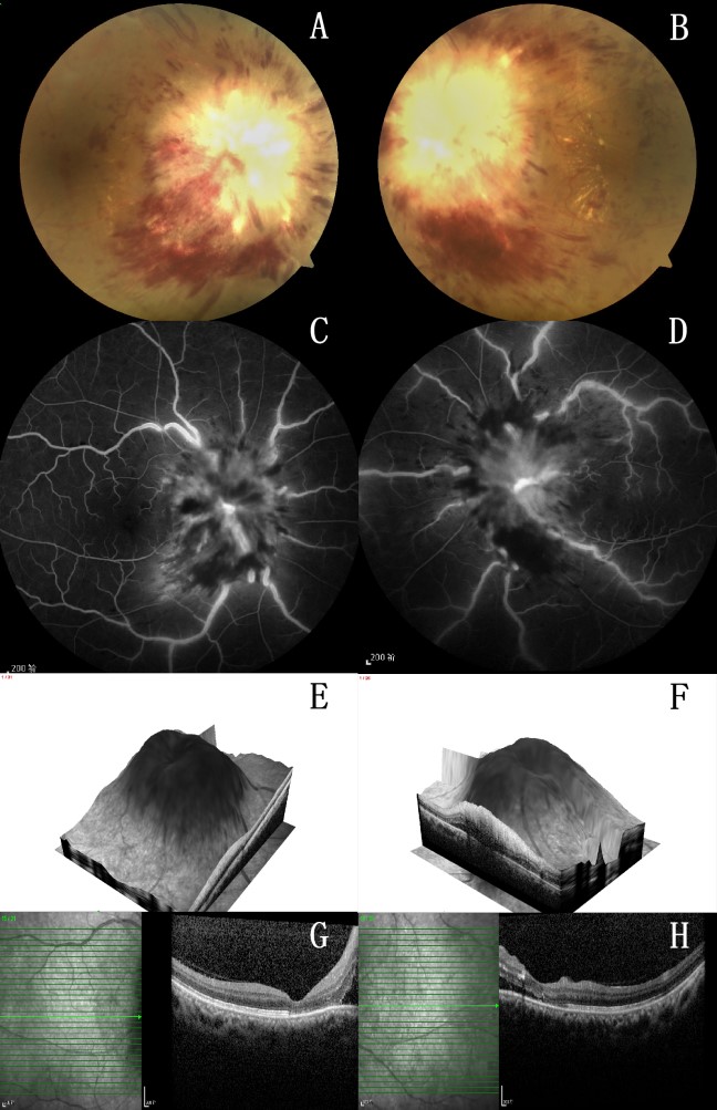
The patient has no history of smoking or alcohol consumption. Upon ophthalmologic examination, her best-corrected visual acuity score was 0.25 in the right eye and hand motion at 30 cm in the left eye. The pupillary light reflex was sluggish, and a general physical examination revealed no significant findings. Funduscopy showed a severely swollen optic disc (Frisen grade 5) with no visible optic disc margin and splinter hemorrhages, and the retinal vessels were obscured, suggesting intracranial hypertension. Additionally, numerous yellow spots were observed around the macular region (Figure 1). The patient was admitted for further evaluation and started on medications to reduce intracranial pressure (ICP), support nerve function, and improve circulation. Fundus fluorescence angiography (FFA) demonstrated a delayed arm-retinal arterial filling time of 14 s, early juxtapapillary capillary dilatation and leakage, late optic disc fluorescence staining, and dilation and staining of posterior pole retinal vessels (Figure 1). Spectral-domain optical coherence tomography (OCT) revealed papilledema and retinal nerve fiber layer (RNFL) detachment in both eyes. Reflective clusters with varying degrees of intensity were noted in the RNFL of the left eye, and the retinal surface appeared rough with multiple nodular projections (Figure 1). Considering treatment failure with hormone therapy and the peripapillary fluid accumulation rather than RNFL thickening on OCT, optic neuritis was excluded.

Figure 1 Fundus examination
Funduscopy showed a severely swollen optic disc (Frisen grade 5) with no visible optic disc margin and splinter hemorrhages (A: right eye; B: left eye.). FFA showed late disc fluorescence staining, and dilation and staining of retinal vessels of the posterior pole (C: right eye; D: left eye.). OCT showed papilledema (E: right eye; F: left eye.) and RNFL detachment (G: right eye; H: left eye.). Clusters of reflection points with varying degrees of intensity existed in the RNFL of left eye.

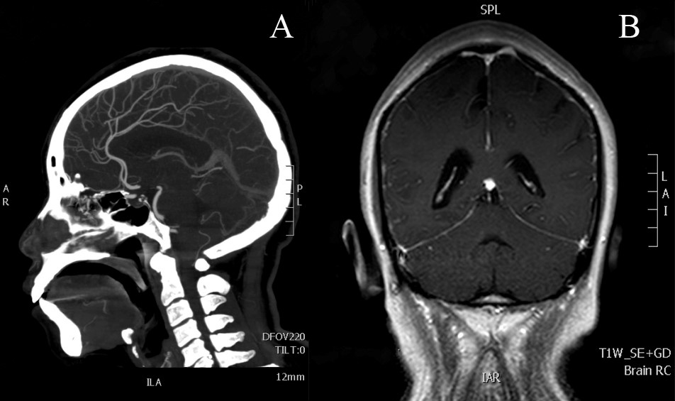
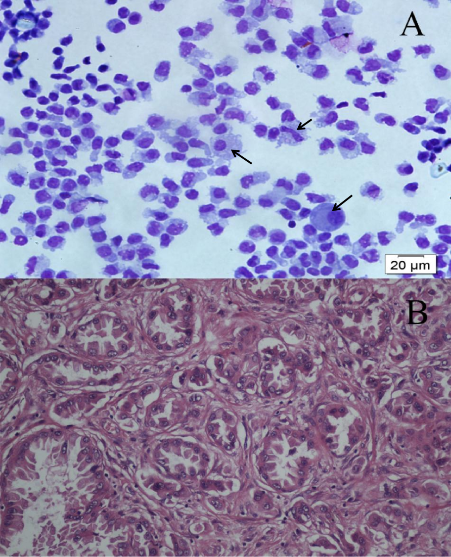
To investigate the cause of increased ICP, gadolinium-enhanced brain magnetic resonance imaging (MRI) revealed diffuse meningeal enhancement. Moreover, computed tomography angiography revealed no significant abnormalities (Figure 2). These findings ruled out intracranial tumors, cerebral pseudotumors, and intracranial venous sinus thrombosis as causes of intracranial hypertension. A lumbar puncture was subsequently performed, and CSF flow analysis revealed a pressure of 28 cm H2O with normal glucose and protein levels. CSF cytopathological analysis revealed malignant cells with karyoplasm ratio imbalance, double nuclei, and abnormal division (Figure 5).

Figure 2 Brain neuroimaging examination
(A): Computed tomography angiography (CTA). (B): Gadolinium-enhanced brain magnetic resonance imaging (MRI). CTA showed no significant abnormalities. MRI revealed diffuse meningeal enhancement.

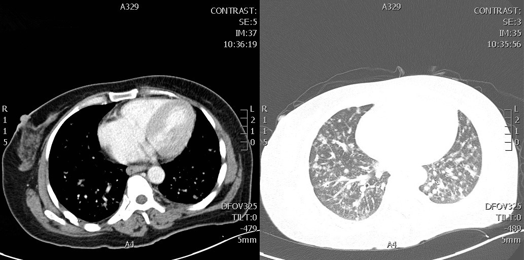
Moreover, the routine chest X-rays revealed scattered, diffuse shadows in both bilateral lungs, while  a contrast-enhanced chest computed tomography (CT) scan revealed pulmonary nodules at the apex of the left lung, consistent with lung cancer with multiple double- pulmonary metastases (Figure 3). In the serum tumor markers, the carcinoembryonic antigen is elevated to 19.19 ng/ml, and the carbohydrate antigen 153 is elevated to 69.99 U/ml. Positron emission tomography-CT (PET-CT), a more sensitive tool for detecting meningeal dissemination, confirmed diffuse malignant lesions in the lung, with suspected lymphatic metastasis involving the left supraclavicular, inferior thoracic, and mediastinal lymph nodes. No abnormalities were found in other regions (Figure 4). A biopsy of the left supraclavicular lymph nodes revealed invasive adenocarcinoma cells (Figure 5).

Figure 3 Enhanced computed tomography (CT) scan of the chest

The CT scan detected pulmonary nodules in the apex of the left lung.

Figure 4 Positron emission tomography-CT (PET-CT)

The PET-CT prompted the diffuse and varied size malignant lesions of lung (A to I) and revealed nothing remarkable in the meninges, brain parenchyma and brain ventricles ( J to M).

Figure 5 Cerebrospinal fluid (CSF) cytopathology and biopsy of lymph nodes
(A): CSF Cytopathology. (B): Biopsy of Lymph Nodes. The CSF cytopathology, using the Wright Staining method, showed the presence of malignant cells with caryoplasm ratio imbalance, double nuclei, and abnormal division (black arrow). Biopsy of the left supraclavicular lymph nodes demonstrated invasive adenocarcinoma cells.

Based on these findings, the patient was diagnosed with MC secondary to lung adenocarcinoma. Twelve days after admission, she complained of progressive weakness, astasia, non-intense headaches, and episodes of dizziness. Her symptoms worsened the following day, leading to severe headaches, nausea, seizures, and aconuresis. The patient declined further medical therapy and opted out of hospice care. Follow-up phone calls revealed that the patient passed away 2 months post-discharge, without no post-mortem examinations.

DISCUSSION

Although MC is a well-documented condition, this case describes unique features of meningeal metastases resulting from advanced lung cancer, accompanied by retinal signs of optic neuritis and resistance to methylprednisolone that have not been reported previously. Although MC is typically linked to advanced stages of systemic cancer, it can occur at any stage of neoplastic disease. A study indicates that in the United States, 1% to 8% of cancer patients are diagnosed with meningeal carcinomatosis, with approximately 110,000 new cases reported annually.[3] Among cancers, melanoma has the highest incidence rate of MC at 23%, followed by lung cancer ( 9%–25%) and breast cancer (5%).[4] Non-small-cell lung cancer (NSCLC) has the highest meningeal spread rates, with approximately 11% of patients with NSCLC developing MC.[5-6] Neurological symptoms are often more prominent than other manifestations and typically involve multiple regions of the neuraxis. Current diagnostic techniques are limited in detecting MC in its early stages, leading to significant neurological damage. Therefore, MC should be considered in the differential diagnosis of multifocal diseases and isolated syndromes, including intracranial hypertension, cranial neuropathy, or cauda equina syndrome. In this case, the patient initially presented with vision loss as the sole symptom, with no apparent abnormalities on initial examinations. She was initially diagnosed with optic neuritis and treated with methylprednisolone, which was ineffective. This prompted further examination of other major organs. As the disease progressed, the patient experienced symptoms such as progressive weakness, astasia, non-intense headaches, and episodes of dizziness, which became more serious, suggesting MC may be the cause of these symptoms. A contrast-enhanced chest CT revealed pulmonary nodules in the apex of the left lung, diagnosed as lung cancer with multiple double-pulmonary metastases. Diffuse thoracic lesions are easily misinterpreted as lung disease (metastatic or primary lung cancer), particularly when the primary lesion is atypical; therefore, PET-CT combined with local lymph node biopsy should be employed to characterize primary lesions for early diagnosis and treatment.

In 2021, the Consensus of Chinese MC Experts recommended prioritizing CSF cytology for diagnosing MC, as it demonstrates greater sensitivity and specificity compared to methods based on neuroimaging or clinical symptoms. Notably, MC should be considered in patients presenting with unusual neuro-ophthalmological features, with a definitive diagnosis made upon cytological analysis of CSF. Lumbar puncture should be performed in all suspected cases of MC to assess cell count, chemistry, and cytopathology.[7] Abnormal, non-specific CSF findings are observed in over 90% of MC cases, including pleocytosis (60%), elevated ICP (50%), elevated protein levels (80%), and low glucose levels (25%).[8] The presence of malignant cells in the CSF is the diagnostic hallmark, but initial lumbar punctures detect these cells in only 50% of patients with MC, necessitating repeated testing. By the third lumbar puncture, CSF cytopathology confirms the diagnosis in 94% of cases.[9] Testing for circulating tumor DNA (ctDNA) in CSF is emerging as a molecular diagnostic tool. Non-tumorous diseases and benign tumors do not produce ctDNA, making CSF ctDNA test findings more representative of the tumor burden status in MC. This is helpful for diagnosing MC in patients who present with negative CSF cytology and imaging findings.[10-12] MRI, although commonly used, has a high false-negative rate of approximately 30%.[13] Patients suspected of MC should undergo neuroimaging examination prior to CSF lumbar puncture. MRI has a sensitivity of 20–90%[14-15] and lacks the specificity to be solely diagnostic. This case illustrates the role of systemic examination in diagnosing MC. MRI revealed diffuse meningeal enhancement, and CSF analysis confirmed the presence of malignant cells. Based on these findings and the symptoms presented, an initial diagnosis of MC was made.

Imaging revealed no meningeal abnormalities, suggesting early meningeal involvement with potential extension to the optic nerve and retina. The mechanism by which malignant cells infiltrate the meninges remains unclear, but proposed pathways, depending on the histological characteristics of the primary tumor, include: (1) hematogenous dissemination first to the choroid plexus, then to the meninges, (2) primary hematogenous metastases through the meningeal vessels, and (3) centripetal extension along perivascular and perineural lymphatics, extending from axial lymphatic nodes and vessels, passing through intervertebral, and possibly from the cranial, foramina to the meninges.[8,14,16-17] Under normal circumstances, the subdural space is a potential space that does not directly communicate with the subarachnoid space. Such communication typically occurs when the barrier between the dura mater and arachnoid mater is disrupted by trauma or certainpathological conditions.[18] Therefore, it is hypothesized that the subdural space could become a factor in the pathogenesis of MC in cases where meningeal disease leads to anatomical abnormalities. In this patient, hematogenous spread was unlikely, and we hypothesized a potential direct communication between the lung and vertebral canal via blood or lymph vessels or direct spread from the spine.

Clinical manifestations of MC are typically due to the obstruction of CSF flow, leading to increased ICP or hydrocephalus. Symptoms can vary depending on the affected region. Intracranial hypertension may cause headaches, nausea, and vomiting, while parenchymal involvement can lead to consciousness disturbances, seizures, cognitive impairment, and gait abnormalities. Cranial nerve involvement may result in diplopia, vision loss, hearing loss, facial muscle paralysis, and difficulty swallowing. Spinal cord and spinal nerve involvement can present as weakness, bowel and bladder dysfunction, and lower back or leg pain. Additionally, direct tumor invasion within the brain or spinal cord can cause cranial-nerve palsies or radiculopathies. Ocular symptoms, such as vision loss, ocular motility deficit, pupillary abnormalities, and papilledema, have been reported in 91% of patients with MC.[8] Furthermore, 30–40% of the patients experience blindness, either in one or both eyes.[19] The exact mechanism of vision loss remains unclear, but proposed mechanisms to explain its occurrence include: 1) Direct involvement of the optic nerve: Malignant cells can directly infiltrate the optic nerve or its sheath, leading to compression, ischemia, and destruction of the optic nerve fibers, and causing vision loss. Moreover, inflammation of the optic nerve due to tumor-associated immune responses or paraneoplastic syndromes can impair signal transmission, leading to impaired vision or  blindness. 2) Increased ICP: Elevated ICP can result in papilledema, which can induce irreversible optic nerve damage and vision loss. In addition, elevated ICP can induce venous congestion, further compromising the optic nerve’ s blood supply and exacerbating damage. 3) Ischemic optic neuropathy: MC may involve occlusion or compression of blood vessels supplying the optic nerve or retina. Ischemia can result in sudden or progressive vision loss. 4) Tumor involvement of the visual pathways: Malignant infiltration of cranial nerves III, IV, and VI may impair eye movement, causing diplopia (double vision), while the involvement of cranial nerve II leads directly to vision loss. 5) Retinal and choroidal metastases: Direct metastases to the retina or choroid can cause localized damage, leading to vision distortion or loss. 6) Treatment-related causes: Radiation therapy (RT) and intrathecal or systemic chemotherapy can damage the optic nerve, manifesting as delayed vision loss. [20]The nature of the reflective points observed in the RNFL of the left eye was uncertain. At the same site, funduscopy revealed yellow spots, which were likely hard exudates; however, no leakage was detected on FFA. Therefore, these speckles were hypothesized to represent hemorrhage or cancer metastasis. Thus, this highlights the potential utility of fundus appearance in the differential diagnosis of MC and optic neuritis.

Despite recent advancements in anticancer therapies, only a limited improvement has been achieved in the treatment of MC. Current treatments primarily focus on alleviating neurological symptoms, improving quality of life, and extending patient survival, but many of these approaches remain largely palliative. The treatment of MC is guided by two primary principles: reducing ICP in the brain caused by CSF buildup and minimizing the number of malignant cells.[21] RT plays an important palliative role in symptom management. The commonly used types of involved-field RT include whole-brain irradiation, lumbosacral irradiation, and skull-base RT. Intra-CSF (intrathecal) therapy is also a frequently utilized treatment approach for MC, providing a statistically significant benefit regarding survival.[8,22] Currently, the four primary agents for intra-CSF therapy include methotrexate, cytarabine, thiotepa, and liposomal cytarabine, with methotrexate being the most commonly used.[23] Recently, the application of immune checkpoint inhibitors (ICIs) in MC has increased. ICIs enhance the immune system's ability to identify and target cancer cells, offering a new therapeutic approach for treating immune-evading cancer cells. Targeted therapies, such as epidermal growth factor receptor inhibitors and gene therapy, have demonstrated efficacy in specific cases but require further clinical validation. Additionally, treatment strategies should be tailored to the patient's overall health and disease progression, integrating chemotherapy, immunotherapy, supportive care, and surgical interventions as appropriate to improve quality of life and extend survival. The prognosis for MC remains generally poor. Without targeted intervention, the median survival time is limited to a few weeks. However, with comprehensive treatment approaches, the median survival can extend to several months.[9,19,24]

CONCLUSION

MC is a severe complication of systemic cancer, often linked to a poor prognosis and limited survival. Treatment typically focuses on alleviating symptoms and prolonging life, making early detection crucial. As demonstrated in our patient, ocular symptoms can occasionally serve as the initial or primary complaint. In such cases, ophthalmologists play a vital role in identifying the condition by conducting appropriate diagnostic tests and providing timely referrals. When a patient presents with acute vision loss without an intraocular cause and exhibits signs of multifocal neurological dysfunction, particularly in the context of suspected systemic cancer, MC should be considered.

Correction notice

None

Acknowledgement

None

Author Contributions

(Ⅰ) Conception and design: Jianqiao Li
(Ⅱ) Administrative support: Jianqiao Li
(Ⅲ) Provision of study materials or patients: Chao Huang
(Ⅳ) Collection and assembly of data: Chao Huang, Ying Li
(Ⅴ) Data analysis and interpretation: Helei Wang, Shanshan Li
(Ⅵ) Manuscript writing: Helei Wang, Ying Li
(Ⅶ) Final approval of manuscript: All authors

Funding

None

Conflict of Interests

None of the authors has any conflicts of interest to disclose.

Patient consent for publication

None

Ethical Statement

None

Provenance and Peer Review

This article was a standard submission to our journal. The article has undergone peer review with our anonymous review system.

Data Sharing Statement

None

Open Access Statement

This is an Open Access article distributed in accordance with the Creative Commons Attribution-NonCommercial-NoDerivs 4.0 International License (CC BY-NC-ND 4.0), which permits the non-commercial replication and distribution of the article with the strict proviso that no changes or edits are made and the original work is properly cited (including links to both the formal publicationthrough the relevant DOI and the license).
1、Kaplan JG, DeSouza TG, Farkash A, et al. Leptomeningeal metastases: comparison of clinical features and laboratory data of solid tumors, lymphomas and leukemias. J Neuro Oncol. 1990, 9(3): 225-229. DOI: 10.1007/BF02341153.Kaplan JG, DeSouza TG, Farkash A, et al. Leptomeningeal metastases: comparison of clinical features and laboratory data of solid tumors, lymphomas and leukemias. J Neuro Oncol. 1990, 9(3): 225-229. DOI: 10.1007/BF02341153.
2、Kesari S, Batchelor TT. Leptomeningeal metastases. Neurol Clin. 2003, 21(1): 25-66. DOI: 10.1016/s0733- 8619(02)00032-4.Kesari S, Batchelor TT. Leptomeningeal metastases. Neurol Clin. 2003, 21(1): 25-66. DOI: 10.1016/s0733- 8619(02)00032-4.
3、Nayar G, Ejikeme T, Chongsathidkiet P, et al. Leptomeningeal disease: current diagnostic and therapeutic strategies. Oncotarget. 2017, 8(42): 73312-73328. DOI: 10.18632/oncotarget.20272.Nayar G, Ejikeme T, Chongsathidkiet P, et al. Leptomeningeal disease: current diagnostic and therapeutic strategies. Oncotarget. 2017, 8(42): 73312-73328. DOI: 10.18632/oncotarget.20272.
4、Thakkar JP, Kumthekar P, Dixit KS, et al. Leptomeningeal metastasis from solid tumors. J Neurol Sci. 2020,411 : 116706. DOI:10.1016/j.jns.2020.116706.Thakkar JP, Kumthekar P, Dixit KS, et al. Leptomeningeal metastasis from solid tumors. J Neurol Sci. 2020,411 : 116706. DOI:10.1016/j.jns.2020.116706.
5、El Rassy E, Botticella A, Kattan J, et al. Non-small cell lung cancer brain metastases and the immune system: from brain metastases development to treatment. Cancer Treat Rev. 2018, 68: 69-79. DOI: 10.1016/j.ctrv.2018.05.015.El Rassy E, Botticella A, Kattan J, et al. Non-small cell lung cancer brain metastases and the immune system: from brain metastases development to treatment. Cancer Treat Rev. 2018, 68: 69-79. DOI: 10.1016/j.ctrv.2018.05.015.
6、Nayak L, Lee EQ, Wen PY. Epidemiology of brain metastases. Curr Oncol Rep. 2012, 14(1): 48-54. DOI: 10.1007/s11912-011-0203-y.Nayak L, Lee EQ, Wen PY. Epidemiology of brain metastases. Curr Oncol Rep. 2012, 14(1): 48-54. DOI: 10.1007/s11912-011-0203-y.
7、Chamberlain MC, Glantz M, Groves MD, et al. Diagnostic tools for neoplastic meningitis: detecting disease, identifying patient risk, and determining benefit of treatment. Semin Oncol. 2009,36(4 Suppl 2): S35-S45. DOI:10.1053/j.seminoncol.2009.05.005Chamberlain MC, Glantz M, Groves MD, et al. Diagnostic tools for neoplastic meningitis: detecting disease, identifying patient risk, and determining benefit of treatment. Semin Oncol. 2009,36(4 Suppl 2): S35-S45. DOI:10.1053/j.seminoncol.2009.05.005
8、Wa l z J . O c u l a r m a n i f e s t a t i o n s o f m e n i n g e a l carcinomatosis: a case report and literature review. Optometry. 2011, 82(7): 408-412. DOI: 10.1016/ j.optm.2010.12.015.Wa l z J . O c u l a r m a n i f e s t a t i o n s o f m e n i n g e a l carcinomatosis: a case report and literature review. Optometry. 2011, 82(7): 408-412. DOI: 10.1016/ j.optm.2010.12.015.
9、Balm M, Hammack J. Leptomeningeal carcinomatosis. Presenting features and prognostic factors. Arch Neurol. 1996, 53(7): 626-632. DOI: 10.1001/ archneur.1996.00550070064013.Balm M, Hammack J. Leptomeningeal carcinomatosis. Presenting features and prognostic factors. Arch Neurol. 1996, 53(7): 626-632. DOI: 10.1001/ archneur.1996.00550070064013.
10、Li YS, Jiang BY, Yang JJ, et al. Unique genetic profiles from cerebrospinal fluid cell-free DNA in leptomeningeal metastases of EGFR-mutant non-small-cell lung cancer: a new medium of liquid biopsy. Ann Oncol..2018, 29(4): 945-952. DOI:10.1093/annonc/mdy009.Li YS, Jiang BY, Yang JJ, et al. Unique genetic profiles from cerebrospinal fluid cell-free DNA in leptomeningeal metastases of EGFR-mutant non-small-cell lung cancer: a new medium of liquid biopsy. Ann Oncol..2018, 29(4): 945-952. DOI:10.1093/annonc/mdy009.
11、Zhao Y, He JY, Zou YL, et al. Evaluating the cerebrospinal fluid ctDNA detection by next-generation sequencing in the diagnosis of meningeal Carcinomatosis. BMC Neurol. 2019, 19(1): 331. DOI: 10.1186/s12883-019-1554-5.Zhao Y, He JY, Zou YL, et al. Evaluating the cerebrospinal fluid ctDNA detection by next-generation sequencing in the diagnosis of meningeal Carcinomatosis. BMC Neurol. 2019, 19(1): 331. DOI: 10.1186/s12883-019-1554-5.
12、Zhao Y, He JY, Cui JZ, et al. Detection of genes mutations in cerebrospinal fluid circulating tumor DNA from neoplastic meningitis patients using next generation sequencing. BMC Cancer. 2020, 20(1): 690. DOI: 10.1186/ s12885-020-07172-x.Zhao Y, He JY, Cui JZ, et al. Detection of genes mutations in cerebrospinal fluid circulating tumor DNA from neoplastic meningitis patients using next generation sequencing. BMC Cancer. 2020, 20(1): 690. DOI: 10.1186/ s12885-020-07172-x.
13、Chamberlain MC, Sandy AD, Press GA. Leptomeningeal metastasis: a comparison of gadolinium-enhanced MR and contrast-enhanced CT of the brain. Neurology. 1990, 40(3 Pt 1): 435-438. DOI: 10.1212/wnl.40.3_part_1.435.Chamberlain MC, Sandy AD, Press GA. Leptomeningeal metastasis: a comparison of gadolinium-enhanced MR and contrast-enhanced CT of the brain. Neurology. 1990, 40(3 Pt 1): 435-438. DOI: 10.1212/wnl.40.3_part_1.435.
14、Le Rhun E, Taillibert S, Chamberlain MC. Carcinomatous meningitis: Leptomeningeal metastases in solid tumors. Surg Neurol Int. 2013, 4(Suppl 4): S265-S288. DOI: 10.4103/2152-7806.111304.Le Rhun E, Taillibert S, Chamberlain MC. Carcinomatous meningitis: Leptomeningeal metastases in solid tumors. Surg Neurol Int. 2013, 4(Suppl 4): S265-S288. DOI: 10.4103/2152-7806.111304.
15、Lee SJ, Lee JI, Nam DH, et al. Leptomeningeal carcinomatosis in non–small-cell lung cancer patients: impact on survival and correlated prognostic factors. J Thorac Oncol. 2013, 8(2): 185-191. DOI: 10.1097/ jto.0b013e3182773f21.Lee SJ, Lee JI, Nam DH, et al. Leptomeningeal carcinomatosis in non–small-cell lung cancer patients: impact on survival and correlated prognostic factors. J Thorac Oncol. 2013, 8(2): 185-191. DOI: 10.1097/ jto.0b013e3182773f21.
16、Kokkoris%20CP.%20Leptomeningeal%20carcinomatosis.%20How%20does%20%0Acancer%20reach%20the%20pia-arachnoid%3F%20Cancer.%201983%2C%2051(1)%3A%20154-%0A160.%20DOI%3A%2010.1002%2F1097-0142(19830101)51%3A%201%3C154%3A%20aid%02cncr2820510130%3E3.0.co%3B2-k.Kokkoris%20CP.%20Leptomeningeal%20carcinomatosis.%20How%20does%20%0Acancer%20reach%20the%20pia-arachnoid%3F%20Cancer.%201983%2C%2051(1)%3A%20154-%0A160.%20DOI%3A%2010.1002%2F1097-0142(19830101)51%3A%201%3C154%3A%20aid%02cncr2820510130%3E3.0.co%3B2-k.
17、Scanlon, E F, and S Murthy. The process of metastasis. CA: a cancer journal for clinicians vol. 41,5 (1991): 301-5. DOI:10.3322/canjclin.41.5.301.Scanlon, E F, and S Murthy. The process of metastasis. CA: a cancer journal for clinicians vol. 41,5 (1991): 301-5. DOI:10.3322/canjclin.41.5.301.
18、Lin MS. Subdural lesions linking additional intracranial spaces and chronic subdural hematomas: a narrative review with mutual correlation and possible mechanisms behind high recurrence. Diagnostics. 2023, 13(2): 235. DOI: 10.3390/diagnostics13020235.Lin MS. Subdural lesions linking additional intracranial spaces and chronic subdural hematomas: a narrative review with mutual correlation and possible mechanisms behind high recurrence. Diagnostics. 2023, 13(2): 235. DOI: 10.3390/diagnostics13020235.
19、Wasserstrom, W R et al. Diagnosis and treatment of leptomeningeal metastases from solid tumors: experience with 90 patients. Cancer. 1982 49 (4): 759-772. DOI:10.1002/1097-0142(19820215)49:4<759::aidcncr2820490427>3.0.co;2-7.Wasserstrom, W R et al. Diagnosis and treatment of leptomeningeal metastases from solid tumors: experience with 90 patients. Cancer. 1982 49 (4): 759-772. DOI:10.1002/1097-0142(19820215)49:4<759::aidcncr2820490427>3.0.co;2-7.
20、McFadzean R, Brosnahan D, Doyle D, et al. A diagnostic quartet in leptomeningeal infiltration of the optic nerve sheath. J Neuroophthalmol. 1994,14(3),: 175-182. DOI:10.3109/01658109409024045.McFadzean R, Brosnahan D, Doyle D, et al. A diagnostic quartet in leptomeningeal infiltration of the optic nerve sheath. J Neuroophthalmol. 1994,14(3),: 175-182. DOI:10.3109/01658109409024045.
21、Corbin, Zachary A, and Seema Nagpal. Leptomeningeal Metastases. JAMA. 2016, 2(6): 839. DOI: 10.1001/jamaoncol.2015.3502.Corbin, Zachary A, and Seema Nagpal. Leptomeningeal Metastases. JAMA. 2016, 2(6): 839. DOI: 10.1001/jamaoncol.2015.3502.
22、Gauthier H, Guilhaume MN, Bidard FC, et al. Survival of breast cancer patients with meningeal carcinomatosis. Ann Oncol. 2010,21(11) : 2183-2187. DOI:10.1093/annonc/ mdq232.Gauthier H, Guilhaume MN, Bidard FC, et al. Survival of breast cancer patients with meningeal carcinomatosis. Ann Oncol. 2010,21(11) : 2183-2187. DOI:10.1093/annonc/ mdq232.
23、Mack%20F%2C%20Baumert%20BG%2C%20Sch%C3%A4fer%20N%2C%20et%20al.%20Therapy%20of%20%0Aleptomeningeal%20metastasis%20in%20solid%20tumors.%20Cancer%20Treat%20%0ARev.%202016%2C%2043%3A%2083-91.%20DOI%3A%2010.1016%2Fj.ctrv.2015.12.004.Mack%20F%2C%20Baumert%20BG%2C%20Sch%C3%A4fer%20N%2C%20et%20al.%20Therapy%20of%20%0Aleptomeningeal%20metastasis%20in%20solid%20tumors.%20Cancer%20Treat%20%0ARev.%202016%2C%2043%3A%2083-91.%20DOI%3A%2010.1016%2Fj.ctrv.2015.12.004.
24、Rudnicka H, Niwińska A, Murawska M. Breast cancer leptomeningeal metastasis: the role of multimodality treatment. J Neuro Oncol. 2007, 84(1): 57-62. DOI: 10.1007/s11060-007-9340-4.Rudnicka H, Niwińska A, Murawska M. Breast cancer leptomeningeal metastasis: the role of multimodality treatment. J Neuro Oncol. 2007, 84(1): 57-62. DOI: 10.1007/s11060-007-9340-4.
上一篇
下一篇
出版者信息
目录