您的位置: 首页 > 2025年9月 第2卷 第3期 > 文字全文
2023年7月 第38卷 第7期11
目录

Bilateral diffuse uveal melanocytic proliferation: a case report and literature review

Bilateral diffuse uveal melanocytic proliferation: a case report and literature review

来源期刊: Eye Science | 2025年9月 第2卷 第3期 276-282 发布时间:2025-09-20 收稿时间:2025/9/25 15:51:16 阅读量:142
作者:
关键词:
BDUMP choroid adrenocortical carcinoma
DOI:
10.12419/ykxb25021501
Received date:
2025-02-27 
Revised date:
2025-03-26 
Accepted date:
2025-04-30 
Published online:
2025-09-20 
Bilateral diffuse uveal melanocytic proliferation (BDUMP) is a rare paraneoplastic ocular syndrome, often associated with significant vision loss and poor prognosis[1].  BDUMP is typically linked to systemic malignancies, including ovarian, lung, pancreatic, and colorectal cancers[1]. Here, we reported a case of BDUMP in a 59-year-old male patient who had been misdiagnosed with a nonfunctional adrenal adenoma. This man, with a history of diabetes mellitus and high blood pressure, complained of progressive visual loss in both eyes. He was diagnosed with BDUMP after undergoing comprehensive ophthalmic assessments at our eye center. After being referred to the urology department, he was initially diagnosed with a right nonfunctional adrenal adenoma because his renin-aldosterone levels and serum potassium levels were normal. However, three months later, the adrenal mass rapidly increased in size, and liver metastasis was confirmed. Despite normal tumor marker levels, a histopathological diagnosis ultimately confirmed adrenocortical carcinoma. This case underscores the importance of considering underlying malignancies in patients with BDUMP and highlights the necessity for early systemic evaluation, including imaging studies and multidisciplinary consultations. Timely diagnosis of underlying cancers may improve patient outcomes. It is worth noting that normal hormone levels and a tumor small-sized cannot be used as conclusive evidence that the tumor is a nonfunctional adrenal adenoma; patients with BDUMP should be closely followed up to identify the primary malignancy.

HIGHLIGHTS

· This is the first report of bilateral diffuse uveal melanocytic proliferation (BDUMP) associated with adrenocortical carcinoma, whereas BDUMP was previously mostly linked to ovarian, lung, pancreatic, and colorectal cancers.
· The combination of multimodal imaging (fundus photography, fluorescein angiography, spectral-domain optical coherence tomography, B-scan and PET-CT) enhances the diagnostic accuracy of BDUMP and the underlying malignancies.
· A surveillance protocol for BDUMP patients is proposed, which includes initial whole-body imaging (CT/PET-CT, abdominal/pelvic MRI) along with regular ophthalmic and systemic evaluations every 3-6 months to facilitate timely detection of underlying malignancies.

INTRODUCTION

Bilateral diffuse uveal melanocytic proliferation (BDUMP), first described by Machemer, is a rare paraneoplastic syndrome that primarily affects both eyes[2]. BDUMP was once regarded as extremely rare, but its increased incidence in recent years is probably attribute to greater awareness of the condition and the longer life expectancy of oncology patients[3]. The most typical manifestations of BDUMP include exudative retinal detachment, progressive cataract, diffuse thickening of the choroid with pigmented choroidal plaques, and mottled fluorescence on fluorescein angiography (FA). These manifestations are caused by the degeneration and hyperplasia of retinal pigment epithelium (RPE)[4,5]. The underlying cause of BDUMP is the diffuse proliferation of benign melanocytes within the uvea, predominantly affecting the choroid[1,4]. Histopathologically, these proliferations are not associated with the primary non-ocular malignancy[1]. BDUMP is generally considered a premetastatic lesion of malignant tumor and associated with remarkable vision loss and a generally poor prognosis.

Epidemiologically, BDUMP mainly affects middle-aged and elderly individuals, with over 80% of cases showing concurrent systemic cancers6,7. BDUMP is commonly observed in female patients, with ovarian carcinoma being prevalent, and in male patients, with lung carcinoma[8,9]. Additionally, BDUMP has been reported in association with gastric adenocarcinoma, colonic adenocarcinoma, bladder cancer, pancreatic carcinoma, and primary vitreoretinal lymphoma[10-12].

In this report, we presented a case of BDUMP in a middle-aged man. He first visited  the ophthalmic clinic and initially misdiagnosed with nonfunctional adrenal adenoma at urology department, but was ultimately found to have adrenocortical carcinoma (ACC).

CASE REPORT

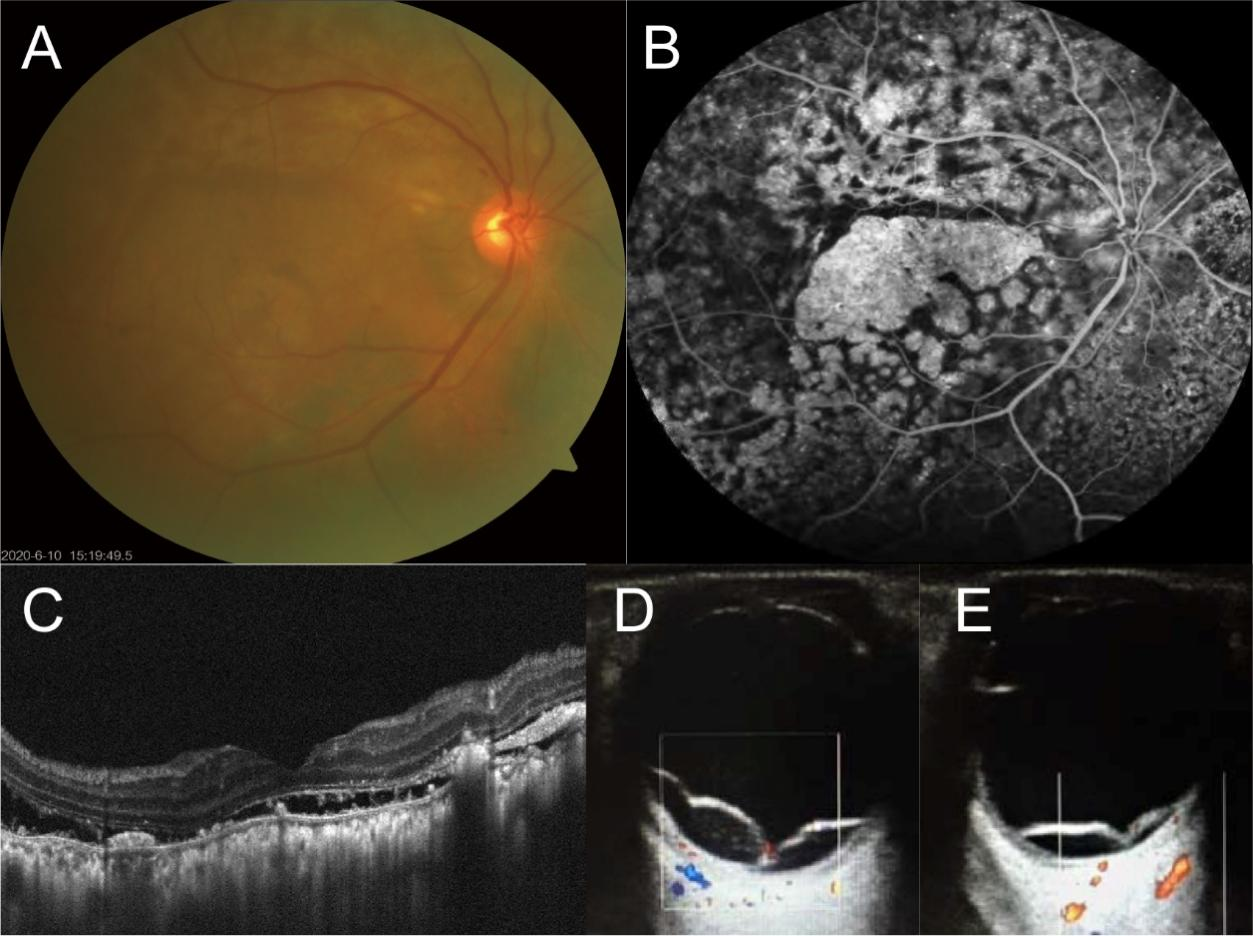
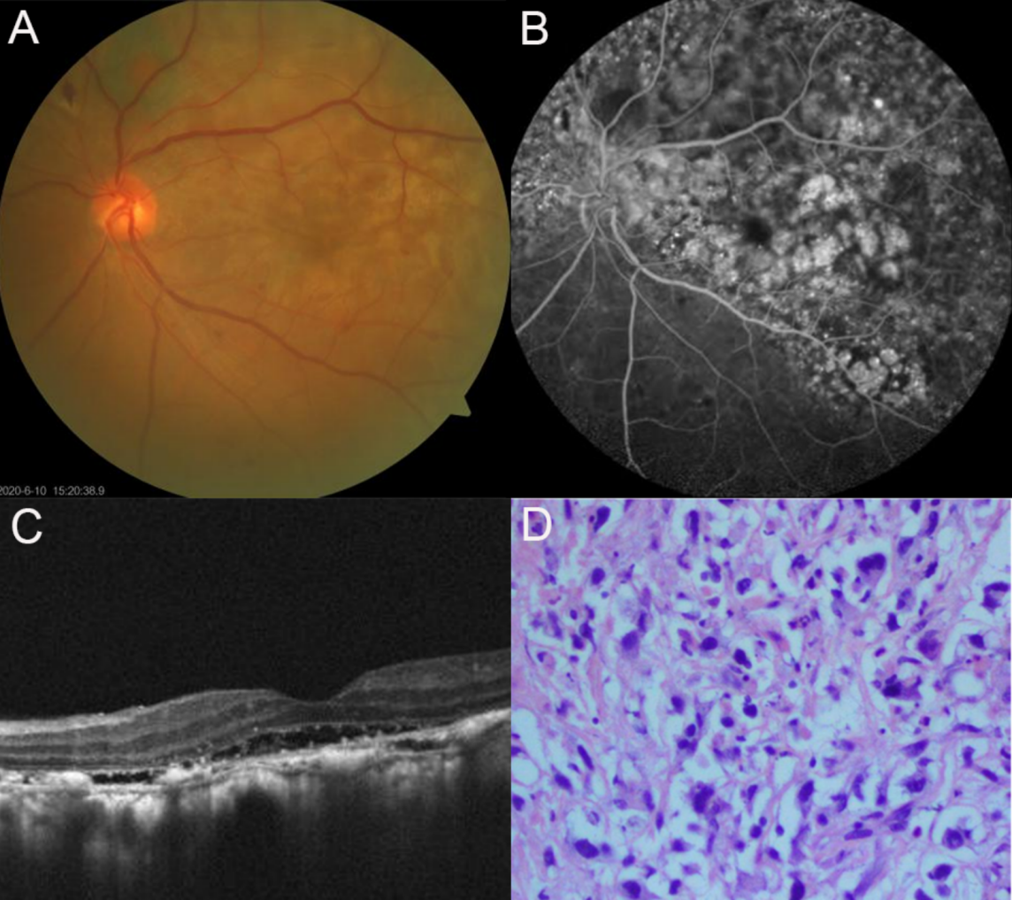
A 59-year-old male patient presented with a 3-month history of progressive visual loss in both eyes. His best corrected visual acuity (BCVA) was 20/300 in the right eye (OD) and 20/200 in the left eye (OS). The intraocular pressure was 10.3 mmHg OD and 9.7 mmHg OS. Fundus photograph exhibited multiple choroidal nevi-like melanocytic tumors and subtle round, orange-red subretinal patches at the level of retinal pigment epithelium in both eyes (Figure 1A, 2A). FA displayed pathognomonic mottled fluorescence patterns, alternating between hypofluorescent plaques and hyperfluorescent window defects (Figure 1B, 2B). Spectral-domain optical coherence tomography revealed bilateral exudative retinal detachment with subretinal fluid accumulation, hyperreflective material deposition in the outer retinal layers and RPE layers, and significant choroidal thickening (Figure 1C, 2C). B-scan images taken one month later reveled more severe retinal detachment (Figures 1D, 1E). The absence of  signs of infectious disease or lymphadenopathy, along with an IL-10/IL-6 ratio <1.0 in the aqueous humor, ruled out infectious granulomatous choroiditis and lymphoma. Based on these findings, this patient, who also had diabetes mellitus, was diagnosed with BDUMP at our eye center.

BDUMP is often considered a precursor to metastatic malignancy[10]. Given the patient's critically elevated blood pressure (204/118 mmHg), we recommended an immediate referral to the cardiology department for blood pressure management and a comprehensive systemic examination. Abdominal computed tomography (CT) identified a well-circumscribed, homogeneous, and isodense nodule (2.6×1.6×3.4 cm) in the right adrenal limb of the patient (Figure 3A, 3B). The patient was then referred to the urology department for further evaluation. After excluding other abnormalities in the renin-angiotensin-aldosterone system and normal potassium levels, a diagnosis of a nonfunctional right adrenal adenoma was made.

Three months later, during follow-up at an outside facility, CT images showed that the adrenal mass had rapidly enlarged to 56 x 46 x 55 mm, become irregular and inhomogeneous with an unclear boundary with the lower margin of the liver (Figure 3C, 3D). 18F-fluorodeoxyglucose positron emission tomography-CT (PET-CT) demonstrated increased metabolic activity in the right adrenal gland, raising suspicion of a malignant tumor (Figure 3E). An adrenal biopsy was performed, and histopathological examination (Figure 2D) along with immunohistochemical analysis of the adrenal and liver tissue confirmed the diagnosis of ACC with liver metastasis (Figure 3A, 3C). Despite normal tumor markers (including carcinoembryonic antigen, alpha-fetoprotein, CA125, CA15-3, and CA19-9), the patient was diagnosed with ACC with liver metastasis. Unfortunately, by the time of definitive diagnosis, the patient was no longer a candidate for surgery due to extensive hepatic involvement. He succumbed to disease progression five months post-diagnosis, with an overall survival of 12 months from the onset of ocular symptoms.

DISCUSSION

BDUMP is a rare paraneoplastic syndrome commonly observed in patients with systemic malignancies, which are often diagnosed after the ocular manifestations appear[13]. Pathophysiologically, tumor-secreted factors induce functional abnormalities in distant organs, including the eye[14]. BDUMP was originally characterized by multifocal reddish-orange subretinal plaques with early hyperfluorescence on FA, diffuse uveal thickening, exudative retinal detachment, and rapid cataract progression[3,14]. In our case, the primary malignancy, ACC, was detected more than four months after the onset of BDUMP and was found to have originated from what was initially thought to be a nonfunctional adrenal adenoma. No systemic malignancy was identified during the initial ophthalmic management.

BDUMP is associated with significant vision loss and poor prognosis[13,15,16]. The vision impairment in BDUMP is primarily due to uveal tract thickening, serous retinal detachment, and cataract formation[17]. Some studies suggest that glaucoma may precede BDUMP due to pigment dispersion, highlighting the importance of examining the iridocorneal angle pigment in suspected cases[18,19].

Although the pathogenesis of BDUMP remains incompletely understood, current evidence supports a paracrine mechanism involving tumor-derived melanocyte growth factors[1,20]. Serum from affected patients contains a melanocyte-specific IgG-bound factor (CMEP) that induces in vitro melanocyte proliferation[12,21]. Specifically, human melanocytes exposed to serum or plasma from BDUMP patients showed increased growth, whereas fibroblasts, keratinocytes, or ovarian cancer cells were not stimulated12. Given the role of CMEP in disease development, therapeutic strategies focus on treating primary malignancies or metastases. Interventions including surgical resection, radiotherapy, and chemotherapy have demonstrated efficacy in improving ocular outcomes[3,12]. Adjunctive therapies such as anti-VEGF agents and corticosteroids may reduce subretinal fluid but show variable results[22,23]. Regrettably, in our case, we could not assess this patient's eye condition further due to his death.

While BDUMP has been linked to several malignancies, including lung, ovarian, pancreatic, and colorectal cancers, this is the first report of its association with ACC. ACC is an aggressive malignancy, with approximately 80% of patients being  asymptomatic at diagnosis and 40% presenting with metastasis[24,25]. ACC tumors are typically heterogeneous, with lesions often greater than 6 cm in diameter, whereas benign adrenal adenomas are typically smaller than 4 cm. In this case, initial imaging revealed a 2.6×1.6×3.4 cm nodule in the right adrenal gland with smooth borders and no signs of hormone secretion[26,27]. Based on these findings, the diagnosis of a nonfunctional adrenal adenoma was made in our case. However, three months after initial diagnosis, follow-up imaging revealed rapid tumor enlargement and the detection of hepatic metastasis. This progression highlights the fact that even when early imaging does not show clear signs of malignancy, BDUMP may be early sign of cancer, such as ACC, especially in small tumors that lack obvious hormonal abnormalities. This emphasizes the need for heightened clinical vigilance during the early stages of ACC, especially in patients with BDUMP.

Therefore, for patients with BDUMP, it is crucial to maintain heightened clinical vigilance for underlying primary malignancies. Since BDUMP is typically first detected by ophthalmologists, it may be indicative of a serious systemic malignancy. Practical surveillance protocols for patients with BDUMP should integrate imaging modalities, biomarker monitoring, and interdisciplinary collaboration, which are essential to ensure early diagnosis and timely intervention. First, an initial comprehensive workup is recommended: whole-body CT/PET-CT should be performed at diagnosis to identify occult malignancies. MRI of the abdomen/pelvis should be used to rule out visceral or retroperitoneal tumors. Meanwhile, laboratory tests include tumor markers based on patient demographics and serum protein electrophoresis to exclude hematologic malignancies. Second, monitoring of the ophthalmic and systemic symptom and biomarkers is necessary: it is recommended that patients with BDUMP undergo ophthalmologic examinations every 3–6 months to monitor disease progression and visual function1. If new symptoms emerge, prompt imaging, targeted biopsies of suspicious lesions, and integration of molecular biomarker analysis are necessary. Third, a follow-up imaging schedule should be established: given the high association of BDUMP with rapidly progressing cancers, whole-body CT or PET-CT should be carried out every 3-6 months in the first year and annual imaging in the subsequent years to monitor for emerging tumors6. Continuous monitoring of tumor dynamics is critical to prevent diagnostic oversight and therapeutic delays. Clinicians should not only focus on ophthalmic manifestations in patients with BDUMP but also remain alert to the possibility of concurrent malignancies to optimize survival outcomes.

Figure 1 Multimodal images of the patient’s right eye and follow-up B-scans of both eyes of the patient

Figure 1 Multimodal images of the patient’s right eye and follow-up B-scans of both eyes of the patient
A) The fundus photography showed diffuse patchy yellowish-white lesions with subretinal pigmented circular lesions and pigment disorder in the right eye. B) FA demonstrated patchy window defect fluorescence with punctate hyperfluorescence in the right eye. C) The optic coherence tomography image of the right eye revealed exudative retinal detachment with subretinal fluid accumulation, hyperreflective material deposition in outer retinal layers and RPE layers, and significant choroidal thickening. D), E) The B-scan images showed more severe retinal detachment in both eyes one month later.

Figure 2 Multimodal images of the patient’s left eye and the histopathological finding of his adrenal tissue

Figure 2 Multimodal images of the patient’s left eye and the histopathological finding of his adrenal tissue
A) The fundus photography showed nummular RPE clumping, pigment disorder in left eye. B) The fluorescence angiography of the left eye indicated the multiple mottled fluorescence patterns, alternating between hypofluorescent plaques and hyperfluorescent window defects. C) The optic coherence tomography images of left eye revealed exudative retinal detachment, subretinal fluid, hyperreflective materials in the outer retina and RPE layer, and thickening of the choroid. D) The hematoxylin and eosin staining of adrenal tissue for histopathological examination revealed that there were significantly pathological cellular pleomorphism and high mitotic activity, and a large number of tumor cells with cytomorphologic features of ACC, such as nuclear shrinkage, nuclear atypia, multiple nuclei and large nucleoli (black arrows).

Figure 3 Initial and follow-up diagnostic CT images and follow-up PET-CT image of the right adrenal gland of this patient

Figure 3. Initial and follow-up diagnostic CT images and follow-up PET-CT image of the right adrenal gland of this patient
A) On initial diagnostic plain scan CT image, a 26 x 16 x 34 mm uniform and isodense nodule with smooth margin was observed in the inner limb of the right adrenal gland. B) The nodule showed moderate and homogeneous enhancement on the enhanced scanning. C) On follow-up diagnostic plain scan CT image, an approximately 56 x 46 x 55 mm irregular and inhomogeneous mass shadow was seen in the right adrenal gland, and the boundary with the lower margin of the liver was unclear. D) The mass had obvious inhomogeneous enhancement on the enhanced scan. E, An irregular mass in the right adrenal area of the retroperitoneum showed increased glucose metabolism on the follow-up PET-CT image.

Correction Notice

None

Acknowledgements

None.

Author Contributions

(I) Conception and design: Weiyi Zheng, Yan Luo
(II) Administrative support: Yan Luo
(III) Provision of study materials or patients: Weiyi Zheng
(IV) Collection and assembly of data: Weiyi Zheng, Aoxiang Wang
(V) Data analysis and interpretation: Aoxiang Wang, Yan Luo
(VI) Manuscript writing: All authors
(VII) Final approval of manuscript: All authors

Funding

This work was supported by the National Natural Science Foundation of China  (81770971).

Conflict of Interests

None of the authors has any conflicts of interest to disclose. All authors have declared in the completed the ICMJE uniform disclosure form.

Patient Consent for Publication

None

Ethics Statement

None

Data Sharing Statement

None

Open Access Statement

This is an Open Access article distributed in accordance with the Creative Commons AttributionNonCommercial-NoDerivs 4.0 International License (CC BY-NC-ND 4.0), which permits the non-commercial replication and distribution of the article with the strict proviso that no changes or edits are made and the original work is properly cited (including links to both the formal publication through the relevant DOI and the license).
1、Lavine JA, Ramos MS, Wolk AM, et al. Heterogeneity of cultured melanocyte elongation and proliferation factor in bilateral diffuse uveal melanocytic proliferation. Exp Eye Res. 2019, 184: 30-37. DOI: 10.1016/j.exer.2019.04.006Lavine JA, Ramos MS, Wolk AM, et al. Heterogeneity of cultured melanocyte elongation and proliferation factor in bilateral diffuse uveal melanocytic proliferation. Exp Eye Res. 2019, 184: 30-37. DOI: 10.1016/j.exer.2019.04.006
2、Tong N, Wang L, Wang N, et al. Bilateral diffuse uveal melanocytic proliferation secondary to rectal adenocarcinoma: a case report and literature review. Front Med (Lausanne). 2021, 8: 691686. DOI: 10.3389/fmed.2021.691686Tong N, Wang L, Wang N, et al. Bilateral diffuse uveal melanocytic proliferation secondary to rectal adenocarcinoma: a case report and literature review. Front Med (Lausanne). 2021, 8: 691686. DOI: 10.3389/fmed.2021.691686
3、Rafei-Shamsabadi D, Schneider J, Trefzer L, et al. Case report: blurred vision and eruptive nevi - bilateral diffuse uveal melanocytic proliferation with mucocutaneous involvement in a lung cancer patient. Front Oncol. 2021, 11: 658407. DOI: 10.3389/fonc.2021.658407.Rafei-Shamsabadi D, Schneider J, Trefzer L, et al. Case report: blurred vision and eruptive nevi - bilateral diffuse uveal melanocytic proliferation with mucocutaneous involvement in a lung cancer patient. Front Oncol. 2021, 11: 658407. DOI: 10.3389/fonc.2021.658407.
4、Guo J, Tang W, Liu W, et al. A case report of ultrasonographic findings in bilateral diffuse Uveal melanocytic proliferation. BMC Ophthalmol. 2020, 20(1): 476. DOI: 10.1186/s12886-020-01720-6.Guo J, Tang W, Liu W, et al. A case report of ultrasonographic findings in bilateral diffuse Uveal melanocytic proliferation. BMC Ophthalmol. 2020, 20(1): 476. DOI: 10.1186/s12886-020-01720-6.
5、Weppelmann TA, Khalil S, Zafrullah N, et al. Ocular paraneoplastic syndromes: a critical review of diffuse uveal melanocytic proliferation and autoimmune retinopathy. Cancer Control. 2022, 29: 10732748221144458. DOI: 10.1177/10732748221144458.Weppelmann TA, Khalil S, Zafrullah N, et al. Ocular paraneoplastic syndromes: a critical review of diffuse uveal melanocytic proliferation and autoimmune retinopathy. Cancer Control. 2022, 29: 10732748221144458. DOI: 10.1177/10732748221144458.
6、Parakh S, Maheshwari S, Das S, et al. Presumed bilateral diffuse uveal melanocytic proliferation - A case report and review of literature. Am J Ophthalmol Case Rep. 2022, 27: 101582. DOI: 10.1016/j.ajoc.2022.101582. Parakh S, Maheshwari S, Das S, et al. Presumed bilateral diffuse uveal melanocytic proliferation - A case report and review of literature. Am J Ophthalmol Case Rep. 2022, 27: 101582. DOI: 10.1016/j.ajoc.2022.101582.
7、 Ryu C, Havens SJ, Chen J, et al. A case of bilateral diffuse uveal melanocytic proliferation followed by massive unilateral uveal proliferation. Ocul Immunol Inflamm. 2024, 32(10): 2605-2611. DOI: 10.1080/09273948.2024.2355577. Ryu C, Havens SJ, Chen J, et al. A case of bilateral diffuse uveal melanocytic proliferation followed by massive unilateral uveal proliferation. Ocul Immunol Inflamm. 2024, 32(10): 2605-2611. DOI: 10.1080/09273948.2024.2355577.
8、Ayachit G, Ayachit A, Joshi S, et al. A rare case of unilateral diffuse melanocytic proliferation. Indian J Ophthalmol. 2018, 66(4): 588-590. DOI: 10.4103/ijo.IJO_979_17.Ayachit G, Ayachit A, Joshi S, et al. A rare case of unilateral diffuse melanocytic proliferation. Indian J Ophthalmol. 2018, 66(4): 588-590. DOI: 10.4103/ijo.IJO_979_17.
9、 Joseph A, Rahimy E, Sarraf D. Bilateral diffuse uveal melanocytic proliferation with multiple iris cysts. JAMA Ophthalmol. 2014, 132(6): 756-760. DOI: 10.1001/jamaophthalmol.2014.311. Joseph A, Rahimy E, Sarraf D. Bilateral diffuse uveal melanocytic proliferation with multiple iris cysts. JAMA Ophthalmol. 2014, 132(6): 756-760. DOI: 10.1001/jamaophthalmol.2014.311.
10、 Kalogeropoulos D, Afshar F, De Salvo G, et al. Diagnostic and therapeutic considerations in patients with bilateral diffuse uveal melanocytic proliferation. Int Ophthalmol. 2024, 44(1): 149. DOI: 10.1007/s10792-024-03087-9. Kalogeropoulos D, Afshar F, De Salvo G, et al. Diagnostic and therapeutic considerations in patients with bilateral diffuse uveal melanocytic proliferation. Int Ophthalmol. 2024, 44(1): 149. DOI: 10.1007/s10792-024-03087-9.
11、De Salvo G, Prakash P, Rennie CA, et al. Long-term survival in a case of bilateral diffuse uveal melanocytic proliferation. Eye (Lond), 2011, 25(10): 1385-1386. DOI: 10.1038/eye.2011.180. De Salvo G, Prakash P, Rennie CA, et al. Long-term survival in a case of bilateral diffuse uveal melanocytic proliferation. Eye (Lond), 2011, 25(10): 1385-1386. DOI: 10.1038/eye.2011.180.
12、Jansen JCG, Van Calster J, Pulido JS, et al. Early diagnosis and successful treatment of paraneoplastic melanocytic proliferation. Br J Ophthalmol. 2015, 99(7): 943-948. DOI: 10.1136/bjophthalmol-2014-305893. Jansen JCG, Van Calster J, Pulido JS, et al. Early diagnosis and successful treatment of paraneoplastic melanocytic proliferation. Br J Ophthalmol. 2015, 99(7): 943-948. DOI: 10.1136/bjophthalmol-2014-305893.
13、Sarkar P, Mehtani A, Gandhi HC, et al. Paraneoplastic ocular syndrome: a Pandora’s box of underlying malignancies. Eye (Lond), 2022, 36(7): 1355-1367. DOI: 10.1038/s41433-021-01676-x. Sarkar P, Mehtani A, Gandhi HC, et al. Paraneoplastic ocular syndrome: a Pandora’s box of underlying malignancies. Eye (Lond), 2022, 36(7): 1355-1367. DOI: 10.1038/s41433-021-01676-x.
14、 Borruat FX, Othenin-Girard P, Uffer S, et al. Natural history of diffuse uveal melanocytic proliferation. Case report. Ophthalmology. 1992, 99(11): 1698-1704. DOI: 10.1016/s0161-6420(92)31743-9. Borruat FX, Othenin-Girard P, Uffer S, et al. Natural history of diffuse uveal melanocytic proliferation. Case report. Ophthalmology. 1992, 99(11): 1698-1704. DOI: 10.1016/s0161-6420(92)31743-9.
15、Mihic Góngora L, Santalla C, Contreras Toledo D, et al. Bilateral diffuse uveal melanocytic proliferation (BDUMP) as ocular toxicity from durvalumab in advanced renal carcinoma. Eur J Cancer. 2022, 171: 10-12. DOI: 10.1016/j.ejca.2022.04.037. Mihic Góngora L, Santalla C, Contreras Toledo D, et al. Bilateral diffuse uveal melanocytic proliferation (BDUMP) as ocular toxicity from durvalumab in advanced renal carcinoma. Eur J Cancer. 2022, 171: 10-12. DOI: 10.1016/j.ejca.2022.04.037.
16、 Jabbarpoor Bonyadi MH, Ownagh V, Rahimy E, et al. Giraffe or leopard spot chorioretinopathy as an outstanding finding: case report and literature review. Int Ophthalmol. 2019, 39(6): 1405-1412. DOI: 10.1007/s10792-018-0948-5. Jabbarpoor Bonyadi MH, Ownagh V, Rahimy E, et al. Giraffe or leopard spot chorioretinopathy as an outstanding finding: case report and literature review. Int Ophthalmol. 2019, 39(6): 1405-1412. DOI: 10.1007/s10792-018-0948-5.
17、 Leskov I, Lyon AT, Jampol LM. Diffuse uveal melanocytic proliferation with primary vitreoretinal lymphoma. JAMA Ophthalmol. 2019, 137(7): 834-837. DOI: 10.1001/jamaophthalmol.2019.0905. Leskov I, Lyon AT, Jampol LM. Diffuse uveal melanocytic proliferation with primary vitreoretinal lymphoma. JAMA Ophthalmol. 2019, 137(7): 834-837. DOI: 10.1001/jamaophthalmol.2019.0905.
18、Da Silva MV, Rose K, Krema H. Pigment-induced glaucoma secondary to bilateral diffuse uveal melanocytic proliferation. Eur J Ophthalmol. 2022, 32(1): NP62-NP65. DOI: 10.1177/1120672120953076. Da Silva MV, Rose K, Krema H. Pigment-induced glaucoma secondary to bilateral diffuse uveal melanocytic proliferation. Eur J Ophthalmol. 2022, 32(1): NP62-NP65. DOI: 10.1177/1120672120953076.
19、Zhao F, Hu L, Guo R, et al. Bilateral diffuse uveal melanocytic proliferation initially presented as glaucoma. Ocul Immunol Inflamm. 2023, 31(3): 601-608. DOI: 10.1080/09273948.2022.2032201. Zhao F, Hu L, Guo R, et al. Bilateral diffuse uveal melanocytic proliferation initially presented as glaucoma. Ocul Immunol Inflamm. 2023, 31(3): 601-608. DOI: 10.1080/09273948.2022.2032201.
20、Duong HQ, McLean IW, Beahm DE. Bilateral diffuse melanocytic proliferation associated with ovarian carcinoma and metastatic malignant amelanotic melanoma. Am J Ophthalmol. 2006, 142(4): 693-695. DOI: 10.1016/j.ajo.2006.04.059. Duong HQ, McLean IW, Beahm DE. Bilateral diffuse melanocytic proliferation associated with ovarian carcinoma and metastatic malignant amelanotic melanoma. Am J Ophthalmol. 2006, 142(4): 693-695. DOI: 10.1016/j.ajo.2006.04.059.
21、Miles SL, Niles RM, Pittock S, et al. A factor found in the IgG fraction of serum of patients with paraneoplastic bilateral diffuse uveal melanocytic proliferation causes proliferation of cultured human melanocytes. Retina. 2012, 32(9): 1959-1966. DOI: 10.1097/IAE.0b013e3182618bab. Miles SL, Niles RM, Pittock S, et al. A factor found in the IgG fraction of serum of patients with paraneoplastic bilateral diffuse uveal melanocytic proliferation causes proliferation of cultured human melanocytes. Retina. 2012, 32(9): 1959-1966. DOI: 10.1097/IAE.0b013e3182618bab.
22、Alrashidi S, Aziz AA, Krema H. Bilateral diffuse uveal melanocytic proliferation: a management dilemma. BMJ Case Rep. 2014, 2014: bcr2014204387. DOI: 10.1136/bcr-2014-204387. Alrashidi S, Aziz AA, Krema H. Bilateral diffuse uveal melanocytic proliferation: a management dilemma. BMJ Case Rep. 2014, 2014: bcr2014204387. DOI: 10.1136/bcr-2014-204387.
23、 Schelvergem KV, Wirix M, Nijs I, et al. Bilateral diffuse uveal melanocytic proliferation with good clinical response to plasmapheresis and treatment of the primary tumor. Retin Cases Brief Rep. 2015, 9(2): 106-108. DOI: 10.1097/ICB.0000000000000104. Schelvergem KV, Wirix M, Nijs I, et al. Bilateral diffuse uveal melanocytic proliferation with good clinical response to plasmapheresis and treatment of the primary tumor. Retin Cases Brief Rep. 2015, 9(2): 106-108. DOI: 10.1097/ICB.0000000000000104.
24、Kenney L, Hughes M. Adrenocortical carcinoma: role of adjuvant and neoadjuvant therapy. Surg Oncol Clin N Am. 2023, 32(2): 279-287. DOI: 10.1016/j.soc.2022.10.005. Kenney L, Hughes M. Adrenocortical carcinoma: role of adjuvant and neoadjuvant therapy. Surg Oncol Clin N Am. 2023, 32(2): 279-287. DOI: 10.1016/j.soc.2022.10.005.
25、Puglisi S, Calabrese A, Basile V, et al. New perspectives for mitotane treatment of adrenocortical carcinoma. Best Pract Res Clin Endocrinol Metab. 2020, 34(3): 101415. DOI: 10.1016/j.beem.2020.101415. Puglisi S, Calabrese A, Basile V, et al. New perspectives for mitotane treatment of adrenocortical carcinoma. Best Pract Res Clin Endocrinol Metab. 2020, 34(3): 101415. DOI: 10.1016/j.beem.2020.101415.
26、Hasebe M, Shibue K, Honjo S, et al. Adrenocortical carcinoma. QJM. 2022, 115(1): 43-44. DOI: 10.1093/qjmed/hcab276.Hasebe M, Shibue K, Honjo S, et al. Adrenocortical carcinoma. QJM. 2022, 115(1): 43-44. DOI: 10.1093/qjmed/hcab276.
27、De Filpo G, Mannelli M, Canu L. Adrenocortical carcinoma: current treatment options. Curr Opin Oncol. 2021, 33(1): 16-22. DOI: 10.1097/CCO.0000000000000695. De Filpo G, Mannelli M, Canu L. Adrenocortical carcinoma: current treatment options. Curr Opin Oncol. 2021, 33(1): 16-22. DOI: 10.1097/CCO.0000000000000695.
1、This work was supported by the National Natural Science Foundation of China (81770971).This work was supported by the National Natural Science Foundation of China (81770971). ( )
下一篇
目录

点击右上角菜单,浏览器打开下载

我知道了