您的位置: 首页 > 2024年1月 第1卷 第1期 > 文字全文
2023年7月 第38卷 第7期11
目录

Repositioning of the complete prolapsed silicone tube with modified suture-probe and silk thread traction method

Repositioning of the complete prolapsed silicone tube with modified suture-probe and silk thread traction method

来源期刊: Eye Science | 2024年1月 第1卷 第1期 80-87 发布时间:2024-03-28 收稿时间:2024/3/21 10:27:12 阅读量:3756
作者:
关键词:
Suture-probe Silk thread traction Complete prolapsed silicone tube Reposition
DOI:
10.12419/es24012401
Received date:
2024-01-24 
Accepted date:
2024-03-28 
Published online:
2024-03-28 

The whole lacrimal passage intubation is widely used in lacrimal surgery. However, one of the most typical complications is the prolapse of the silicone tube from the medial canthus. In case, the bicanalicular silicone tube after whole lacrimal duct intubation has completely prolapsed from the medial canthus before extubation, then cannot be found in the opening of the nasolacrimal duct, and it would be a challenge to reposition or removal. A novel approach to employ a modified suture-probe and silk thread traction technique has been developed, and it is not only safe and effective, but also cost-effective.

The whole lacrimal passage intubation is widely used in lacrimal surgery. However, one of the most typical complications is the prolapse of the silicone tube from the medial canthus. In case, the bicanalicular silicone tube after whole lacrimal duct intubation has completely prolapsed from the medial canthus before extubation, then cannot be found in the opening of the nasolacrimal duct, and it would be a challenge to reposition or removal. A novel approach to employ a modified suture-probe and silk thread traction technique has been developed, and it is not only safe and effective, but also cost-effective.

INTRODUCTION

The whole lacrimal passage intubation is widely used in lacrimal surgery, including puncta, lacrimal canaliculi, nasolacrimal duct stenosis, inflammatory diseases and nasolacrimal duct obstruction.[1-3] Two ends of the bicanalicular silicone tube reach the inferior nasal meatus through puncta, canaliculi, lacrimal sac and nasolacrimal duct, and the free ends of the silicone tube will be fixed by a knot.[4] The role of the whole lacrimal passage intubation is to maintain the patency of newly formed channels, and prevent anastomotic scar formation and stenosis.[5-6] However, the prolapse of the silicone tube from the medial canthus is one of the most common complicationsin the whole lacrimal passage in tubation.[2-3,7]

When the bicanalicular silicone tube is completely prolapsed from medial canthus, the distal part of the silicone tube and the enter suck into the nasolacrimal duct or is embedded in the lacrimal sac, and the silicone tube cannot be found in the inferior meatus and the opening of nasolacrimal duct.[5] In this case, it would be a challenge to reposition or remove. A new method with modified suture-probe and silk thread traction is developed to reposition the bicanalicular silicone tube that has completely prolapsed from the medial canthus.

CASE HISTORY

From 2018 to 2023, 2 patients with complete prolapsed of bicanalicular silicone tube from medial canthus were treated using asuture-probe and silk thread traction method at our hospital. In Case 1, the patient experienced obstruction of the upper and lower lacrimal canaliculus in the right eye. Three months after undergoing an operation involving anastomosis of the upper and lower lacrimal canaliculus combined with silicone tube intubation of the entire lacrimal passage, the silicone tube completely prolapsed from the medial canthus. No silicone tube was observed in the inferior meatus or the nasolacrimal duct opening. Case 2 involved a patient with left eye lacrimal puncta atresia. Six months after undergoing lacrimal canaliculus and nasolacrimal duct laser plasty, along with silicone tube intubation of entire lacrimal passage, the silicone tube prolapsed completely from the medial canthus. In two cases, the bicanalicular silicone tube prolapsed completely from the medial canthus (Figure 1), and with examination, no silicone tube was found in the inferior meatus. The prolapse of the silicone tube occurred because both patients wiped the medial canthus, causing the tube to prolapse, after which they inadvertently pulled the tube out. Using the modifed suture-probe and silk thread traction method, the silicone tubes were successfully repositioned in both cases, and a three-month follow-up was conducted. Throughout the follow-up period until the silicone tubes could be removed, the patients maintained good positioning of silicone tubes without requiring with new ones, and no complications associated with the modifed suture-probe and silk thread traction method were encountered.
20240425091857_1927.png
Figure 1  The prolapsed silicone tube

A. Case 1, the right eye presents with a completely prolapsed silicone tube from the medial canthus. B. Case 2, the left eye presents with a completely prolapsed silicone tube from the medial canthus.

SURGICAL TECHNIQUE

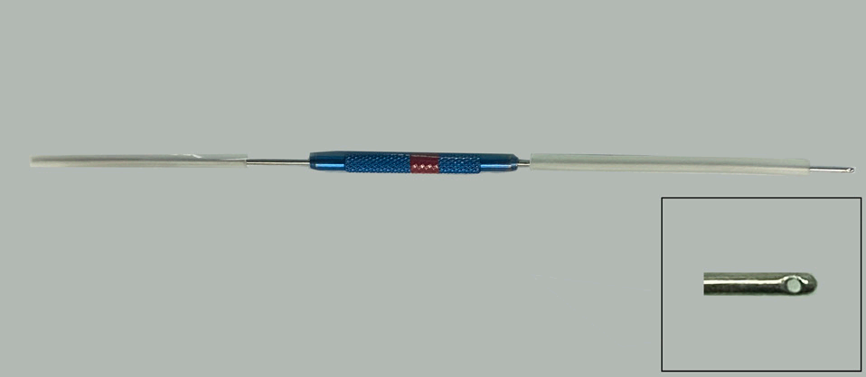
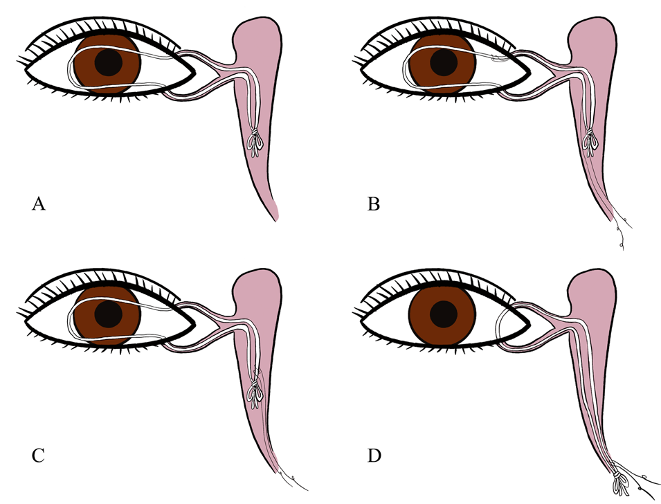
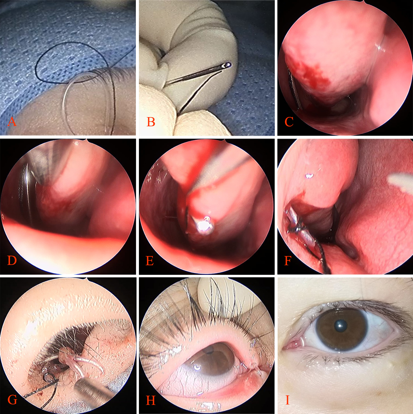
In Modified Suture-probe and silk thread traction method, a 7-8 gauge stainless steel lacrimal suture-probe with a side opening at one end is used (Figure 2). The surgical procedures are performed as follows (Figure 3, Figure 4). 1. Patients assume the supine position. After topical anesthesia with proxymetacaine, a cotton pad soaked in a 0.01% epinephrine lidocaine solution was placed in the lower nasal meatus to contrict the nasal mucosa. 2. The upper silicone tube is gently pulled from the upper punctum towards the temporal side. At this time, the knot should have reached the widest part of the dacryocyst, facilitating the subsequent probe insertion. 3. The 5-0 suture is lubricated with recombinant bovine fbroblast growth factor eye gel. One end of the suture is wrapped around the prolapsed tubetwice(Figure 3 B). The two ends of the suture are then passed through the suture-probetogether (Figure 4 B). It is important to ensure that the ends of the suture are not excessively long or short relative to one end of the probe with the side hole, as this may complicate the retrieval of the suture from the probe and nasal cavity. After lubricating the suture-probewith recombinant bovine fibroblast growth factor eye gel, the suture-probe, along with a 5-0 suture, is inserted into the upper canaliculi, lacrimal sac, nasolacrimal duct and inferior meatus through the upper punctum (Figure 4 C). 4. The suture at the medial canthus is fixed, after which the broken ends of the suture are carefully drawn out from the suture-probe using a nasal cavity hook. Subsequently, the suture thread is pulled out from the suture-probe, and the probe is withdrawn through the upper punctum (Figure 4 E). 5. The suture is loosened, and the proximal end of the silicone tube at the medial canthus is firmly secured, the suture is then pulled from the nasal cavity, causing it to slide towards the distal end of the silicone tube and form a knot(Figure 3 C). 6. Continuing to pull the suture from the nasal cavity, the distal end and the knot of the silicone tube are drawn out to the inferior meatus (Figure 4 F-G). Then, the prolapsed completely bicanalicular silicone tube within the whole lacrimal passage, originating from the medial canthus, was successfully repositioned (Figure 3 D, Figure 4 H-I) . 7. Local application of antibiotic eye drops is administered, and the inferior meatus is filled with a mixture of tobramycin and triamcinolone acetonide.
20240425101644_4770.png
Figure 2  7-8 gauge stainless steel lacrimal suture-probe with side opening at one end
20240425101839_5244.png
Figure 3   A simple diagram of using a 5-0 suture to reposition a complete prolapsed bicanalicular silicone tube after whole lacrimal duct intubation

(A) Completely prolapsed silicone tube from medial canthus, the distal part of the silicone tube and the knot goes deep into the nasolacrimal duct or is embedded in the lacrimal sac. No silicone tube was found in the inferior meatus and the opening of the nasolacrimal duct.(B) After wrapping the prolapsed silicone tube two times with a 5-0 suture, the suture was inserted into the inferior meatus from the superior lacrimal point with a suture probe. (C) The silicone tube at the medial canthus was fixed, and then the suture was pulled from the nasal cavity so that it slipped to the distal end and the knot of the silicone tube. (D) Continue to pull the suture so that the distal end and the knot of the silicone tube are pulled out to the inferior meatus. Then, the prolapsed completely bicanalicular silicone tube of the whole lacrimal passage from the medial canthus was successfully repositioned.
20240425102039_0884.png
Figure 4   The surgical procedures

(A) Patients take the supine position. After topical anesthesia with proxymetacaine, the 0.01% epinephrine lidocaine solution cotton pad was filled in the lower nasal meatus to contract the nasal mucosa. Pull the upper silicone tube from the upper punctum in the medial canthus to the temporal side until it can not be pulled, and pull the lower silicone tube from the lower punctum to the temporal side. At this time, the knot has entered the widest part of the dacryocyst. These facilitate the entry of subsequent probe. Lubricate the 5-0 suture with recombinant bovine fibroblast growth factor eye gel. One end of the suture is wound around the prolapsed tube two times. (B) The two ends of the suture are passed through the suture-probe together. (C) After the suture-probe is lubricated with recombinant bovine fibroblast growth factor eye gel, the suture-probe with a 5-0 suture is inserted into the upper canaliculi, lacrimal sac, nasolacrimal duct and inferior meatus from the upper punctum. (D) No silicone tube was found in the opening of the nasolacrimal duct. (E) The suture at the medial canthus is fixed, then the broken ends of the suture are hooked out from the suture-probe by using a hook in the nasal cavity, the suture thread is pulled out from the suture-probe, and the probe exited from the upper punctum. (F) Pull the suture from the nasal cavity so that the distal part of the silicone tube is pulled out. (G) The knot of the silicone tube is pulled out. (H-I) The silicone tubes in Case 1 and Case 2 were successfully repositioned

DISCUSSION

The complete prolapse of the bicanalicular silicone tube from the whole lacrimal passage, originating from the medial canthus, was successfully repositioned with no complications, using the modified suture-probe and silk thread traction method in this study. The results unequivocally showed that the effectiveness and safty of the modified approach in treating complete dislocations of the bicanalicular silicone tune from the medial canthus. From 2018 to 2023, our hospital encountered only two such cases, indicating the rarity of this condition. This finding contrasts with previous research.[4] To prevent such complete prolapses during whole lacrimal passage intubation surgery, a unique double tube knot technique for the silicone tube was introduced in our hospital. This knot is deliberately designed to be too large to fit through the nasolacrimal duct opening, thus preventing accidental dislocation. Additionally, after the operation, our nursing team will educate patients carefully, emphasing silicone tube care measures, such as keeeping eyes closed during face washing avoiding vigorous eye rubbing, and refraining from forceful nose blowing.[8] These measures, combined with our surgical techniques, contribute to the low incidence of complete prolapses in our hospital.

The dislocation of the bicanalicular silicone tube after whole lacrimal duct intubation can vary from partial to complete, depending on the extent of the displacement.[4] In cases of partial dislocation, where only a small portion of the silicone tube from the inner canthus with, the distal end of the tube and knot remaining visible in the nasal cavity, the tube can be easily repositioned by pulling the distal end down with forceps under the nose or gently pushing it back in place using forceps around  the inner canthus. The management of complete dislocation of the silicone tube, where the tube knot becomes lodged in the nasolacrimal duct or dacryocyst with no visible portion in the nasal cavity, poses a signifcant changeing in terms of repositioning.[4] Fortunately, the incidence of such complete prolapse of silicone tube is relatively are. Byun Z et al.[9] reported 11 cases of complete prolapse of bicanalicular silicone tube from medial canthus in 2 years, and Chu Z et al.[4] reported 12 cases in 5 years. The protrusion of the silicone tube from the medal canthus can readily lead to corneal trauma and conjunctivitis,[4] therefore, it is imerative to promptly reposition or remove the prolapsed silicone tube to prevent any further complications.

There are several ways to take out the silicone tube. Yeh H et al.[10] described an appraoch involving removal the a skin dacryocystorhinostomy,[10] but this method can enlarge the wound, leave behind a visible skin, and increase the cost. Additionally, they also discussed the technique of removing the tube through the transnasal endoscopic dacryocystorhinostomy, but this method can also result in additional trauma and expenses. Patel BC et al.[11] reported clipping the silicone tubes and gently pulling the knot out of the lacrimal puncta for tube removal. However, we found that the forcibly pulling the prolapsed silicone tube from the lacrimal puncta can easily lead to tears in the lacrimal passage and residual lacrimal duct.

There are various approaches to reposition a completely prolapsed silicone tube. Byun Z et al.[9] reported a method that they obliquely cut the prolapsed tube with scissors, creating a small semicircular hole. A probe is then inserted into this hole, entered into the inferior nasal meatus, and the silicone tube is clamped with pliers. Subsquently, the probe is withdraw from the puncta. However, this method is only applicable for hollow silicone tubes. He J et al.[5] reported a different method where the probe with memory wire is passing through the lacrimal puncta and into the inferior nasal meatus. The suture is threaded through the head of the memory wire. After removing the probe from the puncta, the suture is cut and looped around the silicone tube. The two ends of the suture were then knotted. Finally, by pulling the suture from the nasal cavity, the prolapsed tube can be repositioned. Chu Z et al.[4] introduced another method utilizing a suture-probe. They cut the suture, wrapped one end around the silicone tube, and tied it to the other end of the thread to create  a suture knot. Pulling the suture from the nasal cavity resets the prolapsed tube. Additionally, they also suggested attaching 30 mm sutures to the distal end of the silicone tube as a preventive measure against re-prolapse. However, the suture knot formed by the two ends of the sutures poses a risk of loosening as it is withdrawn from the nasal cavity. Additionally, the sutures reserved in the inferior meatus may cause irritation and promote the depostion of secretions.

In this study, patients who experienced complete dislocation of the bicanalicular silicone tube from medial canthus following whole lacrimal duct intubation were treated with modified suture-probe and silk thread traction method. This innovative method addresses the limitation assoicated with previous surgical methods. It is not only simple and safe, but also cost-effective, effectively preventing surgical failures or the need for silicone tube replacement due to premature removal of the prolapsed tube. The modified suture-probe and silk thread traction method introduced in this study offers a novel and effective solution for managing the complete prolapse of the bicanalicular silicone tube from the medial canthus after intubatiion of the entire lacrimal passage. Futhermore, the choice of suture is discretionary. While 5-0 black silk thread is thick and lacks smoothness, resulting in greater friction against the lacrimal duct mucosa, it exhibits a lesser cutting force. Conversely, the 6-0 prolene suture is thinner and smoother, minimizing friction with the lacrimal duct mucosa but possessing a higher cuttting force. The safety of employing various suture tyepes for repositining the completely prolapsed silicone tube remains to be thoroughly investigated within the context of  suture-probe and silk thread traction method. Given the infrequent occurrence of silicone tube prolapse and the limited sample size in this study, additional research is warranted to establish the safety of this method and identify potiential intraoperative complications.

Correction notice

None

Acknowledgement

None

Author Contributions

(I) Conception and design: Xuanwei Liang and Rongxin Chen
(II)Administrative support: None
(III) Provision of study materials or patients: Xuanwei Liang
(IV) Collection and assembly of data: Jing Li
(V) Data analysis and interpretation: Jing Li and Xuanwei Liang
(VI) Manuscript writing: Jing Li
(VII) Final approval of manuscript:All authors

Funding

This study is supported by the Natural Science Foundation of Guangdong Province of China (No. 2021A1515012043). The funding organizations had no role in the following aspects: design and conduct of the study; the collection, management, analysis, and interpretation of the data; preparation, review, or approval of the manuscript; and the decision to submit the manuscript for publication.

Confict of Interests

None of the authors has any conflicts of interest to disclose. All authors have declared in the completed the ICMJE uniform disclosure form.

Patient consent for publication

None

Ethical Statement

This study was approved by the Institutional Review Board of the Zhongshan Ophthalmic Center, Sun Yat-sen University, China (No.2021KYPJ100) and adhered to the tenets of the Helsinki Declaration. 

Informed consent was obtained from all subjects.

Provenance and Peer Review

This article was a standard submission to our journal. The article has undergone peer review with our anonymous review system

Data Sharing Statement

None

OpenAccess Statement

This is an Open Access article distributed in accordance with the Creative Commons Attribution-NonCommercial-NoDerivs 4.0 International License (CC BY-NC-ND 4.0), which permits the non-commercial replication and distribution of the article with the strict proviso that no changes or edits are made and the original work is properly cited (including links to both the formal publication through the relevant DOI and the license). See: https://creativecommons.org/licenses/by-nc-nd/4.0/.
1、Meduri A, Inferrera L, Tumminello G, et al. The use of a venous catheter as a stent for treatment of acquired punctal and canalicular stenosis[J]. J Craniofacial Surg, 2019, 30(8): 2544-2545. DOI: 10.1097/scs.0000000000005743.Meduri A, Inferrera L, Tumminello G, et al. The use of a venous catheter as a stent for treatment of acquired punctal and canalicular stenosis[J]. J Craniofacial Surg, 2019, 30(8): 2544-2545. DOI: 10.1097/scs.0000000000005743.
2、Kasaee A, Eshraghi B, Ameli K, et al. Pulled versus pushed monocanalicular silicone intubation in adults with lacrimal drainage system stenosis: a comparative case series[J]. J Ophthalmol, 2021, 2021: 5592039. DOI: 10.1155/2021/5592039.Kasaee A, Eshraghi B, Ameli K, et al. Pulled versus pushed monocanalicular silicone intubation in adults with lacrimal drainage system stenosis: a comparative case series[J]. J Ophthalmol, 2021, 2021: 5592039. DOI: 10.1155/2021/5592039.
3、Alsulaiman N, Alsuhaibani AH. Bicanalicular silicone intubation for the management of punctal stenosis and obstruction in patients with allergic conjunctivitis[J]. Ophthalmic Plast Reconstr Surg, 2019, 35(5): 451-455. DOI: 10.1097/IOP.0000000000001315.Alsulaiman N, Alsuhaibani AH. Bicanalicular silicone intubation for the management of punctal stenosis and obstruction in patients with allergic conjunctivitis[J]. Ophthalmic Plast Reconstr Surg, 2019, 35(5): 451-455. DOI: 10.1097/IOP.0000000000001315.
4、Chu Z, Lu G, Tan Y, et al. Repositioning a prolapsed tube after bicanalicular intubation of the lacrimal system[J]. Ophthalmic Plast Reconstr Surg, 2019, 35(6): 623-627. DOI: 10.1097/IOP.0000000000001400.Chu Z, Lu G, Tan Y, et al. Repositioning a prolapsed tube after bicanalicular intubation of the lacrimal system[J]. Ophthalmic Plast Reconstr Surg, 2019, 35(6): 623-627. DOI: 10.1097/IOP.0000000000001400.
5、He J, Gong J, Zheng Q, et al. Repositioning of the severe prolapsed silicone tubes after bicanalicular nasal intubation: a novel technique[J]. J Ophthalmol, 2021, 2021: 6669717. DOI: 10.1155/2021/6669717.He J, Gong J, Zheng Q, et al. Repositioning of the severe prolapsed silicone tubes after bicanalicular nasal intubation: a novel technique[J]. J Ophthalmol, 2021, 2021: 6669717. DOI: 10.1155/2021/6669717.
6、Demirci H, Elner VM. Double silicone tube intubation for the management of partial lacrimal system obstruction[J]. Ophthalmology, 2008, 115(2): 383-385. DOI: 10.1016/j.ophtha.2007.03.078.Demirci H, Elner VM. Double silicone tube intubation for the management of partial lacrimal system obstruction[J]. Ophthalmology, 2008, 115(2): 383-385. DOI: 10.1016/j.ophtha.2007.03.078.
7、DeParis SW, Hougen CJ, Rajaii F, et al. Bicanalicular intubation with the kaneka lacriflow for proximal lacrimal drainage system stenosis[J]. Clin Ophthalmol, 2020, 14: 915-920. DOI: 10.2147/OPTH.S248423.DeParis SW, Hougen CJ, Rajaii F, et al. Bicanalicular intubation with the kaneka lacriflow for proximal lacrimal drainage system stenosis[J]. Clin Ophthalmol, 2020, 14: 915-920. DOI: 10.2147/OPTH.S248423.
8、Liu Y, Tian B, Xiao H, Su L. Cause analysis and nursing strategy of accidental lacrimal duct decannulation in 37 eyes [in Chinese]. Qilu Journal of Nursing. 2016,22(21): 68-70.Liu Y, Tian B, Xiao H, Su L. Cause analysis and nursing strategy of accidental lacrimal duct decannulation in 37 eyes [in Chinese]. Qilu Journal of Nursing. 2016,22(21): 68-70.
9、Byun ZY, Lee BR, Kim SC. Successful reposition of prolapsed silicone tube using hole and lacrimal probe method[J]. Korean J Ophthalmol, 2021, 35(3): 231-234. DOI: 10.3341/kjo.2021.0033.Byun ZY, Lee BR, Kim SC. Successful reposition of prolapsed silicone tube using hole and lacrimal probe method[J]. Korean J Ophthalmol, 2021, 35(3): 231-234. DOI: 10.3341/kjo.2021.0033.
10、eh H, Berlin AJ, Price RL. Silicone intubation of the lacrimal system: loop retraction as a complication[J]. Ann Ophthalmol, 1979, 11(4): 665-666.eh H, Berlin AJ, Price RL. Silicone intubation of the lacrimal system: loop retraction as a complication[J]. Ann Ophthalmol, 1979, 11(4): 665-666.
11、Patel BC, Anderson RL. Transcanalicular removal of Silastic nasolacrimal tubes[J]. Ophthalmic Plast Reconstr Surg, 1997, 13(1): 74-75. DOI: 10.1097/00002341-199703000-00017.Patel BC, Anderson RL. Transcanalicular removal of Silastic nasolacrimal tubes[J]. Ophthalmic Plast Reconstr Surg, 1997, 13(1): 74-75. DOI: 10.1097/00002341-199703000-00017.
上一篇
下一篇
出版者信息
目录