您的位置: 首页 > 2024年6月 第1卷 第2期 > 文字全文
2023年7月 第38卷 第7期11
目录

Large-Scale Proteome-Wide Mendelian Randomization Identifies Novel Proteins for Glaucoma and Related Traits

Large-Scale Proteome-Wide Mendelian Randomization Identifies Novel Proteins for Glaucoma and Related Traits

来源期刊: Eye Science | 2024年6月 第1卷 第2期 93-114 发布时间:2024-06-28 收稿时间:2024/7/15 9:55:05 阅读量:1429
作者:
关键词:
Proteome Mendelian Randomization Primary Open-Angle Glaucoma GCIPL Thickness RNFL Thickness Intraocular Pressure
DOI:
10.12419/es24042802
Received date:
2024-04-01 
Accepted date:
2024-06-28 
Published online:
2024-06-28 
Purpose: To identify plasma proteins that are causally related to primary open-angle glaucoma (POAG) for potential therapeutic targeting. Methods: Summary statistics of plasma protein quantitative trait loci (pQTL) were derived from two extensive genome-wide analysis study (GWAS) datasets and one systematic review, with over 100 thousand participants covering thousands of plasma proteins. POAG data were sourced from the largest FinnGen study, comprising 8,530 DR cases and 391,275 European controls. A two-sample MR analysis, supplemented by bidirectional MR, Bayesian co-localization analysis, and phenotype scanning, was conducted to examine the causal relationships between plasma proteins and POAG. The analysis was validated by identifying associations between plasma proteins and POAG-related traits, including intraocular pressure (IOP), retinal nerve fibre layer (RNFL), and ganglion cell and inner plexiform layer (GCIPL). By searching druggable gene lists, the ChEMBL database, and the ClinicalTrials.gov database, the druggability and clinical development activity of the identified proteins were systematically evaluated. Results: Eighteen proteins were identified with significant associations with POAG risk after multiple comparison adjustments. The ORs per standard deviation increase in protein levels ranged from 0.39 (95% CI: 0.24–0.62; P = 7.70×10⁻⁵) for phospholipase C gamma 1 (PLCG1) to 1.29 (95% CI: 1.16–1.44; P = 6.72×10⁻⁶) for nidogen-1 (NID1). Bidirectional MR indicated that reverse causality did not interfere with the results of the main MR analyses. Five proteins exhibited strong co-localization evidence (PH4 ≥ 0.8): protein sel-1 homolog 1 (SEL1L), tyrosine-protein kinase receptor UFO (AXL), nidogen-1 (NID1) and FAD-linked sulfhydryl oxidase ALR (GFER) were negatively associated with POAG risk, while roundabout homolog 1 (ROBO1) showed a positive association. The phenotype scanning did not reveal any confounding factors between pQTLs and POAG. Further, validation analyses identified nine proteins causally related to POAG traits, with five proteins including interleukin-18 receptor 1 (IL18R1), interleukin-1 receptor type 1 (IL1R1), phospholipase C gamma 1 (PLCG1), ribonuclease pancreatic (RNASE1), serine protease inhibitor Kazal-type 6 (SPINK6) revealing consistent directional associations. In addition, 18 causal proteins were highlighted for their druggability, of which 5 proteins are either already approved drugs or in clinical trials and 13 proteins are novel drug targets. Conclusions: This study identifies 18 plasma proteins as potential therapeutic targets for POAG, particularly emphasizing the role of genomic and proteomic integration in drug discovery. Future experimental and clinical studies should be conducted to validate the efficacy of these proteins and to conduct more comprehensive proteomic explorations, thus taking a significant leap toward innovative POAG treatments.
Purpose: To identify plasma proteins that are causally related to primary open-angle glaucoma (POAG) for potential therapeutic targeting. Methods: Summary statistics of plasma protein quantitative trait loci (pQTL) were derived from two extensive genome-wide analysis study (GWAS) datasets and one systematic review, with over 100 thousand participants covering thousands of plasma proteins. POAG data were sourced from the largest FinnGen study, comprising 8,530 DR cases and 391,275 European controls. A two-sample MR analysis, supplemented by bidirectional MR, Bayesian co-localization analysis, and phenotype scanning, was conducted to examine the causal relationships between plasma proteins and POAG. The analysis was validated by identifying associations between plasma proteins and POAG-related traits, including intraocular pressure (IOP), retinal nerve fibre layer (RNFL), and ganglion cell and inner plexiform layer (GCIPL). By searching druggable gene lists, the ChEMBL database, and the ClinicalTrials.gov database, the druggability and clinical development activity of the identified proteins were systematically evaluated. Results: Eighteen proteins were identified with significant associations with POAG risk after multiple comparison adjustments. The ORs per standard deviation increase in protein levels ranged from 0.39 (95% CI: 0.24–0.62; P = 7.70×10⁻⁵) for phospholipase C gamma 1 (PLCG1) to 1.29 (95% CI: 1.16–1.44; P = 6.72×10⁻⁶) for nidogen-1 (NID1). Bidirectional MR indicated that reverse causality did not interfere with the results of the main MR analyses. Five proteins exhibited strong co-localization evidence (PH4 ≥ 0.8): protein sel-1 homolog 1 (SEL1L), tyrosine-protein kinase receptor UFO (AXL), nidogen-1 (NID1) and FAD-linked sulfhydryl oxidase ALR (GFER) were negatively associated with POAG risk, while roundabout homolog 1 (ROBO1) showed a positive association. The phenotype scanning did not reveal any confounding factors between pQTLs and POAG. Further, validation analyses identified nine proteins causally related to POAG traits, with five proteins including interleukin-18 receptor 1 (IL18R1), interleukin-1 receptor type 1 (IL1R1), phospholipase C gamma 1 (PLCG1), ribonuclease pancreatic (RNASE1), serine protease inhibitor Kazal-type 6 (SPINK6) revealing consistent directional associations. In addition, 18 causal proteins were highlighted for their druggability, of which 5 proteins are either already approved drugs or in clinical trials and 13 proteins are novel drug targets. Conclusions: This study identifies 18 plasma proteins as potential therapeutic targets for POAG, particularly emphasizing the role of genomic and proteomic integration in drug discovery. Future experimental and clinical studies should be conducted to validate the efficacy of these proteins and to conduct more comprehensive proteomic explorations, thus taking a significant leap toward innovative POAG treatments.

INTRODUCTION

Glaucoma is the leading cause of irreversible blindness worldwide, characterized by the degeneration of the retinal ganglion cell and optic nerve that is commonly associated with elevated intraocular pressure (IOP).[1] Primary open-angle glaucoma (POAG), the most prevalent form, is projected to affect 80 million individuals by 2040.[2] Despite the potential for conventional treatment methods (medication, laser, or surgery) to lower IOP, it does not offer universal protection against disease progression.[3] This underscores the critical need for early detection and innovative therapies to combat the growing prevalence of glaucoma among an aging demographic worldwide.[4]

Proteins play a crucial role in both health and disease and serve as primary targets for numerous drugs. The plasma proteome, consisting of a wide range of circulating molecules, serves as a reflection of an individual’s systemic health, shaped by genetics, lifestyle, and environmental factors.[5] Advances in proteomics have deepened our understanding of the role of plasma proteome in diseases such as dementia, heart failure, and cancer, thus solidifying its importance as a vital resource for drug discovery.[6-8] Remarkably, in 2017, three-quarters of newly approved drugs targeted human proteins, underscoring the critical role of proteomics in driving innovation for future therapies.[9]

Mendelian randomization (MR), which simulates the conditions of randomized controlled trials, is instrumental in identifying causal relationships between variables such as plasma proteins and POAG, while minimizing confounding factors.[10] Although MR has been used to explore the causality of specific biomarkers in POAG,[11-13] there is a notable scarcity of proteome-wide studies for identifying new POAG-related molecules. Recent genome-wide association studies (GWAS) have profiled approximately 4,800 plasma proteins,[5, 14-19] thereby providing a basis for proteome-wide MR (PW-MR) studies aimed at investigating disease etiology. Therefore, this study aims to identify potential therapeutic targets for POAG, explore novel therapeutic mechanisms, and systematically rank these proteins for the advancement of POAG therapies.

METHODS

Study Design

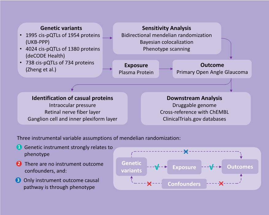
This study employed a two-sample PW-MR approach using protein quantitative trait loci (pQTL) data from seven extensive proteomics investigations to explore their correlation with POAG (Figure 1). Rigorous sensitivity analyses were conducted to reinforce the causal link between plasma proteins and POAG. To validate the association of proteins with POAG, their causal connections with various POAG-related traits were established. The study culminated in a comprehensive evaluation of the druggability of these proteins, with a specific focus on their potential as therapeutic agents. Ethical clearances from the original studies, which relied on publicly available summary data, obviated the need for additional ethical approval or patient consent for this study.
20240715111856_2872.pdf
Figure 1 A flow chart of the study design and a schematic illustration of cis-MR

Data Sources

Instrumental variables (IVs) for plasma proteins, specifically cis-pQTLs, were primarily derived from two large-scale genome-wide association study (GWAS) datasets: the UK Biobank Pharma Proteomics Project (UKB-PPP) and the deCODE Health study.
[15- 20] A total of 734 plasma proteins associated with cis-pQTLs were identified from Zheng et al.'s research.[21] The selection criteria included genome-wide significance (P < 5×10⁻⁸), independence (linkage disequilibrium [LD] clump r 2 < 0.001), and proximity, with cis-pQTLs located within 1 Mb of their protein-coding genes. Following these guidelines, we cataloged 1,995 cis-pQTLs for 1,954 plasma proteins from the UK Biobank Pharma Proteomics Project (UKB-PPP) (Olink platform, n = 54,306) and 4,024 cis-pQTLs for 1,380 circulating proteins from the deCODE Health study (Somascan platform, n = 35,559). In addition, 738 cis-pQTLs for 734 plasma proteins based on the Olink and Somascan  platform reported by Zheng et al., included studies by Sun et al., Emilsson et al., Suhre et al., Yao et al., and Folkersen et al. (eTable 1 in the supplement).

Further, data on single nucleotide polymorphisms (SNPs) linked to plasma proteins and their associations with POAG were obtained from the FinnGen R10 study. This study comprised 8,530 cases of European descent and 391,275 controls.[22] We also integrated the largest GWAS dataset for three established POAG-related phenotypes: intraocular pressure (IOP),[23] retinal nerve fiber layer (RNFL) thickness, and ganglion cell-inner plexiform layer (GCIPL) thickness,[24] thereby ensuring racial homogeneity in the exposure and outcome datasets.

STATISTICAL ANALYSIS

Two-sample MR Analysis

Leveraging cis-pQTLs for plasma proteins sourced from three comprehensive databases, we undertook a meticulous evaluation of genetic instrument reliability. This involved calculating the variance proportion (R ²) explained by the genetic IVs for each protein as well as evaluating the association strength between these IVs and risk factors utilizing F-statistics. Proteins without available IVs were excluded to ensure data consistency. By conducting thorough screening, we found 5,722 cis-pQTLs for 2,412 proteins with an R² greater than 0.1% to be valid genetic instruments for our analysis.

In our primary analysis, we used plasma proteins as the exposure and POAG as the outcome, utilizing the “TwoSampleMR” software package for computation. We derived association estimates between plasma proteins and POAG utilizing the Wald ratio method (for proteins associated with a single cis-pQTL) and the inverse variance-weighted (IVW) MR method (for proteins linked to multiple cis-pQTLs). Confidence intervals (CIs) were determined through the delta method, with results expressed as MR odds ratios (ORs) and corresponding 95% CIs for an increase of one standard deviation (SD) in gene-predicted plasma protein levels.[25] To mitigate the risk of false positives due to multiple testing, we applied the Benjamini–Hochberg correction, targeting a false discovery rate (FDR) of 0.05 and a more stringent Bonferroni correction, setting significance at P < 2.07 × 10⁻⁵ (0.05 / 2,412).

Reverse Causality Detection

To address the possibility of reverse causation, we utilized bidirectional MR to assess the relationship between POAG risk and the levels of causal plasma proteins. Effect estimates were derived using the MR-inverse variance weighted (MR-IVW) method, and were further validated using MR-Egger, weighted median, simple mode, and weighted mode techniques. Statistical significance was established at P < 0.05, based on comprehensive GWAS data from three previous plasma protein studies.

Bayesian Colocalization Analysis

We conducted colocalization analysis to discern the impact of horizontal pleiotropy from genetic IVs on the identified causal links.[26] For this analysis, we utilized a Bayesian framework to evaluate posterior probabilities for five scenarios regarding the genetic overlap between plasma proteins and POAG. The scenarios ranged from no shared causal variants (H0) to specific causal variants for proteins (H1) or POAG (H2), independent causal variants affecting both (H3), and a common causal variant for both (H4). A posterior probability of (PH4) ≥ 0.8 indicates strong colocalization, whereas 0.8 > PH4 ≥ 0.5 suggestes moderate support. This was performed using the “coloc” package in R (version 4.4.1).

Phenotype Scanning

Utilizing the “phenoscanner” R package, we explored large-scale genetic association studies for links between our identified pQTLs and POAG-related traits.[27] SNPs that showed pleiotropy met stringentcriteria: genome-wide significance (P < 5×10⁻⁸), association studies in European populations, and links to established POAG risk factors, including metabolic traits, proteins, or clinical features.

Identification in POAG-related Traits

To validate identified plasma proteins and investigate their potential as therapeutic targets, we conducted two-sample MR analyses to assess the relationship between plasma proteins and POAG-related phenotypes. Utilizing SNPs from an extensive database of plasma protein cis-pQTLs as IVs, we focused on three major GWAS datasets for POAG traits: intraocular pressure (IOP), retinal nerve fiber layer (RNFL) thickness, and ganglion cell-inner plexiform layer (GCIPL) thickness. Through comprehensive MR analyses, we estimated the associations between these proteins and POAG-related phenotypes using both the Wald ratio and inverse variance-weighted MR methods. The results are reported as MR ORs with 95% CIs for an increase in plasma protein levels by one standard deviation, with significance set at P < 0.05.

Downstream Analysis of Drug Target Proteins

We explored plasma proteins as potential drug targets, and examined the druggability of MR-prioritized proteins against an updated list of drug-gene candidates.[28] The ChEMBL database (version 33) was consulted for drug molecule classifications, approved indications, and clinical trial outcomes,[29] alongside a detailed review through the https://www.ClinicalTrials.gov website for additional relevant information. This druggability assessment categorized proteins into four groups: approved (for proteins with at least one approved drug), in development (for proteins under clinical investigation), druggable (for proteins identified as potential targets but not yet listed in the drug database), and not currently listed as druggable (for proteins not listed as potential targets in the drug database).

Data Availability

Summary-level statistics for cis-pQTLs are available from the original publications. POAG GWAS summary statistics can be accessed via the FinnGen consortium (https://www.finngen.fi). Summary statistics for IOP, RNFL, and GCIPL can be retrieved from the GWAS Catalog (https://www.ebi.ac.uk/gwas).

RESULTS

Mendelian Randomization Between Plasma Proteins and POAG

The study incorporated a large number of plasma proteins and genetic IVs as the basis for MR analysis (Figure 1). We aggregated cis-pQTLs for plasma proteins from three independent GWAS studies, resulting in 6,362 cis-pQTLs that corresponded to 2,602 proteins. Following a thorough selection process for genetic robustness (R² > 0.1%) and excluding proteins without appropriate genetic IVs, we narrowed our focuse to 2,412 proteins identified in the FinnGen R10 (Figure 2).
20240715113301_2473.pdf
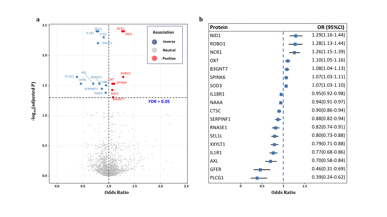
Figure 2 Result summary of MR and colocalization analysis on the associations between plasma proteins and the risk of primary open-angle glaucoma.
OR=odds ratio; FDR=false discovery rate
In the MR analysis, utilizing these 2,412 plasma proteins as study exposures and implementing FDR correction, we identified 18 plasma proteins significantly associated with POAG risk (Table 1). For each protein, an increment of one SD in genetically predicted levels altered the odds of POAG, with ORs ranging from 0.39 (95% CI: 0.24–0.62; P = 7.70×10⁻⁵) for phospholipase C gamma 1 (PLCG1) to 1.29 (95% CI: 1.16–1.44; P = 6.72 ×10⁻⁶) for nidogen-1 (NID1). Among these proteins, 7 exhibited positive associations with POAG risk, while 11 displayed negative associations (Table 1, Figure 2). After applying a Bonferroni correction for multiple testing (P < 2.07×10⁻⁵), six proteins remained significantly linked to POAG. Notably, all analyzed proteins demonstrated F-statistic values above 10, thereby effectively eliminating the possibility of weak instrumental variable bias.

Table 1 Plasma proteins significantly associated with primary open-angle glaucoma after false discovery rate (FDR) correction


Protein

Chr

Position

rs number cis-pQTL

EAF

EA

MR analysis

Source


β (SE)

OR

95%CI

P value

R2

F statistics


AXL

19

41233275

rs66841352

0.40

C

-0.36(0.09)

0.70

0.58-0.84

1.62×10⁻⁴

0.014

473.03

UKB-PPP

B3GNT7

2

231398416

rs2290130

0.23

A

0.08(0.02)

1.08

1.04-1.13

3.77×10⁻⁴

0.169

6888.59

UKB-PPP

GFER

16

1973321

rs61516948

0.13

T

-0.77(0.20)

0.46

0.31-0.69

1.50×10⁻⁴

0.003

89.38

UKB-PPP

PLCG1

20

41168825

rs753381

0.47

C

-0.95(0.24)

0.39

0.24-0.62

7.70×10⁻⁵

0.002

70.03

deCODE

SEL1Lb

14

81506097

rs11499034

0.01

C

-0.22(0.05)

0.80

0.73-0.88

5.05×10⁻⁶

0.044

1538.02

UKB-PPP

SERPINF1

17

1666253

rs62088172

0.35

T

-0.13(0.04)

0.88

0.82-0.94

2.54×10⁻⁴

0.088

316.65

Sun

CTSCa, b

11

88337746

rs11600158

0.10

G

-0.10(0.02)

0.90

0.86-0.94

1.06×10⁻⁵

0.146

5787.36

UKB-PPP

88066714

rs55897509

0.92

A

0.085

509.91

Emilsson

IL18R1a

2

102377596

rs10190555

0.77

G

-0.05(0.01)

0.95

0.92-0.98

1.86×10⁻⁴

0.228

9955.02

UKB-PPP

101625575

rs115232861

0.03

C

0.006

226.86

deCODE

103035044

rs1420106

0.78

G

0.270

1221.57

Sun

102877724

rs183611009

0.02

G

0.006

227.53

deCODE

102030778

rs187572594

0.02

G

0.002

63.20

deCODE

101829431

rs55715763

0.06

G

0.004

133.11

deCODE

102264346

rs55871806

0.15

C

0.088

3399.96

deCODE

102549866

rs72995641

0.21

A

0.031

1126.2

deCODE

101525580

rs75094400

0.02

C

0.003

101.34

deCODE

102971459

rs80339564

0.03

C

0.003

116.82

deCODE

IL1R1a, b

2

102156623

rs3917238

0.30

T

-0.27(0.06)

0.77

0.68-0.86

4.22×10⁻⁶

0.015

528.97

UKB-PPP

102138349

rs6706048

0.28

C

0.012

440.56

deCODE

102746276

rs7588201

0.72

A

0.007

39.29

Emilsson

NAAAa

4

75932965

rs112197434

0.23

T

-0.06(0.02)

0.94

0.91-0.97

3.00×10⁻⁴

0.141

5525.12

UKB-PPP

76102346

rs12331871

0.17

G

0.016

568.16

deCODE

75906793

rs1394919

0.29

A

0.148

6148.47

deCODE

76209167

rs76229059

0.17

G

0.005

190.32

deCODE

76848231

rs9996608

0.30

T

0.119

445.59

Sun

NCR1a, b

19

55419632

rs143981324

0.90

T

0.23(0.05)

1.26

1.15-1.39

1.99×10⁻⁶

0.013

74.26

Emilsson

54904812

rs7255591

0.09

C

0.031

1073.76

UKB-PPP

NID1a, b

1

236099073

rs17554536

0.18

A

0.26(0.06)

1.29

1.16-1.44

6.72×10⁻⁶

0.002

67.92

deCODE

236104574

rs58074293

0.02

A

0.011

379.94

deCODE

236067017

rs76183323

0.01

A

0.012

402.08

UKB-PPP

OXTa

20

3067658

rs6115776

0.40

C

0.10(0.03)

1.10

1.05-1.16

1.61×10⁻⁴

0.059

2208.44

deCODE

3100953

rs857244

0.46

C

0.001

41.77

deCODE

3049890

rs877172

0.66

T

0.125

776.73

Emilsson

RNASE1a

14

20789957

rs11624082

0.26

T

-0.20(0.05)

0.82

0.74-0.91

1.16×10⁻⁴

0.001

50.25

deCODE

20814883

rs12897030

0.30

T

0.016

592.52

deCODE

21280678

rs17254387

0.69

A

0.022

72.52

Sun

20828671

rs188152481

0.02

C

0.002

63.19

deCODE

20722147

rs2002078

0.41

T

0.004

158.96

deCODE

20627658

rs75124405

0.01

A

0.002

66.29

deCODE

ROBO1a

3

78946828

rs331172

0.49

G

0.25(0.06)

1.28

1.13-1.44

6.78×10⁻⁵

0.003

88.58

deCODE

78725134

rs3773233

0.21

T

0.014

485.44

deCODE

78735620

rs3773244

0.20

A

0.020

687.56

UKB-PPP

79787885

rs62256944

0.10

A

0.002

58.34

deCODE

77848463

rs73103884

0.02

G

0.001

45.09

deCODE

78741405

rs73111654

0.01

A

0.002

81.48

deCODE

79369917

rs7639769

0.46

T

0.003

100.95

deCODE

SOD3a

4

24800212

rs1799895

0.01

G

0.06(0.02)

1.07

1.03-1.10

2.42×10⁻⁴

0.133

5170.20

UKB-PPP

24802616

rs2695234

0.08

G

0.031

1145.21

deCODE

25235769

rs313548

0.30

A

0.003

109.25

deCODE

25664005

rs76930256

0.02

T

0.005

190.96

deCODE

24444429

rs77514718

0.01

C

0.014

505.01

deCODE

24639073

rs80234081

0.03

C

0.021

751.96

deCODE

SPINK6a

5

148263754

rs116664727

0.13

A

0.07(0.02)

1.07

1.03-1.11

1.41×10⁻⁴

0.006

223.76

deCODE

148186561

rs12717962

0.55

G

0.072

2630.07

UKB-PPP

147294723

rs137864680

0.02

A

0.001

50.58

deCODE

148310721

rs139120114

0.01

C

0.014

511.80

deCODE

147603178

rs1432688

0.92

G

0.155

605.02

Sun

149047717

rs148402978

0.02

T

0.003

113.83

deCODE

147734214

rs148632877

0.01

T

0.002

70.11

deCODE

148183699

rs74765295

0.01

T

0.007

248.34

deCODE

147882756

rs75666474

0.05

A

0.003

93.34

deCODE

148453118

rs75666559

0.04

T

0.007

259.13

deCODE

148057440

rs7719473

0.03

A

0.057

2144.61

deCODE

XXYLT1a, b

3

195287123

rs1538767

0.46

C

-0.23(0.05)

0.79

0.71-0.88

1.58×10⁻⁵

0.016

564.92

deCODE

195619664

rs534994266

0.09

A

0.002

80.40

deCODE

194783033

rs55947051

0.84

T

0.010

56.46

Emilsson

195072715

rs7635512

0.16

C

0.007

240.77

deCODE

Source indicates the protein GWAS providing the estimate of the effect of the cis-pQTL on protein level. Results express changes in primary open angle glaucoma risk per 1-SD increase in protein level. EA=effect allele; EAF=effect allele frequency; β=effect on diabetic retinopathy; OR=odds ratio.

a Mendelian randomization using the inverse variance weighted methods.

b Plasma protein passed Bonferroni correction (P < 2.12×10⁻⁵).

Multiple Sensitivity Analyses

To verify the reliability of the causal connections between the 18 identified proteins and POAG, we conducted a series of sensitivity analyses. Bidirectional MR analyses enabled the elimination of reverse causality in our initial MR findings (Table 2).

Table 2. Summary of reverse causality detection, Bayesian co-localization analysis and phenotype scanning on 18 potential causal proteins.


Protein

Bidrectional MR

Co-localization

Previously reported associations

OR(95%CI)

P value

PPH3

PPH4

SEL1L

1.00(0.98-1.02)

0.80

0.002

0.995

lipid metabolism

ROBO1

1.00(0.98-1.03)

0.92

0.065

0.865

NA

AXL

1.00(0.99-1.00)

0.12

0.052

0.858

NA

NID1

1.00(0.97-1.02)

0.73

0.039

0.806

NA

GFER

1.01(0.99-1.03)

0.34

0.125

0.804

NA

NCR1

1.00(0.97-1.02)

0.81

0.335

0.505

NA

SERPINF1

0.96(0.91-1.02)

0.20

0.366

0.476

Body measurement

CTSC

0.99(0.97-1.01)

0.36

0.323

0.431

NA

SPINK6

1.00(0.98-1.02)

0.94

0.399

0.260

NA

B3GNT7

1.01(0.99-1.04)

0.25

0.322

0.210

Body measurement

RNASE1

0.97(0.93-1.02)

0.29

0.117

0.193

Cervix uteri disease

NAAA

0.99(0.97-1.01)

0.36

0.181

0.116

NA

IL1R1

1.00(0.97-1.02)

0.75

0.354

0.076

Blood cell, asthma, hayfever, rhinitis, eczema, bronchitis, emphysema

IL18R1

1.02(1.00-1.03)

0.07

0.340

0.026

Blood cell, inflammatory bowel disease

PLCG1

1.01(0.96-1.06)

0.68

0.092

0.025

Blood cell, lipid metabolism

XXYLT1

0.96(0.91-1.02)

0.20

0.520

0.022

NA

SOD3

0.99(0.97-1.01)

0.42

0.506

0.009

Vascular dementia

OXT

1.02(0.96-1.08)

0.54

0.018

0.004

NA

                                                                        OR=odds ratio; NA=not applicable.

Colocalization analysis was employed to address potential confounding from linkage disequilibrium (LD), thereby determining whether the levels of causal plasma proteins shared genetic origins with POAG. This analysis confirmed strong co-localization evidence (PH4 ≥ 0.8) for SEL1L, ROBO1, AXL, NID1, and GFER, with NCR1 showing moderately strong evidence (0.8 > PH4 ≥ 0.5), thereby suggesting shared genetic determinants between these proteins and POAG (eFigure in the supplement).

Furthermore, phenotypic scanning of cis-pQTLs for POAG-associated proteins revealed no direct links between the IVs and POAG or its risk factors. However, we identified associations of cis-pQTLs for certain proteins with other health conditions: IL1R1 (rs3917238, rs6706048, and rs7588201) and IL18R1 (rs10190555 and rs1420106) with blood components; SEL1L (rs11499034) and PLCG1 (rs753381) with specific diseases; RNASE1 (rs75124405) with cervical diseases; SOD3 (rs80234081) with vascular dementia; IL18R1 with inflammatory bowel disease; and IL1R1 with immune disorders such as asthma, hay fever, allergic rhinitis, and eczema. Modifying the significance threshold to P < 1 × 10⁻⁵ did not identify any cis-pQTLs for plasma proteins related to POAG or its risk factors.

Identification of Plasma Proteins in IOP, RNFL, and GCIPL

The MR validation for 18 plasma proteins in relation to POAG traits was processed (eTable2 in the supplement). Among these, nine proteins demonstrated genetic associations with at least one POAG-related phenotype. Notably, pigment epithelium-derived factor (SERPINF1) was the only protein found to be significantly associated with IOP outcomes. Four proteins namely dipeptidyl peptidase 1 (CTSC), serine protease inhibitor Kazal-type 6 (SPINK6), interleukin-1 receptor type 1 (IL1R1), and ribonuclease pancreatic (RNASE1) were found to show significant causal relationships with RNFL thickness. Moreover, five proteins including interleukin-18 receptor 1 (IL18R1), namely N-acylethanolamine-hydrolyzing acid amidase (NAAA), IL1R1, phospholipase C gamma 1 (PLCG1), and sel-1 homolog 1 (SEL1L) were significantly associated with GCIPL thickness.

Directional consistency was observed for IL18R1, IL1R1, PLCG1, RNASE1, and SPINK6 across their associations with POAG-related traits and POAG itself. Remarkably, an increase of one standard deviation in genetically predicted levels of IL1R1 revealed a significant positive causal correlation with both RNFL (OR = 1.42; 95% CI, 1.11–1.83; P = 0.01) and GCIPL (OR = 1.42; 95% CI, 1.02–1.84; P = 0.04), thereby indicating IL1R1’s protective effect on these eye structures and its beneficial role in POAG, as highlighted in the primary MR analysis.

The Druggability and Development Status of POAG-causal Protein

To assess the druggability and advancement of plasma protein targets associated with POAG, we undertook a comprehensive review of druggable genes, the ChEMBL drug discovery database, and ClinicalTrials.gov. We identified 18 drug targets and sorted them into four categories based on their development stage: approved (targets with drugs already , authorized for use), in development (targets undergoing clinical trials), druggable (potential targets), and not currently listed as druggable (not potential targets) (Table 3). This analysis revealed five plasma proteins as prime candidates for future drug development efforts: tyrosine-protein kinase receptor UFO (AXL), dipeptidyl peptidase 1 (CTSC), interleukin-18 receptor 1 (IL18R1), oxytocin (OXT), and interleukin-1 receptor type 1 (IL1R1).

Drugs targeting AXL, such as gilteritinib, have received approval for specific neoplasms, including acute myeloid leukemia. Similarly, IL1R1 has played a key role in treating immune disorders, such as cryopyrin- associated periodic syndromes and rheumatoid arthritis, with anakinra being a notable example. Apart from these, the other three highlighted proteins are associated with drugs in Phase I–III clinical trials; however, there is no direct evidence of their use in developing treatment for POAG. The drug development status for the remaining 13 plasma proteins remains unclear. Our findings suggest AXL, GFER, SERPINF1, CTSC, IL18R1, IL1R1, NAAA, NCR1, NID1, OXT, RNASE1, SOD3, and SPINK6 as significant druggable targets that warrant further investigation for drug development.

Table 3. Summary of druggability and clinical development activity for primary open angle glaucoma associated with causal associations on MR analysis.


Target

Compound name

Clinical development activities

Approved

AXL

GILTERITINIB (CHEMBL3301622)b

Approved for neoplasms, acute myeloid leukemia, myeloid leukemia; Phase II: myelodysplastic syndromes, blast crisis; Phase I: non-smal-cell lung carcinoma, kidney diseases, liver diseases

IL1R1

ANAKINRA (CHEMBL1201570)b

Approved: immune system diseases; cryopyrin-associated periodic syndromes, rheumatoid arthritis; Phase III: metabolic syndrome, giant cell arteritis, pneumonia, severe acute respiratory syndrome, myocardial infarction, mucocutaneous lymph node syndrome, familial mediterranean fever, influenza, dermatitis, allergic contact; Phase II: brain injuries, lymphoma, follicular, multiple myeloma, pericarditis, diabetes mellitus, Still's disease, uveitis, hvidradenitis suppurativa, myositis, alcoholic hepatitis, macrophage activation syndrome, gout, dermatomyositis, sarcoidosis, amyotrophic lateral sclerosis, infections, hypoglycemia, type 1 diabetes mellitus, osteoarthritis, juvenile arthritis, heart failure, cerebral hemorrhage, polycystic ovary syndrome, myocarditis, labyrinth diseases, chronic fatigue syndrome, chronic renal insufficiency; Phase I: castration-resistant prostatic neoplasms, pancreatic neoplasms, knee injuries, blepharitis, HIV infections, multiple sclerosis, breast neoplasms, pulmonary hypertension, pain, smoldering multiple, myeloma, asthma, rectal neoplasms, chronic B-cell leukemia lymphocytic, hearing loss, neoplasms

In development

AXL

BEMCENTINIB (CHEMBL3809489)b

Phase II: adenocacinoma of lung; mesothelioma; severe acute respiratory; inflammatory breast neoplasms; Phase I: myelodysplastic syndromes; acute leukemia myeloid; pancreatic neoplasms; non-small-cell lung carcinoma; melanoma

AXL

ENAPOTAMAB VEDOTIN (CHEMBL4297987)b

Phase I: non-small-cell lung carcinoma

AXL

BPI-9016 (CHEMBL3545236)b

Phase I: non-small-cell lung carcinoma; neoplasms

CTSC

BRENSOCATIB (CHEMBL3900409)b

Phase III: cystic fibrosis; COVID-19; Phase II: bronchiectasis

IL18R1

IBOCTADEKIN (CHEMBL2108034)b

Phase II: melanoma; Phase I: neoplasms; lymphoma non-hodgin; lymphoma

IL1R1

MEDI-8968 (CHEMBL2109607)b

Phase II: hidradenitis suppurativa; chronic obstructive pulmonary disease

IL1R1

AMG-108 (CHEMBL2109458)b

Phase II: rheumatoid arthritis; diabetes mellitus; osteoarthritis

OXT

OXYTOCINc

Phase III: Prader-Willi syndrome (NCT02804373), socially adaptive mirroring (NCT03640156); Phase II: central diabetes insipidus (NCT06036004), schizophrenia (NCT01699997)

Druggablea

AXL, GFER, SERPINF1, CTSC, IL18R1, IL1R1, NAAA, NCR1, NID1, OXT, RNASE1, SOD3, SPINK6

Not currently listed as druggablea

B3GNT7, PLCG1, SEL1L, ROBO1, XXYLT1

                                                                                a Data from druggable gene list.

                                                                                b Data from ChEMBL release 33 (compound ID in brackets).

                                                                                c Data from ClinicalTrials.gov.

DISCUSSION

Summary of Findings

This PW-MR study reveals 18 plasma proteins as potential therapeutic targets for POAG, thereby effectively addressing concerns related to pleiotropy and reverse causality. Co-localization analysis reveals that elevated genetic expressions of SEL1L, AXL, and GFER decrease POAG risk, while higher levels of ROBO1, NID1, and NCR1 increase it. Validation confirms that nine proteins are causally associated with POAG related phenotypes, with IL18R1, IL1R1, PLCG1, RNASE1, and SPINK6 consistently linked to POAG. The assessment of durability and clinical development activities prioritized 18 causal plasma proteins. These findings provide novel insights into POAG drug development strategies and emphasize the potential of PW-MR in unraveling the etiological biology of complex diseases.

MR-derived novel biomarkers and potential targets for POAG

Our study identified 18 proteins as novel POAG targets, with specific emphasis on SEL1L, ROBO1, AXL, NID1, and GFER for their strong co-localization evidence. SEL1L, as the protein most significantly associated with POAG risk—is integral to the endoplasmic reticulum-associated degradation (ERAD) system, which mitigates ER stress by eliminating misfolded proteins.[30-32] This feature proposes the capacity of SEL1L to impede the advancement of POAG, thus emphasizing the cruciality of additional assessment via meticulous experimental and clinical investigations to substantiate these proteins as potential therapeutic objectives for POAG.

In addition, ROBO1, which is integral to neurogenesis and angiogenesis, may be associated with POAG risk through its involvement in the SLIT/ ROBO1 pathway and SRGAP2 expression.[33-36] Studies have demonstrated that an increase in ROBO1 levels is closely linked to POAG, which may lead to retinal neovascularization and the possible onset of secondary glaucoma.[37-38] These results underscore the imperative for additional investigations into the involvement of ROBO1 in POAG neuroprotection and angiogenesis. 

Nidogen-1 (NID1), a key basement membrane glycoprotein, has been found to be as positively associated with POAG risk.[39-40] Increased nidogen presence in glaucoma patients suggests its involvement in extracellular matrix dynamics and aqueous efflux obstruction.[41] The specific mechanisms by which NID1 contributes to POAG remain to be clarified, thereby highlighting a crucial area for future research. Our study found that higher levels of GFER, which is a protein vital for mitochondrial integrity, is correlated with reduced POAG risk.[42-44] This suggests GFER’s protective role in preventing oxidative stress and mitochondrial dysfunction, both of which contributes in glaucoma progression.[44-45] The potential of GFER as a therapeutic target for POAG highlights the need for additional research into its mechanisms of action.

AXL, known for its regulatory functions in cancer and inflammation,[46] emerges as a neuroprotective factor in POAG through its interaction with the APOE gene and microglial regulation.[47-48] This study proposed the involvment of AXL in reducing neurodegeneration in glaucoma, thus advocating for further investigation to validate AXL as a promosing therapeutic approach in the management of POAG.

Multiple Sensitivity Analysis for MR Assumptions

To ensure the scientific validity and robustness of our results, we conducted a rigorous sensitivity analysis to confirm the assumptions of the MR. Initially, our focus was on cis-pQTLs within 1 Mb of protein-coding genes, specifically selected for their direct impact on protein expression. We adhered to stringent selection criteria including linkage disequilibrium (LD) clustering (20.001), a significance threshold (P ≤ 5.0 × 10⁻⁸), and F-statistics over 10 to ensure the relevance of genetic IVs. Further, we guaranteed MR independence by examining associations between specific proteins (e.g., IL1R1, SERPINF1, and SOD3) and various traits, but we found no confounding influences on our MR analysis. To address the exclusion restriction assumption, we uitilized the Bayesian co-localization analysis, identifying shared variants of five proteins (SEL1L, ROBO1, AXL, NID1, and GFER) with POAG, while acknowledging the method’s high false-negative rate (~60%), which did not detract from our main conclusions. Moreover, a bidirectional MR analysis was conducted to eliminate the possibility of reverse causation, ultimately confirming the absence of it among the studied proteins. This comprehensive approach emphasizes our commitment to maintaining the accuracy of MR assumptions, even though we acknowledge the ongoing potential impact of reverse causation.

Validation of Causal Proteins

The primary aim of this study was to establish a causative correlation between particular proteins and POAG-associated characteristics, with the intention of confirming potential pharmaceutical targets and elucidating the fundamental mechanisms of operation. By analyzing data from the most extensive GWAS database for IOP, RNFL, and GCIPL, our MR study revealed significant causal links between various plasma proteins and POAG-related traits, further validating them as potential targets for medication. This holds particular significance as the field pivots its attention towards not only decreasing IOP, but also delving into neuroprotective treatments that combat the apoptosis of retinal ganglion cells (RGCs), encompassing factors such as inflammation, neuroglial activation, neurotrophic deficiencies, and oxidative stress. Notably, IL1R1—part of the IL-1receptor family known for its role in inflammation and microglia activation—was associated with an increase in RNFL and GCIPL thickness, thereby suggesting a protective effect against POAG.[49-51] This positions IL1R1 as a promising candidate for neuroprotective therapeutic strategies in POAG, although further investigation is required to fully understand the roles of these proteins in POAG pathogenesis.

Scientific and Clinical Implications

This study provides critical insights that have profound implications for both medical research and clinical practice, emphasizing the value of a comprehensive approach in biomarker testing for identifying novel drug targets. Our analysis spanned 2,412 plasma proteins related to POAG, which is merely a fraction of the total proteome and, thus, emphasizes the untapped potential in the vast protein landscape. The findings from the TwoSampleMR (PW-MR) analysis not only shed light on potential drug mechanisms associated with these proteins but also validate the causal influence of certain proteins on POAG and its related traits. This correlation lays a genetic groundwork for developing neuroprotective therapies tailored to the molecular mechanisms of POAG.
Furthermore, our results indicate that proteins causally associated with POAG might also influence susceptibility to other diseases, thereby indicating the possibility of shared pathological pathways across different conditions. Interestingly, certain identified proteins are already recognized in the treatment of other diseases or are in clinical trials, thereby hinting at the potential for repurposing existing drugs for POAG treatment. Moreover, PW-MR analysis emerges as a powerful preclinical tool for drug target prioritization and the anticipation of possible adverse effects, thereby facilitating the discovery of novel therapeutic agents with superior efficacy and safety profiles. This study emphasizes the necessity of exploring the proteome more comprehensively to unveil novel pathways and therapeutic targets for POAG and beyond.

Strengths and Limitations

This study has several key strengths. First, it leverages the latest comprehensive GWAS data from European populations, thereby effectively reducing population stratification bias and enhancing
the identification of causal associations. Rigorous validation through a suite of sensitivity analyses, including two-way MR, co-localization analyses, and phenotypic scanning, was instrumental. In particular, co-localization analyses were crucial in identifying shared genetic variations between exposures and outcomes. And MR validation analysis reinforced the causal relationships between plasma proteins and POAG-related phenotypes as well as affirming the viability of potential drug targets.

However, this study is not without its limitations. While single IV MR analysis may prompt concerns over incidental findings, our approach mitigated this by selecting strong, highly correlated cis-pQTLs, backed by robust SNPs, thereby ensuring reliability. We also employed multiple protein quantification techniques, such as Olink and Somascan, to address technique-specific biases.[52] Although our plasma protein data predominantly originated from the UK, the diverse origins of most cis-pQTLs bolster the generalizability of our findings, despite the potential for sample overlap. Further, this study's focus on individuals of European descent (due to data availability) highlights the necessity for future GWAS on biomarkers in non-European populations to enable more inclusive cross-ethnic MR analyses.

CONCLUSION

In conclusion, our systematic MR analysis of the plasma proteome has identified 18 promising drug targets for POAG, with robust co-localization support for five (SEL1L, ROBO1, AXL, NID1, GFER). These findings provide an unprecedented genetic insight into the etiology of POAG, thereby highlighting potential therapeutic targets and avenues. Our findings also emphasize the vast potential for future research in population genomics, proteomics, and their interplay with disease to discover new drug candidates. The next steps involve experimental and clinical studies to validate the functions and therapeutic efficacy of these proteins and, thus, mark a significant leap toward innovative POAG treatments.

Correction notice

None

Acknowledgement

None

Author Contributions

(Ⅰ) Conception and design: W.W., W.H
(Ⅱ) Administrative support: S.Y., Z.Z., W.H
(Ⅲ) Provision of study materials or patients: All authors
(Ⅳ) Collection and assembly of data: W.W., S.Y., Z.Z
(Ⅴ) Data analysis and interpretation: S.Y., Z.Z., X.Z.
(Ⅵ) Manuscript writing: All authors
(Ⅶ) Final approval of manuscript: All authors

Fundings

This work was supported by the Hainan Province Clinical Medical Center, the National Natural Science Foundation of China (82171084, 82371086).

Conflict of Interests

None of the authors has any conflicts of interest to disclose. All authors have declared in the completed the ICMJE uniform disclosure form.

Patient consent for publication

None

Ethical Statement

None

Provenance and Peer Review

This article was a standard submission to our journal. The article has undergone peer review with our anonymous review system.

Data Sharing Statement

None

Open Access Statement

This is an Open Access article distributed in accordance with the Creative Commons Attribution-NonCommercial-NoDerivs 4.0 International License (CC BY-NC-ND 4.0), which permits the non-commercial replication and distribution of the article with the strict proviso that no changes or edits are made and the original work is properly cited (including links to both the formal publication through the relevant DOI and the license). See: https://creativecommons.org/licenses/by-nc-nd/4.0/.


1、Stein JD, Khawaja AP, Weizer JS. Glaucoma in adultsscreening, diagnosis, and management: a review. JAMA. 2021, 325(2): 164-174. DOI: 10.1001/jama.2020.21899.Stein JD, Khawaja AP, Weizer JS. Glaucoma in adultsscreening, diagnosis, and management: a review. JAMA. 2021, 325(2): 164-174. DOI: 10.1001/jama.2020.21899.
2、Tham YC, Li X, Wong TY, et al. Global prevalence of glaucoma and projections of glaucoma burden through 2040: a systematic review and meta-analysis. Ophthalmology. 2014, 121(11): 2081-2090. DOI: 10.1016/ j.ophtha.2014.05.013.Tham YC, Li X, Wong TY, et al. Global prevalence of glaucoma and projections of glaucoma burden through 2040: a systematic review and meta-analysis. Ophthalmology. 2014, 121(11): 2081-2090. DOI: 10.1016/ j.ophtha.2014.05.013.
3、Gedde%20SJ%2C%20Vinod%20K%2C%20Wright%20MM%2C%20et%20al.%20Primary%20%0Aopen-angle%20glaucoma%20preferred%20practice%20pattern%C2%AE.%20%0AOphthalmology.%202021%2C%20128(1)%3A%20P71-P150.%20DOI%3A%2010.1016%2F%0Aj.ophtha.2020.10.022.Gedde%20SJ%2C%20Vinod%20K%2C%20Wright%20MM%2C%20et%20al.%20Primary%20%0Aopen-angle%20glaucoma%20preferred%20practice%20pattern%C2%AE.%20%0AOphthalmology.%202021%2C%20128(1)%3A%20P71-P150.%20DOI%3A%2010.1016%2F%0Aj.ophtha.2020.10.022.
4、Jayaram H, Kolko M, Friedman DS, et al. Glaucoma: now and beyond. Lancet. 2023, 402(10414): 1788-1801. DOI: 10.1016/S0140-6736(23)01289-8.Jayaram H, Kolko M, Friedman DS, et al. Glaucoma: now and beyond. Lancet. 2023, 402(10414): 1788-1801. DOI: 10.1016/S0140-6736(23)01289-8.
5、 Emilsson V, Ilkov M, Lamb JR, et al. Co-regulatory networks of human serum proteins link genetics to disease. Science. 2018, 361(6404): 769-773. DOI: 10.1126/science. aaq1327. Emilsson V, Ilkov M, Lamb JR, et al. Co-regulatory networks of human serum proteins link genetics to disease. Science. 2018, 361(6404): 769-773. DOI: 10.1126/science. aaq1327.
6、 Xu JY, Zhang C, Wang X, et al. Integrative proteomic characterization of human lung adenocarcinoma. Cell. 2020, 182(1): 245-261.e17. DOI: 10.1016/j.cell.2020.05.043. Xu JY, Zhang C, Wang X, et al. Integrative proteomic characterization of human lung adenocarcinoma. Cell. 2020, 182(1): 245-261.e17. DOI: 10.1016/j.cell.2020.05.043.
7、Chouliaras L, Thomas A, Malpetti M, et al. Differential levels of plasma biomarkers of neurodegeneration in Lewy body dementia, Alzheimer’s disease, frontotemporal dementia and progressive supranuclear palsy. J Neurol Neurosurg Psychiatry. 2022, 93(6): 651-658. DOI: 10.1136/jnnp-2021-327788.Chouliaras L, Thomas A, Malpetti M, et al. Differential levels of plasma biomarkers of neurodegeneration in Lewy body dementia, Alzheimer’s disease, frontotemporal dementia and progressive supranuclear palsy. J Neurol Neurosurg Psychiatry. 2022, 93(6): 651-658. DOI: 10.1136/jnnp-2021-327788.
8、Chan MY, Efthymios M, Tan SH, et al. Prioritizing candidates of post-myocardial infarction heart failure using plasma proteomics and single-cell transcriptomics. Circulation. 2020, 142(15): 1408-1421. DOI: 10.1161/ CIRCULATIONAHA.119.045158.Chan MY, Efthymios M, Tan SH, et al. Prioritizing candidates of post-myocardial infarction heart failure using plasma proteomics and single-cell transcriptomics. Circulation. 2020, 142(15): 1408-1421. DOI: 10.1161/ CIRCULATIONAHA.119.045158.
9、Santos R, Ursu O, Gaulton A, et al. A comprehensive map of molecular drug targets. Nat Rev Drug Discov. 2017, 16(1): 19-34. DOI: 10.1038/nrd.2016.230.Santos R, Ursu O, Gaulton A, et al. A comprehensive map of molecular drug targets. Nat Rev Drug Discov. 2017, 16(1): 19-34. DOI: 10.1038/nrd.2016.230.
10、Davey Smith G, Hemani G. Mendelian randomization: genetic anchors for causal inference in epidemiological studies. Hum Mol Genet. 2014, 23(R1): R89-R98. DOI: 10.1093/hmg/ddu328.Davey Smith G, Hemani G. Mendelian randomization: genetic anchors for causal inference in epidemiological studies. Hum Mol Genet. 2014, 23(R1): R89-R98. DOI: 10.1093/hmg/ddu328.
11、 Nusinovici S, Li H, Thakur S, et al. High-density lipoprotein 3 cholesterol and primary open-angle glaucoma: metabolomics and Mendelian randomization analyses. Ophthalmology. 2022, 129(3): 285-294. DOI: 10.1016/j.ophtha.2021.09.013. Nusinovici S, Li H, Thakur S, et al. High-density lipoprotein 3 cholesterol and primary open-angle glaucoma: metabolomics and Mendelian randomization analyses. Ophthalmology. 2022, 129(3): 285-294. DOI: 10.1016/j.ophtha.2021.09.013.
12、Hu Z, Zhou F, Kaminga AC, et al. Type 2 diabetes, fasting glucose, hemoglobin A1c levels and risk of primary openangle glaucoma: a Mendelian randomization study. Invest Ophthalmol Vis Sci. 2022, 63(5): 37. DOI: 10.1167/ iovs.63.5.37.Hu Z, Zhou F, Kaminga AC, et al. Type 2 diabetes, fasting glucose, hemoglobin A1c levels and risk of primary openangle glaucoma: a Mendelian randomization study. Invest Ophthalmol Vis Sci. 2022, 63(5): 37. DOI: 10.1167/ iovs.63.5.37.
13、Rajasundaram S, Zebardast N, Mehta P, et al. TIE1 and TEK signalling, intraocular pressure, and primary openangle glaucoma: a Mendelian randomization study. J Transl Med. 2023, 21(1): 847. DOI: 10.1186/s12967-023- 04737-9.Rajasundaram S, Zebardast N, Mehta P, et al. TIE1 and TEK signalling, intraocular pressure, and primary openangle glaucoma: a Mendelian randomization study. J Transl Med. 2023, 21(1): 847. DOI: 10.1186/s12967-023- 04737-9.
14、Pietzner M, Wheeler E, Carrasco-Zanini J, et al. Mapping the proteo-genomic convergence of human diseases. Science. 2021, 374(6569): eabj1541. DOI: 10.1126/science.abj1541.Pietzner M, Wheeler E, Carrasco-Zanini J, et al. Mapping the proteo-genomic convergence of human diseases. Science. 2021, 374(6569): eabj1541. DOI: 10.1126/science.abj1541.
15、Ferkingstad E, Sulem P, Atlason BA, et al. Large-scale integration of the plasma proteome with genetics and disease. Nat Genet. 2021, 53(12): 1712-1721. DOI: 10.1038/s41588-021-00978-w.Ferkingstad E, Sulem P, Atlason BA, et al. Large-scale integration of the plasma proteome with genetics and disease. Nat Genet. 2021, 53(12): 1712-1721. DOI: 10.1038/s41588-021-00978-w.
16、Ursu O, Glick M, Oprea T. Novel drug targets in 2018. Nat Rev Drug Discov. 2019. DOI: 10.1038/d41573-019- 00052-5.Ursu O, Glick M, Oprea T. Novel drug targets in 2018. Nat Rev Drug Discov. 2019. DOI: 10.1038/d41573-019- 00052-5.
17、Yao C, Chen G, Song C, et al. Genome-wide mapping of plasma protein QTLs identifies putatively causal genes and pathways for cardiovascular disease. Nat Commun. 2018, 9(1): 3268. DOI: 10.1038/s41467-018-05512-x.Yao C, Chen G, Song C, et al. Genome-wide mapping of plasma protein QTLs identifies putatively causal genes and pathways for cardiovascular disease. Nat Commun. 2018, 9(1): 3268. DOI: 10.1038/s41467-018-05512-x.
18、Sun BB, Maranville JC, Peters JE, et al. Genomic atlas of the human plasma proteome. Nature. 2018, 558(7708): 73- 79. DOI: 10.1038/s41586-018-0175-2.Sun BB, Maranville JC, Peters JE, et al. Genomic atlas of the human plasma proteome. Nature. 2018, 558(7708): 73- 79. DOI: 10.1038/s41586-018-0175-2.
19、Hulur I, Gamazon ER, Skol AD, et al. Enrichment of inflammatory bowel disease and colorectal cancer risk variants in colon expression quantitative trait loci. BMC Genomics. 2015, 16(1): 138. DOI: 10.1186/s12864-015- 1292-z.Hulur I, Gamazon ER, Skol AD, et al. Enrichment of inflammatory bowel disease and colorectal cancer risk variants in colon expression quantitative trait loci. BMC Genomics. 2015, 16(1): 138. DOI: 10.1186/s12864-015- 1292-z.
20、 Sun BB, Chiou J, Traylor M, et al. Plasma proteomic associations with genetics and health in the UK Biobank. Nature. 2023, 622(7982): 329-338. DOI: 10.1038/s41586- 023-06592-6. Sun BB, Chiou J, Traylor M, et al. Plasma proteomic associations with genetics and health in the UK Biobank. Nature. 2023, 622(7982): 329-338. DOI: 10.1038/s41586- 023-06592-6.
21、Zheng J, Haberland V, Baird D, et al. Phenome-wide Mendelian randomization mapping the influence of the plasma proteome on complex diseases. Nat Genet. 2020, 52(10): 1122-1131. DOI: 10.1038/s41588-020-0682-6.Zheng J, Haberland V, Baird D, et al. Phenome-wide Mendelian randomization mapping the influence of the plasma proteome on complex diseases. Nat Genet. 2020, 52(10): 1122-1131. DOI: 10.1038/s41588-020-0682-6.
22、Kurki MI, Karjalainen J, Palta P, et al. FinnGen provides genetic insights from a well-phenotyped isolated population. Nature. 2023, 613(7944): 508-518. DOI: 10.1038/s41586-022-05473-8.Kurki MI, Karjalainen J, Palta P, et al. FinnGen provides genetic insights from a well-phenotyped isolated population. Nature. 2023, 613(7944): 508-518. DOI: 10.1038/s41586-022-05473-8.
23、Springelkamp H, Iglesias AI, Cuellar-Partida G, et al. ARHGEF12 influences the risk of glaucoma by increasing intraocular pressure. Hum Mol Genet. 2015, 24(9): 2689- 2699. DOI: 10.1093/hmg/ddv027.Springelkamp H, Iglesias AI, Cuellar-Partida G, et al. ARHGEF12 influences the risk of glaucoma by increasing intraocular pressure. Hum Mol Genet. 2015, 24(9): 2689- 2699. DOI: 10.1093/hmg/ddv027.
24、Currant H, Hysi P, Fitzgerald TW, et al. Genetic variation affects morphological retinal phenotypes extracted from UK Biobank optical coherence tomography images. PLoS Genet. 2021, 17(5): e1009497. DOI: 10.1371/journal. pgen.1009497.Currant H, Hysi P, Fitzgerald TW, et al. Genetic variation affects morphological retinal phenotypes extracted from UK Biobank optical coherence tomography images. PLoS Genet. 2021, 17(5): e1009497. DOI: 10.1371/journal. pgen.1009497.
25、Pierce BL, Burgess S. Efficient design for Mendelian randomization studies: subsample and 2-sample instrumental variable estimators. Am J Epidemiol. 2013, 178(7): 1177-1184. DOI: 10.1093/aje/kwt084.Pierce BL, Burgess S. Efficient design for Mendelian randomization studies: subsample and 2-sample instrumental variable estimators. Am J Epidemiol. 2013, 178(7): 1177-1184. DOI: 10.1093/aje/kwt084.
26、Foley CN, Staley JR, Breen PG, et al. A fast and efficient colocalization algorithm for identifying shared genetic risk factors across multiple traits. Nat Commun. 2021, 12(1): 764. DOI: 10.1038/s41467-020-20885-8.Foley CN, Staley JR, Breen PG, et al. A fast and efficient colocalization algorithm for identifying shared genetic risk factors across multiple traits. Nat Commun. 2021, 12(1): 764. DOI: 10.1038/s41467-020-20885-8.
27、Kamat MA, Blackshaw JA, Young R, et al. PhenoScanner V2: an expanded tool for searching human genotypephenotype associations. Bioinformatics. 2019, 35(22): 4851-4853. DOI: 10.1093/bioinformatics/btz469.Kamat MA, Blackshaw JA, Young R, et al. PhenoScanner V2: an expanded tool for searching human genotypephenotype associations. Bioinformatics. 2019, 35(22): 4851-4853. DOI: 10.1093/bioinformatics/btz469.
28、Finan C, Gaulton A, Kruger FA, et al. The druggable genome and support for target identification and validation in drug development. Sci Transl Med. 2017, 9(383): eaag1166. DOI: 10.1126/scitranslmed.aag1166.Finan C, Gaulton A, Kruger FA, et al. The druggable genome and support for target identification and validation in drug development. Sci Transl Med. 2017, 9(383): eaag1166. DOI: 10.1126/scitranslmed.aag1166.
29、 Mendez D, Gaulton A, Bento AP, et al. ChEMBL: towards direct deposition of bioassay data. Nucleic Acids Res. 2019, 47(D1): D930-D940. DOI: 10.1093/nar/gky1075. Mendez D, Gaulton A, Bento AP, et al. ChEMBL: towards direct deposition of bioassay data. Nucleic Acids Res. 2019, 47(D1): D930-D940. DOI: 10.1093/nar/gky1075.
30、Bhattacharya A, Wei J, Song W, et al. SEL1L-HRD1 ER-associated degradation suppresses hepatocyte hyperproliferation and liver cancer. iScience. 2022, 25(10): 105183. DOI: 10.1016/j.isci.2022.105183.Bhattacharya A, Wei J, Song W, et al. SEL1L-HRD1 ER-associated degradation suppresses hepatocyte hyperproliferation and liver cancer. iScience. 2022, 25(10): 105183. DOI: 10.1016/j.isci.2022.105183.
31、Hattori T, Hanafusa K, Wada I, et al. SEL1L degradation intermediates stimulate cytosolic aggregation of polyglutamine-expanded protein. FEBS J. 2021, 288(15): 4637-4654. DOI: 10.1111/febs.15761.Hattori T, Hanafusa K, Wada I, et al. SEL1L degradation intermediates stimulate cytosolic aggregation of polyglutamine-expanded protein. FEBS J. 2021, 288(15): 4637-4654. DOI: 10.1111/febs.15761.
32、Hwang J, Qi L. Quality Control in the Endoplasmic Reticulum: Crosstalk between ERAD and UPR pathways. Trends Biochem Sci. 2018, 43(8): 593-605. DOI: 10.1016/ j.tibs.2018.06.005.Hwang J, Qi L. Quality Control in the Endoplasmic Reticulum: Crosstalk between ERAD and UPR pathways. Trends Biochem Sci. 2018, 43(8): 593-605. DOI: 10.1016/ j.tibs.2018.06.005.
33、Bhosle VK, Tan JM, Li T, et al. SLIT2/ROBO1 signaling suppresses mTORC1 for organelle control and bacterial killing. Life Sci Alliance. 2023, 6(8): e202301964. DOI: 10.26508/lsa.202301964.Bhosle VK, Tan JM, Li T, et al. SLIT2/ROBO1 signaling suppresses mTORC1 for organelle control and bacterial killing. Life Sci Alliance. 2023, 6(8): e202301964. DOI: 10.26508/lsa.202301964.
34、Blockus H, Chédotal A. Slit-robo signaling. Development. 2016, 143(17): 3037-3044. DOI: 10.1242/dev.132829.Blockus H, Chédotal A. Slit-robo signaling. Development. 2016, 143(17): 3037-3044. DOI: 10.1242/dev.132829.
35、Courtney JM, Ye Q, Nesbitt AE, et al. Experimental protein structure verification by scoring with a single, unassigned NMR spectrum. Structure. 2015, 23(10): 1958- 1966. DOI: 10.1016/j.str.2015.07.019.Courtney JM, Ye Q, Nesbitt AE, et al. Experimental protein structure verification by scoring with a single, unassigned NMR spectrum. Structure. 2015, 23(10): 1958- 1966. DOI: 10.1016/j.str.2015.07.019.
36、Gan YJ, Cao Y, Zhang ZH, et al. Srgap2 suppression ameliorates retinal ganglion cell degeneration in mice. Neural Regen Res. 2023, 18(10): 2307-2314. DOI: 10.4103/1673-5374.369122.Gan YJ, Cao Y, Zhang ZH, et al. Srgap2 suppression ameliorates retinal ganglion cell degeneration in mice. Neural Regen Res. 2023, 18(10): 2307-2314. DOI: 10.4103/1673-5374.369122.
37、Huang L, Xu Y, Yu W, et al. Effect of Robo1 on retinal pigment epithelial cells and experimental proliferative vitreoretinopathy. Invest Ophthalmol Vis Sci. 2010, 51(6): 3193-3204. DOI: 10.1167/iovs.09-3779.Huang L, Xu Y, Yu W, et al. Effect of Robo1 on retinal pigment epithelial cells and experimental proliferative vitreoretinopathy. Invest Ophthalmol Vis Sci. 2010, 51(6): 3193-3204. DOI: 10.1167/iovs.09-3779.
38、Rama N, Dubrac A, Mathivet T, et al. Slit2 signaling through Robo1 and Robo2 is required for retinal neovascularization. Nat Med. 2015, 21(5): 483-491. DOI: 10.1038/nm.3849.Rama N, Dubrac A, Mathivet T, et al. Slit2 signaling through Robo1 and Robo2 is required for retinal neovascularization. Nat Med. 2015, 21(5): 483-491. DOI: 10.1038/nm.3849.
39、Balasubramani M, Schreiber EM, Candiello J, et al. Molecular interactions in the retinal basement membrane system: a proteomic approach. Matrix Biol. 2010, 29(6): 471-483. DOI: 10.1016/j.matbio.2010.04.002.Balasubramani M, Schreiber EM, Candiello J, et al. Molecular interactions in the retinal basement membrane system: a proteomic approach. Matrix Biol. 2010, 29(6): 471-483. DOI: 10.1016/j.matbio.2010.04.002.
40、Jayadev R, Sherwood DR. Basement membranes. Curr Biol. 2017, 27(6): R207-R211. DOI: 10.1016/j.cub. 2017.02.006.Jayadev R, Sherwood DR. Basement membranes. Curr Biol. 2017, 27(6): R207-R211. DOI: 10.1016/j.cub. 2017.02.006.
41、Vranka JA, Kelley MJ, Acott TS, et al. Extracellular matrix in the trabecular meshwork: intraocular pressure regulation and dysregulation in glaucoma. Exp Eye Res. 2015, 133: 112-125. DOI: 10.1016/j.exer.2014.07.014.Vranka JA, Kelley MJ, Acott TS, et al. Extracellular matrix in the trabecular meshwork: intraocular pressure regulation and dysregulation in glaucoma. Exp Eye Res. 2015, 133: 112-125. DOI: 10.1016/j.exer.2014.07.014.
42、Ibrahim S, Weiss TS. Augmenter of liver regeneration: essential for growth and beyond. Cytokine Growth Factor Rev. 2019, 45: 65-80. DOI: 10.1016/j.cytogfr.2018.12.003.Ibrahim S, Weiss TS. Augmenter of liver regeneration: essential for growth and beyond. Cytokine Growth Factor Rev. 2019, 45: 65-80. DOI: 10.1016/j.cytogfr.2018.12.003.
43、Verma AK, Sharma A, Subramaniyam N, et al. Augmenter of liver regeneration: Mitochondrial function and steatohepatitis. J Hepatol. 2022, 77(5): 1410-1421. DOI: 10.1016/j.jhep.2022.06.019.Verma AK, Sharma A, Subramaniyam N, et al. Augmenter of liver regeneration: Mitochondrial function and steatohepatitis. J Hepatol. 2022, 77(5): 1410-1421. DOI: 10.1016/j.jhep.2022.06.019.
44、Weinreb RN, Aung T, Medeiros FA. The pathophysiology and treatment of glaucoma: a review. JAMA. 2014, 311(18): 1901-1911. DOI: 10.1001/jama.2014.3192.Weinreb RN, Aung T, Medeiros FA. The pathophysiology and treatment of glaucoma: a review. JAMA. 2014, 311(18): 1901-1911. DOI: 10.1001/jama.2014.3192.
45、Ju WK, Perkins GA, Kim KY, et al. Glaucomatous optic neuropathy: Mitochondrial dynamics, dysfunction and protection in retinal ganglion cells. Prog Retin Eye Res. 2023, 95: 101136. DOI: 10.1016/j.preteyeres.2022.101136.Ju WK, Perkins GA, Kim KY, et al. Glaucomatous optic neuropathy: Mitochondrial dynamics, dysfunction and protection in retinal ganglion cells. Prog Retin Eye Res. 2023, 95: 101136. DOI: 10.1016/j.preteyeres.2022.101136.
46、Zhu C, Wei Y, Wei X. AXL receptor tyrosine kinase as a promising anti-cancer approach: functions, molecular mechanisms and clinical applications. Mol Cancer. 2019, 18(1): 153. DOI: 10.1186/s12943-019-1090-3.Zhu C, Wei Y, Wei X. AXL receptor tyrosine kinase as a promising anti-cancer approach: functions, molecular mechanisms and clinical applications. Mol Cancer. 2019, 18(1): 153. DOI: 10.1186/s12943-019-1090-3.
47、Zhao W, Fan J, Kulic I, et al. Axl receptor tyrosine kinase is a regulator of apolipoprotein E. Mol Brain. 2020, 13(1): 66. DOI: 10.1186/s13041-020-00609-1.Zhao W, Fan J, Kulic I, et al. Axl receptor tyrosine kinase is a regulator of apolipoprotein E. Mol Brain. 2020, 13(1): 66. DOI: 10.1186/s13041-020-00609-1.
48、Margeta MA, Yin Z, Madore C, et al. Apolipoprotein E4 impairs the response of neurodegenerative retinal microglia and prevents neuronal loss in glaucoma. Immunity. 2022, 55(9): 1627-1644.e7. DOI: 10.1016/j.immuni.2022.07.014.Margeta MA, Yin Z, Madore C, et al. Apolipoprotein E4 impairs the response of neurodegenerative retinal microglia and prevents neuronal loss in glaucoma. Immunity. 2022, 55(9): 1627-1644.e7. DOI: 10.1016/j.immuni.2022.07.014.
49、Wooff Y, Man SM, Aggio-Bruce R, et al. IL-1 family members mediate cell death, inflammation and angiogenesis in retinal degenerative diseases. Front Immunol. 2019, 10: 1618. DOI: 10.3389/fimmu.2019. 01618.Wooff Y, Man SM, Aggio-Bruce R, et al. IL-1 family members mediate cell death, inflammation and angiogenesis in retinal degenerative diseases. Front Immunol. 2019, 10: 1618. DOI: 10.3389/fimmu.2019. 01618.
50、Cavalli G, Colafrancesco S, Emmi G, et al. Interleukin 1α: a comprehensive review on the role of IL-1α in the pathogenesis and treatment of autoimmune and inflammatory diseases. Autoimmun Rev. 2021, 20(3): 102763. DOI: 10.1016/j.autrev.2021.102763.Cavalli G, Colafrancesco S, Emmi G, et al. Interleukin 1α: a comprehensive review on the role of IL-1α in the pathogenesis and treatment of autoimmune and inflammatory diseases. Autoimmun Rev. 2021, 20(3): 102763. DOI: 10.1016/j.autrev.2021.102763.
51、Chen M, Rong R, Xia X. Spotlight on pyroptosis: role in pathogenesis and therapeutic potential of ocular diseases. J Neuroinflammation., 2022, 19(1): 183. DOI: 10.1186/ s12974-022-02547-2.Chen M, Rong R, Xia X. Spotlight on pyroptosis: role in pathogenesis and therapeutic potential of ocular diseases. J Neuroinflammation., 2022, 19(1): 183. DOI: 10.1186/ s12974-022-02547-2.
52、Joshi A, Mayr M. In aptamers they trust: the caveats of the SOMAscan biomarker discovery platform from SomaLogic. Circulation. 2018, 138(22): 2482-2485. DOI: 10.1161/CIRCULATIONAHA.118.036823.Joshi A, Mayr M. In aptamers they trust: the caveats of the SOMAscan biomarker discovery platform from SomaLogic. Circulation. 2018, 138(22): 2482-2485. DOI: 10.1161/CIRCULATIONAHA.118.036823.
1、This work was supported by the Hainan Province Clinical Medical Center, the National Natural Science Foundation of China (82171084, 82371086).()
上一篇
下一篇
出版者信息
目录