您的位置: 首页 > 2025年6月 第2卷 第2期 > 文字全文
2023年7月 第38卷 第7期11
目录

Progress and prospects: the escalating significance of VR technology in the field of ophthalmology

Progress and prospects: the escalating significance of VR technology in the field of ophthalmology

来源期刊: Eye Science | 2025年6月 第2卷 第2期 - 发布时间:2025-06-27 收稿时间:2025/6/27 18:28:56 阅读量:70
作者:
关键词:
virtual reality ophthalmic surgery training amblyopia myopia ophthalmic education
DOI:
10.12419/es25022405
Received date:
2025-02-26 
Revised date:
2025-03-18 
Accepted date:
2025-03-27 
Published online:
2025-06-27 
Virtual Reality (VR) technology is widely recognized as a prominent technological paradigm. Its potential and promise in the domain of ophthalmology are substantial, and the evolution of VR technology has significantly influenced the contemporary landscape of ophthalmology. Numerous empirical studies have validated the practical utility of VR technology in domains such as ophthalmic disease treatment and surgery training. This paper offers a comprehensive overview of VR technology's utilization in ophthalmic disease treatment, student education, and surgery training, expands the application of VR technology in ophthalmic evaluation and disease diagnosis, discusses the challenges and limitations of VR technology in ophthalmology, and expounds on emerging trends and future developments of VR technology in ophthalmology. This endeavor aims to provide readers with an in-depth comprehension of the current status and future prospects of VR technology application in ophthalmology, with the ultimate objective of fostering more effective advancements and applications of VR technology in the realm of ophthalmology.

HIGHTLIGHTS

1.Critical Discoveries and Outcomes

    · Leveraging a cohesive fusion of the Internet, big data, artificial intelligence, and cutting-edge computer technologies, VR technology has emerged as a valuable asset with substantial application prospects and untapped horizons, thereby presenting both unprecedented opportunities and intricate challenges for ophthalmologists and patients alike.

2.Methodological Innovations

    · Literature search was conducted on the PubMed, Embase, Web of Science, EBSCO, and Cochrane library databases, and we aimed to search for cutting-edge research on the application of VR technology in the field of ophthalmology. So literature from January 2019 to December 2023 was searched, and as keywords, we considered myopia, amblyopia, strabismus, cataract, retina, ophthalmic education, ophthalmic training, and ophthalmic prospective.
    · A systematic literature search was conducted on the PubMed, Embase, Web of Science, EBSCO, and Cochrane library databases, and we aimed to search for cutting-edge research on the application of VR technology in the field of ophthalmology. So literature from January 2019 to December 2023 was searched, and as keywords, we considered myopia, amblyopia, strabismus, cataract, retina, ophthalmic education, ophthalmic training, and ophthalmic prospective.

3. Prospective Applications and Future Directions

     · The application of virtual reality technology in ophthalmology needs to find a balance between technological innovation and ethical constraints to ensure that the technology truly serves patients.
    · The exploration of novel paradigms and harnessing the inherent advantages of VR technology to align with the ever-evolving requisites of ophthalmology emerge as imperative endeavors. Just like artificial intelligence in ophthalmology, developing intelligent machines that learn, reason, judge, and make decisions like humans can better serve humans

1. Introduction

Virtual Reality (VR) is a technology that leverages an array of sensors, multimedia elements, computer simulation, computer graphics, and allied technologies to engender an immersive 3D virtual environment. Users typically interface with this virtual realm through apparatus such as head-mounted displays, VR headsets, and VR glasses. VR can be broadly classified into two primary categories: non-immersive and immersive. In the former, virtual information is presented on screens surrounding the user, necessitating minimal bodily movement [1]. In contrast, immersive VR meticulously tracks the user's movements and dynamically adjusts the virtual environment accordingly [2]. The fundamental attributes defining VR encompass immersion, sensory feedback, and interactive capabilities. Over time, VR has found extensive applications in diverse domains, including the military [3], gaming [4], tourism [5], education [6], and security training [7]. Employing technologies such as digital image processing, human-machine interface innovations, information sensing systems, and insights from psychology, VR engenders a simulated experience closely resembling real-world interactions. As technological progress marches forward, computers have grown increasingly anthropomorphic, notably with the advent of VR. The nexus between humans and computers is predominantly realized through three-dimensional devices, which constitute the bedrock of VR technology [8].

Within the realm of ophthalmology, VR technology has exhibited remarkable promise. Ophthalmology, being reliant on multifaceted diagnostic procedures, is notably amenable to the integration of VR technology due to its inherent efficiency and portability advantages [9]. VR technology equips ophthalmologists and researchers with novel tools and resources, enabling the precise and visual examination of ocular lesions. In contradistinction to traditional ophthalmic surgery, which largely relies on the experiential acumen of practitioners, VR technology offers highly authentic surgical simulations and training environments, affording practitioners real-time observation and modification of surgical procedures within a virtual milieu. This simulated training environment augments the safety and precision of surgical interventions, ultimately enhancing success rates. Additionally, VR technology can replicate visual symptoms associated with various disease conditions, endowing clinicians with profound insights and diagnostic foundations.

In this paper, we first delve into the application of VR technology in myopia treatment, amblyopia treatment, and strabismus treatment. Subsequently, our investigation extends to its utility in ophthalmic education, mainly focusing on student education and surgery training. We then expand the application of VR technology to ophthalmic evaluation and disease diagnosis. Finally, we discuss the challenges and limitations of VR technology in ophthalmology and expound on emerging trends and future developments of VR technology in ophthalmology. Our overarching objective is to augment the readers' comprehension of VR technology's role in ophthalmology, propel technological advancements in the sphere of ophthalmic care, and proffer more precise and tailored solutions for the diagnosis, treatment, and management of ophthalmic disorders.

2. Methodology

Literature search was conducted on the PubMed, Embase, Web of Science, EBSCO, and Cochrane library databases, and we aimed to search for cutting-edge research on the application of VR technology in the field of ophthalmology. So literature from January 2019 to December 2023 was searched, and as keywords, we considered myopia, amblyopia, strabismus, cataract, retina, ophthalmic education, ophthalmic training, and ophthalmic prospective.

Regarding the application of VR technology in amblyopia, cataract, and retina, in order to improve the accuracy of the search, we mainly searched inside the PubMed database and the Embase database, and the literature searched in other databases will be applied elsewhere in the article. The inclusion criteria for the study were articles that focused on the application of VR technology in amblyopia, cataract surgery training, and retinal surgery training with improved outcomes, forming a complete experimental category. Review articles, systematic evaluations, protocols, and conference articles were excluded from the analysis.

We performed an advanced search using ophthalmology and virtual reality for a uniform period of time from January 2019 to December 2023, and 271 publications were searched in the PubMed database, and 324 publications were searched in the Embase database, in addition to an advanced search by combining virtual reality and keywords in order, and the keywords included myopia, amblyopia, strabismus, cataract, and retina, ophthalmology education, ophthalmology training, ophthalmology prospective, 614 documents were searched in the PubMed database, and 277 documents were searched in the Embase database. Figure 1 mainly shows the flowchart of the design of the literature retrieval grid in PubMed and Embase.

Figure 1 The flowchart of the design of the literature retrieval grid in PubMed and Embase

Figure 1. The flowchart of the design of the literature retrieval grid in PubMed and Embase

3. VR technology in ophthalmic disease treatment

3.1 Application of VR technology in myopia treatment

In recent years, the prevalence of myopia among adolescents has significantly increased due to the widespread use of electronic devices. Myopia-induced visual impairment can lead to complications such as glaucoma, retinal detachment, and myopic macular degeneration [10]. Current treatment approaches mainly involve the use of glasses, corneal contact lenses, or surgery. The emergence of VR technology presents a novel opportunity for myopia prevention and control.

VR vision training systems aim to improve visual acuity and restore stereovision by relieving ciliary muscle spasm, restoring its regulatory function, and enhancing visual focusing ability [11,12]. One proposed application of VR technology is the simulation of an ideal lighting environment to create a virtual outdoor setting, where peripheral defocus can be maintained through fixation point rendering and eye-tracking mechanisms [13,14]. These features enable personalized visual training tailored to individual refractive errors and accommodative responses.

However, certain considerations must be addressed. VR experiences may induce symptoms similar to motion sickness, including nausea and dizziness, which necessitate careful regulation of training duration to prevent discomfort such as dry eyes, reduced concentration, and eye strain in adolescents [15]. Regular follow-ups should be conducted to assess the effectiveness of VR-based myopia interventions and make timely adjustments if necessary.

3.2 Application of VR technology in amblyopia treatment

Amblyopia is a condition characterized by reduced best-corrected visual acuity in one or both eyes due to factors such as monocular strabismus, refractive aberrations, high refractive errors, or form deprivation, without detectable organic pathology [16]. Traditional treatment methods include occlusion therapy, pharmacological intervention, and surgery, but these approaches have limitations. VR technology provides an innovative alternative for amblyopia rehabilitation.

By employing VR head-mounted displays, amblyopic patients engage in interactive visual tasks, such as target tracking, shape recognition, and spatial localization, designed to stimulate visual attention and active participation of the amblyopic eye [17]. Short-term visual perception training in a VR environment has demonstrated significant improvements in visual function [18]. In recent years, the NEIVATECH system has been developed to reconstruct binocular visual function in children with anisometropic amblyopia. This system integrates perceptual learning, auditory training, and gamification in an immersive VR environment, comprehensively assessing parameters such as best-corrected visual acuity (BCVA), binocular vision, stereovision, and fusion function [19].

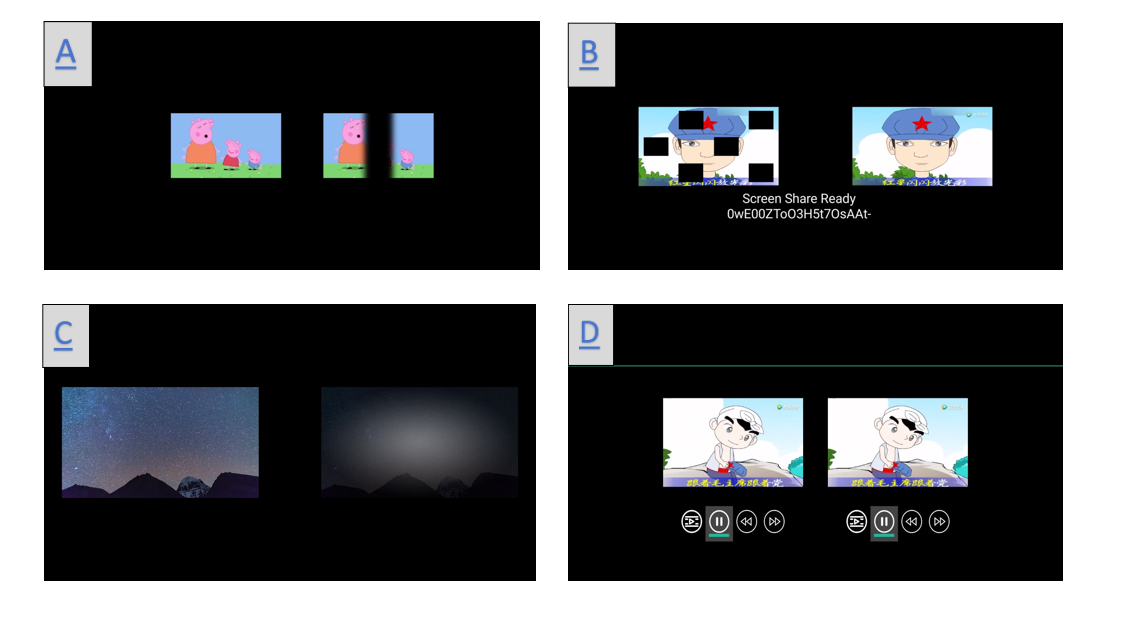
Additionally, the AMBER test employs serious games on tablet-based VR platforms for amblyopia training, aiming to enhance BCVA, binocular vision, stereovision, functional vision, and cerebral visual response [20]. These advancements highlight VR's potential in amblyopia treatment by providing engaging and adaptive training paradigms. Table 1 reviews recent applications of VR technology in amblyopia treatment over the past five years. Figure 2 illustrates different VR-based amblyopia training methods using cartoon simulations to enhance children's engagement in occlusion therapy.

Table 1 Application of VR technology in amblyopia treatment

Year

Authors

Sample Size

Purpose

Results

2021

Elhusseiny AM et al. [21]

20 participants

Evaluation of a prototype virtual reality-based binocular amblyopia treatment for optimal gains in corrected unilateral amblyopia and stereopsis in children and adults aged 7 years and older

Significant improvement in stereoscopic acuity from 7.3 ± 2 at baseline to 6.6 ± 2.3 at 8 weeks and 6.7 ± 2.6 at 16 weeks

2022

Li L et al. [22]

76 cases of children with amblyopia

Comparison of the compliance of children with amblyopia trained with the smartphone VR training method (EYEBIT) with those trained with the traditional method

The EYEBIT method has better adherence than the traditional method. There was a significant correlation between the adherence components of the two methods

2022

Tan F et al. [23]

145 cases of children with amblyopia

Exploring the potential impact of short-term plastic visual perception training based on VR and AR platforms for people with amblyopia

Significant improvements in best-corrected visual acuity and fine stereopsis were observed in both the VR and AR groups after training

2021

Rajavi Z et al. [17]

50 cases of unilateral amblyopia in children aged 4 to 10 years

Comparing the visual effects of masking therapy and VR games for the treatment of amblyopia in children

 Mean best-corrected visual acuity based on logMAR units improved significantly in both groups, with higher changes in BCVA in the VR group than in the masked group

2021

Halicka J et al. [24]

A case of refractive parametric amblyopia in an adult

Exploring whether adult patients with refractive error and amblyopia improve after VR visual training

After 1.5 years, the best corrected distance visual acuity of the amblyopic eye improves from 0.05 to 0.5, and the patient gradually develops stereo vision

2020

Halika J et al. [25]

84 cases of refractive parallax amblyopia

Retrospective analysis of the effectiveness of refractive parametric amblyopia treatment using VR technology in adults with amblyopia

The best corrected visual acuity improved from 0.48 to 0.58

2023

Molina-Martn A et al. [18]

4 children

Exploring the initial experience of using immersive VR devices and prototype software to treat refractive parallax amblyopia

Visual acuity improved after training, amblyopic eye distance VA improved in all three subjects, final stereo vision was 60 seconds arc, and 3 cpd showed an increase in spatial frequency of approximately 0.5 contrast sensitivity units

                                                                                                        VR: Virtual reality; AR: Augmented reality; BCVA: Best-corrected visual acuity.

Figure 2 Images of VR technology training amblyopia under different covering conditions. 

 (A. Strip localized masking mode; B. Multi-region localized masking mode; C. Adjusting contrast sensitivity training mode; D. Fusion training mode under unmasking.)

Figure 2. Images of VR technology training amblyopia under different covering conditions. (A. Strip localized masking mode; B. Multi-region localized masking mode; C. Adjusting contrast sensitivity training mode; D. Fusion training mode under unmasking.)

3.3 Application of VR technology in strabismus treatment

Strabismus is a condition in which the eyes fail to align properly on a target, with one eye fixating while the other deviates. Although surgical correction remains the primary treatment for pediatric strabismus, some patients experience postoperative recurrence, leading to abnormal binocular vision development. VR-based training offers a supplementary approach to enhance postoperative rehabilitation.

Studies have shown that VR-assisted training significantly improves eye position correction and three-tier visual function in children following surgical intervention [26]. In a study of 236 postoperative strabismus patients, 111 received VR-based rehabilitation training, leading to improved 1.5-meter and dynamic stereovision, thereby promoting binocular vision recovery [27]. To enhance diagnostic accuracy, VR technology has also been employed for objective eye deviation measurement through image-processing-based pupil tracking mechanisms, demonstrating strong consistency with clinical assessments performed by ophthalmologists [28].

Furthermore, VR-based simulation training has been utilized to improve ophthalmic residents' clinical diagnostic skills in strabismus management. Most participants reported that VR applications provided a valuable and efficient learning experience for strabismus evaluation and treatment planning [29,30]. These findings emphasize the role of VR technology in both clinical practice and medical education.

3.4 Core VR technologies in vision therapy

Several key VR technologies underpin its application in vision therapy across myopia, amblyopia, and strabismus treatment:

3.4.1 Eye-Tracking Mechanisms: Enables real-time monitoring of ocular movements and fixation stability, crucial for implementing personalized visual training programs.

3.4.2 Fixation Point Rendering: Maintains controlled peripheral defocus to simulate myopia prevention strategies and facilitate amblyopia rehabilitation.

3.4.3 Immersive Perceptual Learning: Integrates gamified visual stimulation to enhance visual attention, spatial orientation, and binocular coordination.

3.4.4 Motion Sickness Mitigation: Optimizes frame rates, latency reduction, and adaptive training durations to prevent discomfort and improve patient adherence .

4. VR technology in ophthalmic education

4.1 Application of VR technology in the education of ophthalmic students

Ophthalmology student education plays a crucial role in training future professionals in the field. With the increasing complexity of clinical practice and the growing responsibilities of medical care, there is a need for innovative solutions. VR technology has emerged as a promising tool to address these challenges. By utilizing VR technology, students can construct 3D models of eye anatomy and access live-streamed surgeries or recorded videos of VR surgical demonstrations [31]. The visual effects provided by VR are perceptive, realistic, and visible, enabling students to observe and learn more effectively. This immersive learning experience allows students to quickly and intuitively grasp knowledge during the learning process, enhancing their ability to apply it in practice [32].

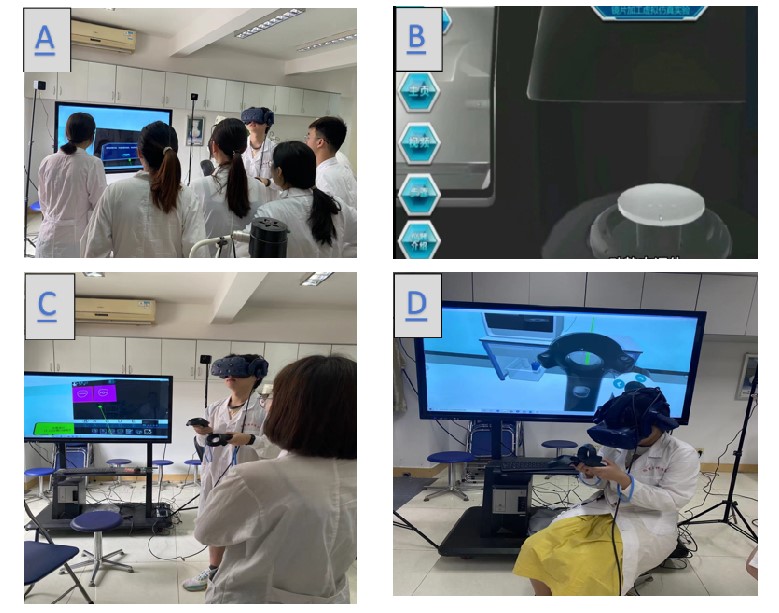
Through immersive visual experiences, students can gain a deeper understanding and analysis of ocular diseases and improve their diagnostic skills. In terms of disease diagnosis [33], the EyesiNet learning platform provides the content that meets the theoretical requirements in the teaching directory. 32 students first learned the theoretical knowledge on the platform and then trained with the Eyesi indirect simulator. After training, it was found that the students' ability to diagnose some retinal diseases has been significantly improved, which can be reflected in the test scores, while the test scores of students without simulator training have not changed [33]. In contrast, the Eyesi indirect simulator can see the real image around the patient's eyes using VR. In this way, the examiner can continue to control his hand under the ophthalmoscope in order to find a stable position for the examination. Through the effective combination of theory and practice, a kind of real and dynamic three-dimensional perception is generated in the mixture of real and virtual situations, which increases students' interest in learning ophthalmology knowledge, which on the one hand improves student learning and on the other hand makes teaching less difficult for teachers. Although there has been a shift in remote learning during the coronavirus disease 2019 (COVID-19) pandemic, starting with pedagogical methods and specific e-learning platforms, this suggests that the importance of virtual learning in ophthalmology has also become more important [34]. VR technology can simulate the real eye surgery scene, provide a more real learning experience, and enable students to receive real-time visual feedback, but it can't completely replace the actual surgery experience and the real patient situation, and the introduction of relevant technologies may have certain technical and financial challenges for schools. Figure 3 mainly shows the application of VR technology in the experimental teaching of ophthalmology. As shown in the figure, in ophthalmology laboratory teaching, we can wear VR glasses to simulate the process of lens production, optometry, so that students are immersed in the hands-on operation, which allows students to quickly understand the content of the experiment and improves learning efficiency.

Figure 3. Application of VR technology in the experimental teaching of ophthalmology.

(A. Students' co-participatory VR teaching situation; B. Virtual operation situation of VR teaching and training; C. Eye-hand operation screen in students' VR teaching; D. Simulated eyeglass lens positioning in VR teaching.)

20250701153802_2620.jpg

4.2 Application of VR technology in the surgery training of ophthalmologists

4.2.1 Application of VR technology in cataract surgery training
Cataract is a visual impairment disease caused by the clouding of the lens. It is most commonly observed in the elderly, particularly individuals over the age of 50, and is currently treated mainly by surgery [35]. Challenges in cataract surgery tend to be greater in the case of concurrent cataracts, such as high myopia with cataract, glaucoma with cataract, and diabetes with cataract [36]. The success rate of surgery heavily relies on the surgeon’s clinical experience and knowledge base. However, due to the lack of structured training courses, hands-on practice, and laboratory facilities, the probability of intraoperative and postoperative complications can be very high. The arrival of VR technology could well optimize the current situation and improve the outcome and safety of cataract surgery [37]. Using a cataract surgery simulator, surgeons can simulate and practice surgery in a virtual environment, become familiar with the surgical steps, and pre-assess the risks and complications of surgery to more effectively target individualized treatment [38]. A prominent example in this context is the deployment of the EyesI simulator, developed by VRmagic in Germany, which finds extensive application in cataract surgery, vitrectomy, and various other ophthalmic surgical procedures [39]. This simulator incorporates a virtual microscope capable of generating binocular imagery. Users can manipulate pan, zoom, and focus operations through foot controls. Additionally, it incorporates a model of the human eye, facilitating realistic simulations of eye surgeries through hands-on training [39]. Furthermore, the Microvistouch simulator, engineered by Immersivetouch in Chicago, IL, USA, simulates the intricate interactions between surgical instruments and ocular tissues in ophthalmic surgeries, employing touch screens and electromagnetic sensors to deliver realistic tactile feedback [40]. This simulator accommodates the emulation of cataract surgery, vitrectomy, retinal surgery, and various other ophthalmic interventions. The HelpMeSee Ophthalmic Surgery Simulator (HelpMeSee Inc., NY, USA) is a high-fidelity VR simulator specifically designed to support training in manual small-incision cataract surgery. The simulator combines high-quality computer graphics with the ability to provide real-time haptic feedback to provide a realistic experience of the performance of surgical tasks required for skills training [41]. In order to evaluate the application value of the HelpMeSee ophthalmic surgery simulator, Nair Ag designed a questionnaire with 11 questions, which was completed by 35 experienced surgeons. The core of the questionnaire was the visual reality in the simulator [42]. In general, 26 doctors believed that the overall visual performance of the eyes and instruments in the simulator was real, and 33 doctors suggested cataract surgery training on the simulator [42]. This study evaluates the value of the simulator in the form of a questionnaire, which can widely collect doctors' ideas, but its participants only include experts, so we can consider expanding the participants, and the research methods can also include some surgical learning and testing. It should be noted that although the development prospect of an ophthalmic surgery simulator is very good, the practical application still needs to undergo strict research, verification, and supervision. In terms of surgical assistance, the current VR technology has been more widely applied in the virtual training of ophthalmic surgeries. In the future, it can be synchronized with ophthalmic surgeries to treat patients, thereby improving the practicality of VR technology. Table 2 mainly provides a review of the application of VR technology in cataract surgery training over the last five years. Table 3 illustrates the technical differences between Eyesi and Microvistouch.

Table 2 Application of VR technology in cataract surgery training

Year

Authors

Sample Size

Purpose

Results

2022

Nair AG et al. [42]

35 MSICS Surgeons

Assessing the facial and content validity of the HelpMeSee cataract surgery simulator

94.3% of the subjects thought the simulator would help develop hand-eye coordination

2020

Ferris JD et al. [43]

265 trainee surgeons, 17,831 cataract operations

Study of the impact of the EyeSi surgical simulator on posterior capsular rupture cataract surgery for first- and second-year trainee surgeons

Complications of posterior capsule rupture cataract surgery fall by 38% for surgeons trained in surgical simulators

2020

Adnane I et al. [44]

12 residents, 300 cataract operations

Assessing the impact of surgical simulation training on residents

With the use of the simulator, the average operating time for the surgeon improved by 17 minutes, and only 6.7% of capsular ruptures occurred ruptures occurred

2022

Eltanamly RM et al. [45]

30 ophthalmologists

Assessing whether virtual reality simulators can help cataract surgeons train their non-dominant hand to perform surgery

After passing the simulator, 26 surgeons scored 90% of the average score of the dominant hand using the non-dominant hand

2022

Mathis T et al. [46]

24 ophthalmology residents

Assessing whether the use of the EyeSi simulator improves the time required for cataract training programs for ophthalmology residents

All doctors completed courses A and B. A total of 22 (91.7%) completed course C, and 5 (20.8%) completed course D

2019

Jacobsen MF et al. [47]

19 Cataract Surgeons

Exploring the correlation between virtual reality simulator performance and realistic cataract surgery performance

 Pearson correlation of 0.65, with simulator performance significantly correlated with actual cataract surgery performance

                                                                                                     MSICS: Manual small-incision cataract surgery.

Table 3. Differences in Eyesi and Microvistouch technology


Eyesi

Microvistouch

Core technology pathways

Binocular stereo vision is predominant, high hardware dependence, real-time priority

AI-driven monocular depth estimation with data-driven optimisation and dynamic environment adaptation

Hardware Architecture and Sensor Fusion

Multi-sensor fusion, high resolution camera

Lightweight hardware design, IR expandability

Algorithms and Data Processing

Traditional algorithms combined with deep learning, online calibration techniques

End-to-end deep learning models, a unified multi-task architecture


4.2.2 Application of VR technology in retinal surgery training
Retina is a crucial component of eye tissue, and untreated retinal diseases like macular degeneration and retinal detachment can lead to a decline in retinal function and even blindness. Performing retinal surgery is complex, and it becomes more challenging when complicated by other eye diseases. In recent years, the introduction of retinal virtual simulators has demonstrated the value of VR technology in this field.
One significant application of VR technology is in teaching retinal surgery [48]. The retinal virtual simulator allows surgeons to experience a simulated surgical scenario prior to the actual procedure. It generates an accurate model of the patient's retina, enabling surgical planning and simulation in a virtual environment. This assists surgeons in determining the most suitable surgical strategy. During VR-assisted surgery, ophthalmologists view the surgical scene through a binocular microscope or a flat-screen display, which provides a three-dimensional surgical space and helps them estimate the distance between instruments and the target structure [49]. VR technology also proves useful for prognostic assessment of retinopathy and postoperative follow-up, providing surgeons with more accurate management and treatment guidance [50]. However, the implementation of VR technology in retinal surgery faces challenges related to cost and accuracy. High-quality VR devices and software are relatively expensive, limiting their widespread adoption and use in clinical practice. Furthermore, newly developed devices require additional clinical trials and long-term follow-up to validate their safety and effectiveness [51]. At present, VR technology has great potential in the field of ophthalmic surgical assistance, but it should be noted that each clinical case is unique. When applying VR technology, doctors need to comprehensively consider factors such as the patient's condition, surgical complexity, and medical resources to make the best decision. Table 4 primarily reviews the application of VR technology in retinal surgery training over the past five years.

Table 4 Application of VR technology in retinal surgery training

Year

Authors

Sample Size

Purpose

Results

2022

Adatia FA et al. [48]

22 participants

Determine whether real-life surgical experience correlates with retinal virtual simulator scores and the impact of various challenges on surgical outcomes

When comparing Baseline 1 to Baseline 2, the overall improvement in scores reached 12.5%

2022

Seddon IA et al. [52]

10 surgical cases

Exploring the potential of real-time, three-dimensional surgical monitoring to improve education in vitreoretinal surgery

Ten surgical cases were successfully transmitted to two surgeons in real time. Case-specific details Visualization of low latency and interaction with the surgical surgeon are possible without compromising the quality of the surgical display

2020

Forslund Jacobsen M. et al. [53]

20 ophthalmologists

Comparing manual and robotically assisted vitreoretinal surgery using a VR surgical simulator

Robotic-assisted vitreoretinal surgery can improve precision and reduce tissue damage

2019

Ermolaev AP et al. [54]

19 patients

Development of a virtual reality device (p-VRD)-based visual field examination method for patients with impaired central vision

The p-VRD visual field examination evaluates the light sensitivity of non-centrally sighted eyes and is well comparable with the Goldmann test (coefficient of conformity K = 73.7%)

p-VRD: Virtual reality device; VR: Virtual reality.

5. VR technology in ophthalmic evaluation and disease diagnosis

VR technology has a wide range of applications in ophthalmic evaluation and disease diagnosis. In addition to visual field measurements, virtual reality is used in the measurement of visual function parameters, such as contrast sensitivity, dynamic visual acuity, determination of retinal neovascularization, and funduscopic examination, which helps to serve as a diagnostic tool for choroidal, retinal, and macular diseases.

In the measurement of the visual field, although the standard automatic perimetry is the gold standard of the current test field of vision, it also has corresponding limitations and shortcomings [55]. First of all, it needs the patient's learning experience and cooperation; otherwise, the measurement results are prone to error. Secondly, the standard automatic perimetry equipment is bulky, occupies space, and is expensive, which largely limits the application of vision in telemedicine, portable care, and home health care. In order to solve these problems, a company has developed a virtual reality headworn device with an eye tracking system that can track the eye movement of patients and instantly adjust the stimulating part according to the current fixation point. At the same time, it is also portable, which makes it more comfortable for patients to wear during the examination [56]. In addition, there are many visual field assessment devices based on virtual reality, such as the Oculus Quest VR headset (Facebook Technologies, LLC, Bern, Switzerland) [57], the VisuALL visual field platform (Olleyes, Inc., Summit, NJ) [58], the Vivid Vision Perimetry (VVP; Vivid Vision, Inc.) [59], and even the perimetry specially measured for children [60]. Secondly, in the measurement of visual function parameters, there is a rapid OKN-based virtual diagnostic tool that automatically estimates contrast sensitivity in 3.5 minutes without the active cooperation of the patient and the physician [61]. In addition, dynamic visual acuity can be measured and evaluated by using a virtual reality head-mounted display [62]. In a patient with chronic central plasma choroidal retinopathy (CSCR) combined with retinal neoangiogenesis (RNV), new diagnostic and therapeutic possibilities are available in combination with 3D reconstructive virtual reality technology and OCT angiography [63]. Of course, whether these devices can really replace the traditional perimeters needs further exploration.

6. Challenges and limitations of VR technology in ophthalmology

VR technology, with its inherent potential for personalized treatment and enhanced convenience, thrives in multiple facets. The creation of precise virtual models representing diverse ophthalmic diseases markedly augments screening and diagnostic capabilities, while the visualization of data and images underpins simulated surgical training, efficacy appraisal, and the mitigation of associated risks [64,65]. Furthermore, VR technology assumes a pivotal role in tracking patients' post-surgical convalescence, offering real-time feedback to foster effective rehabilitation and elevate their overall quality of life [66]. However, alongside these promising prospects, ophthalmic VR technology grapples with a range of challenges and limitations. Firstly, the conundrum of visual adaptation emerges. Prolonged use of VR headsets can induce visual fatigue and discomfort. Factors such as pressure, resolution, and motion blur related to headset usage may impair users' visual adaptability [13]. Secondly, financial and equipment constraints come into focus. Premium-quality VR equipment often entails substantial costs, potentially limiting its accessibility for certain institutions and individuals [51]. Due to financial constraints, it is difficult for primary hospitals to popularise VR technology, resulting in an uneven distribution of healthcare resources. the operation and interpretation of VR technology requires doctors to undergo specialised training, which adds to the time and financial costs. Medical institutions need to have specialised technicians to maintain the VR system, further increasing manpower costs. Thirdly, Ophthalmic surgery is distinguished by its intricate nature, characterized by a confluence of heightened risk and exacting precision. It mandates a meticulous orchestration of hand-eye coordination and demands an extensive comprehension of ocular anatomy. The distinctive challenges encountered in this discipline encompass the diminutive and intricate nature of ocular structures, the delicacy of ocular tissues, restricted visibility during surgical procedures, and the imperative to consider factors such as aesthetic outcomes and the broader health implications for the patient [67-71]. For example, vitrectomy requires precision down to the micron level, which may lead to surgical errors if the VR system has too much latency or insufficient resolution. Eye tracking is a core feature of VR technology in ophthalmic applications, but the limited precision and stability of existing devices may affect the accuracy of diagnostic results (e.g., strabismus assessment) or surgical navigation. Lastly, the matter of standardization and specifications warrants attention. The application of VR technology in ophthalmology is a relatively nascent endeavor, marked by a dearth of unified standards and specifications. This scarcity could lead to issues of compatibility between equipment and applications from distinct manufacturers and disparities across various research, diagnostic, and treatment modalities. For example, VR systems need to be seamlessly integrated with ophthalmic examination equipment (e.g., OCT, fundus camera), but the interfaces and data formats of equipment from different vendors vary widely, increasing the difficulty of technical integration.

7. Emerging trends and future developments of VR technology in ophthalmology

In recent years, the integration of VR technology into the sphere of ophthalmology has witnessed a noteworthy surge, conjoining the domains of computer science, optics, and ophthalmic practice. In a crossover randomized controlled trial, virtual reality-based gaze training improved the ability of retinitis pigmentosa patients to navigate their eyes and increased overall independence in completing daily visual tasks [72]. Traditional research methods have struggled to accurately simulate and evaluate visual correction methods, resulting in time-consuming assessments with limited scope and flexibility. VisionaryVR is an optical simulation tool that can support dynamic visual tasks using a versatile VR environment with integrated comprehensive eye tracking but falls short because of the resolution of its VR glasses, which have not yet reached the level of resolution needed to perfectly mimic real-life scenes with the required fidelity [73]. Even in a recent study, eye movement indices obtained using VR eye trackers could be useful biomarkers for diagnosing and tracking depression—gaze and sweep indices, to be precise [74]. VR technology affords ophthalmologists access to a highly realistic environment for surgical simulation and training, facilitating meticulous preoperative practice and, consequently, promoting enhanced scrutiny and evaluation of ocular anomalies while concurrently mitigating the occurrence of surgical errors [75]. Moreover, VR technology has the capacity to simulate a spectrum of visual symptoms associated with diverse pathological conditions, augmenting medical practitioners' comprehension and furnishing a diagnostic foundation. This simulated training milieu engenders a heightened level of safety and precision in surgical procedures, culminating in elevated surgical success rates and patient well-being [76]. Additionally, VR technology augments visualization and assists in operative procedures through features such as image-based navigation, surgical path planning, and procedural guidance. This comprehensive support empowers surgeons to conduct surgeries with greater precision, thereby amplifying the overall quality and efficacy of ophthalmic interventions [77].

Ophthalmic VR technology stands as an innovative amalgamation of VR technology and the domain of ophthalmic medicine, harboring substantial potential and prospective developments across multifarious domains. First and foremost, it is pivotal in the domain of surgical planning and navigation. Given the exigent precision demanded in ophthalmic surgery, the capacity for meticulous surgical planning and navigation holds paramount importance. VR technology facilitates the emulation of the surgical procedure within a virtual environment, aiding ophthalmologists in strategizing the surgical process, encompassing the delineation of operative pathways, and facilitating immersive training. This, in turn, augments the precision and safety of ophthalmic surgeries [78,79]. Secondly, VR technology has ushered in the realm of remote diagnosis and rehabilitation. It offers a means for patients to engage in ocular examinations from the comfort of their homes through VR equipment, subsequently transmitting the data for remote diagnosis by ophthalmologists. This holds particular value for patients who face physical constraints when visiting medical facilities or reside in remote areas. Additionally, VR technology enables patients to undertake personalized visual rehabilitation regimens, expediting the rehabilitation process [80]. Furthermore, VR technology has been transformative in the realm of ophthalmology education and training. Through realistic simulations and practical experience in VR environments, both ophthalmologists and students can attain a heightened degree of experiential authenticity, thereby enhancing their competencies and skills [33,34]. Finally, AI-integrated virtual reality systems are also promising for the future. FovealNet, a state-of-the-art AI-powered eye-tracking framework, aims to optimise system performance by strategically improving eye-tracking accuracy [81]. AI integration in cataract surgery promises to improve surgical accuracy, enhance patient outcomes and improve the efficiency of healthcare practitioners [82].

The ethical implications of virtual reality technology in ophthalmology can be seen in patient privacy and data security, informed consent and patient autonomy, technological reliability and medical liability definition, social equity and technological accessibility, and psychology and cognition. Regarding the regulatory framework, we can effectively build data protection and privacy regulations, strengthen ethical review and technology validation mechanisms, define responsibility and legal regulation, and promote technology accessibility and fairness policies. The application of virtual reality technology in ophthalmology needs to find a balance between technological innovation and ethical constraints to ensure that the technology truly serves patients.

In tandem with the relentless progression of technology, VR technology assumes an unparalleled role within the field of ophthalmology. Its application in ophthalmic adjunct examinations exemplifies the emerging trend of virtual diagnostic instruments, holding substantial promise in bolstering disease screening endeavors and potentially retarding the progression of ocular conditions over time [83,84]. Notably, the hallmarks of VR technology encompass interactivity and intelligence. The exploration of novel paradigms and harnessing the inherent advantages of VR technology to align with the ever-evolving requisites of ophthalmology emerge as imperative endeavors [85]. Just like artificial intelligence in ophthalmology, developing intelligent machines that learn, reason, judge, and make decisions like humans can better serve humans [86].

8. Conclusion

In conclusion, the integration of VR technology into ophthalmology clinical disease, student education, and surgical training has reached an advanced stage of development. Leveraging a cohesive fusion of the Internet, big data, artificial intelligence, and cutting-edge computer technologies, VR technology has emerged as a valuable asset with substantial application prospects and untapped horizons, thereby presenting both unprecedented opportunities and intricate challenges for ophthalmologists and patients alike. Through an ongoing commitment to research and innovation, VR technology holds the promise of ushering in a transformative paradigm in the future of ophthalmology, thereby contributing to the advancement of ocular health on a global scale.

Correction notice

It has been amended in accordance with regulations

Acknowledgements

None

Author Contributions

(I) Conception and design: Boyuan Wen, Li Li, Yan Huang, Zhuoting Zhu
(II) Administrative support: Yan Huang, Zhuoting Zhu
(III) Provision of study materials or patients: Boyuan Wen, Li Li
(IV) Collection and assembly of data: Boyuan Wen, Li Li, Jiahao Liu, Yunxi Ye, Wenbin Chen, Xierong Wang
(V) Data analysis and interpretation: Boyuan Wen, Li Li, Jiahao Liu, Yunxi Ye, Wenbin Chen, Xierong Wang
(VI) Manuscript writing: Boyuan Wen, Li Li
(VII) Manuscript review and revision: All authors
(VIII)Final approval of manuscript: All authors

Conflict of Interests

The authors declare no conflicts of interest.

Patient consent for publication

None

Ethics approval and consent to participate

None

Data availability statement

None

Open access

This is an Open Access article distributed in accordance with the Creative Commons AttributionNonCommercial-NoDerivs 4.0 International License (CC BY-NC-ND 4.0), which permits the non-commercial replication and distribution of the article with the strict proviso that no changes or edits are made and the original work is properly cited (including links to both the formal publication through the relevant DOI and the license).
1、Salatino A, Zavattaro C, Gammeri R, et al. Virtual reality rehabilitation for unilateral spatial neglect: a systematic review of immersive, semi-immersive and non-immersive techniques. Neurosci Biobehav Rev. 2023, 152: 105248. DOI: 10.1016/j.neubiorev.2023.105248. Salatino A, Zavattaro C, Gammeri R, et al. Virtual reality rehabilitation for unilateral spatial neglect: a systematic review of immersive, semi-immersive and non-immersive techniques. Neurosci Biobehav Rev. 2023, 152: 105248. DOI: 10.1016/j.neubiorev.2023.105248.
2、Wender C. Immersive virtual reality to relieve exercise-induced pain caused by aerobic cycling. Pain Manag. 2022, 12(5): 665-674. DOI: 10.2217/pmt-2021-0095. Wender C. Immersive virtual reality to relieve exercise-induced pain caused by aerobic cycling. Pain Manag. 2022, 12(5): 665-674. DOI: 10.2217/pmt-2021-0095.
3、MacKenzie CF, Harris TE, Shipper AG, et al. Virtual reality and haptic interfaces for civilian and military open trauma surgery training: a systematic review. Injury. 2022, 53(11): 3575-3585. DOI: 10.1016/j.injury.2022.08.003.MacKenzie CF, Harris TE, Shipper AG, et al. Virtual reality and haptic interfaces for civilian and military open trauma surgery training: a systematic review. Injury. 2022, 53(11): 3575-3585. DOI: 10.1016/j.injury.2022.08.003.
4、Yen HY, Chiu HL. Virtual reality exergames for improving older adults' cognition and depression: a systematic review and meta-analysis of randomized control trials. J Am Med Dir Assoc. 2021, 22(5): 995-1002. DOI: 10.1016/j.jamda.2021.03.009. Yen HY, Chiu HL. Virtual reality exergames for improving older adults' cognition and depression: a systematic review and meta-analysis of randomized control trials. J Am Med Dir Assoc. 2021, 22(5): 995-1002. DOI: 10.1016/j.jamda.2021.03.009.
5、Saneinia S, Zhou R, Gholizadeh A, et al. Immersive media-based tourism emerging challenge of VR addiction among generation Z. Front Public Health. 2022, 10: 833658. DOI: 10.3389/fpubh.2022.833658.Saneinia S, Zhou R, Gholizadeh A, et al. Immersive media-based tourism emerging challenge of VR addiction among generation Z. Front Public Health. 2022, 10: 833658. DOI: 10.3389/fpubh.2022.833658.
6、Plotzky C, Lindwedel U, Sorber M, et al. Virtual reality simulations in nurse education: a systematic mapping review. Nurse Educ Today. 2021, 101: 104868. DOI: 10.1016/j.nedt.2021.104868. Plotzky C, Lindwedel U, Sorber M, et al. Virtual reality simulations in nurse education: a systematic mapping review. Nurse Educ Today. 2021, 101: 104868. DOI: 10.1016/j.nedt.2021.104868.
7、Veneziano D, Cacciamani G, Rivas JG, et al. VR and machine learning: novel pathways in surgical hands-on training. Curr Opin Urol. 2020, 30(6): 817-822. DOI: 10.1097/MOU.0000000000000824. Veneziano D, Cacciamani G, Rivas JG, et al. VR and machine learning: novel pathways in surgical hands-on training. Curr Opin Urol. 2020, 30(6): 817-822. DOI: 10.1097/MOU.0000000000000824.
8、Demeco A, Zola L, Frizziero A, et al. Immersive virtual reality in post-stroke rehabilitation: a systematic review. Sensors (Basel), 2023, 23(3): 1712. DOI: 10.3390/s23031712. Demeco A, Zola L, Frizziero A, et al. Immersive virtual reality in post-stroke rehabilitation: a systematic review. Sensors (Basel), 2023, 23(3): 1712. DOI: 10.3390/s23031712.
9、Paul SK, Clark MA, Scott IU, et al. Virtual eye surgery training in ophthalmic graduate medical education. Can J Ophthalmol. 2018, 53(6): e218-e220. DOI: 10.1016/j.jcjo.2018.03.018. Paul SK, Clark MA, Scott IU, et al. Virtual eye surgery training in ophthalmic graduate medical education. Can J Ophthalmol. 2018, 53(6): e218-e220. DOI: 10.1016/j.jcjo.2018.03.018.
10、 Sun MT, Tran M, Singh K, et al. Glaucoma and myopia: diagnostic challenges. Biomolecules. 2023, 13(3): 562. DOI: 10.3390/biom13030562. Sun MT, Tran M, Singh K, et al. Glaucoma and myopia: diagnostic challenges. Biomolecules. 2023, 13(3): 562. DOI: 10.3390/biom13030562.
11、Guo DY, Shen YY, Zhu MM, et al. Virtual reality training improves accommodative facility and accommodative range. Int J Ophthalmol. 2022 Jul 18;15(7):1116-1121. DOI: 10.18240/ijo.2022.07.11.Guo DY, Shen YY, Zhu MM, et al. Virtual reality training improves accommodative facility and accommodative range. Int J Ophthalmol. 2022 Jul 18;15(7):1116-1121. DOI: 10.18240/ijo.2022.07.11.
12、Shi Y. Effect of atropine eye drops combined with VR-based binocular visual function balance training for prevention and control of juvenile myopia. Evid Based Complement Alternat Med. 2022, 2022: 4159996. DOI: 10.1155/2022/4159996.Shi Y. Effect of atropine eye drops combined with VR-based binocular visual function balance training for prevention and control of juvenile myopia. Evid Based Complement Alternat Med. 2022, 2022: 4159996. DOI: 10.1155/2022/4159996.
13、Li Y, Yip MYT, Ting DSW, et al. Artificial intelligence and digital solutions for myopia. Taiwan J Ophthalmol. 2023, 13(2): 142-150. DOI: 10.4103/tjo.TJO-D-23-00032. Li Y, Yip MYT, Ting DSW, et al. Artificial intelligence and digital solutions for myopia. Taiwan J Ophthalmol. 2023, 13(2): 142-150. DOI: 10.4103/tjo.TJO-D-23-00032.
14、Zhao%20F%2C%20Chen%20L%2C%20Ma%20H%2C%20et%20al.%20Virtual%20reality%3A%20a%20possible%20approach%20to%20myopia%20prevention%20and%20control%3F%20Med%20Hypotheses.%202018%2C%20121%3A%201-3.%20DOI%3A%2010.1016%2Fj.mehy.2018.09.021.%20Zhao%20F%2C%20Chen%20L%2C%20Ma%20H%2C%20et%20al.%20Virtual%20reality%3A%20a%20possible%20approach%20to%20myopia%20prevention%20and%20control%3F%20Med%20Hypotheses.%202018%2C%20121%3A%201-3.%20DOI%3A%2010.1016%2Fj.mehy.2018.09.021.%20
15、Lee SH, Kim M, Kim H, et al. Visual fatigue induced by watching virtual reality device and the effect of anisometropia. Ergonomics. 2021, 64(12): 1522-1531. DOI: 10.1080/00140139.2021.1957158. Lee SH, Kim M, Kim H, et al. Visual fatigue induced by watching virtual reality device and the effect of anisometropia. Ergonomics. 2021, 64(12): 1522-1531. DOI: 10.1080/00140139.2021.1957158.
16、Bui Quoc E, Kulp MT, Burns JG, et al. Amblyopia: a review of unmet needs, current treatment options, and emerging therapies. Surv Ophthalmol. 2023, 68(3): 507-525. DOI: 10.1016/j.survophthal.2023.01.001. Bui Quoc E, Kulp MT, Burns JG, et al. Amblyopia: a review of unmet needs, current treatment options, and emerging therapies. Surv Ophthalmol. 2023, 68(3): 507-525. DOI: 10.1016/j.survophthal.2023.01.001.
17、Rajavi Z, Soltani A, Vakili A, et al. Virtual reality game playing in amblyopia therapy: a randomized clinical trial. J Pediatr Ophthalmol Strabismus. 2021, 58(3): 154-160. DOI: 10.3928/01913913-20210108-02. Rajavi Z, Soltani A, Vakili A, et al. Virtual reality game playing in amblyopia therapy: a randomized clinical trial. J Pediatr Ophthalmol Strabismus. 2021, 58(3): 154-160. DOI: 10.3928/01913913-20210108-02.
18、Molina-Martín A, Leal-Vega L, de Fez D, et al. Amblyopia treatment through immersive virtual reality: a preliminary experience in anisometropic children. Vision (Basel), 2023, 7(2): 42. DOI: 10.3390/vision7020042.Molina-Martín A, Leal-Vega L, de Fez D, et al. Amblyopia treatment through immersive virtual reality: a preliminary experience in anisometropic children. Vision (Basel), 2023, 7(2): 42. DOI: 10.3390/vision7020042.
19、Leal%20Vega%20L%2C%20Pi%C3%B1ero%20DP%2C%20Hern%C3%A1ndez%20Rodr%C3%ADguez%20CJ%2C%20et%20al.%20Study%20protocol%20for%20a%20randomized%20controlled%20trial%20of%20the%20NEIVATECH%20virtual%20reality%20system%20to%20improve%20visual%20function%20in%20children%20with%20anisometropic%20amblyopia.%20BMC%20Ophthalmol.%202022%2C%2022(1)%3A%20253.%20DOI%3A%2010.1186%2Fs12886-022-02466-z.%20Leal%20Vega%20L%2C%20Pi%C3%B1ero%20DP%2C%20Hern%C3%A1ndez%20Rodr%C3%ADguez%20CJ%2C%20et%20al.%20Study%20protocol%20for%20a%20randomized%20controlled%20trial%20of%20the%20NEIVATECH%20virtual%20reality%20system%20to%20improve%20visual%20function%20in%20children%20with%20anisometropic%20amblyopia.%20BMC%20Ophthalmol.%202022%2C%2022(1)%3A%20253.%20DOI%3A%2010.1186%2Fs12886-022-02466-z.%20
20、Simon-Martinez C, Antoniou MP, Bouthour W, et al. Stereoptic serious games as a visual rehabilitation tool for individuals with a residual amblyopia (AMBER trial): a protocol for a crossover randomized controlled trial. BMC Ophthalmol. 2023, 23(1): 220. DOI: 10.1186/s12886-023-02944-y. Simon-Martinez C, Antoniou MP, Bouthour W, et al. Stereoptic serious games as a visual rehabilitation tool for individuals with a residual amblyopia (AMBER trial): a protocol for a crossover randomized controlled trial. BMC Ophthalmol. 2023, 23(1): 220. DOI: 10.1186/s12886-023-02944-y.
21、Elhusseiny AM, Bishop K, Staffa SJ, et al. Virtual reality prototype for binocular therapy in older children and adults with amblyopia. J Am Assoc Pediatr Ophthalmol Strabismus. 2021, 25(4): 217.e1-217.e6. DOI: 10.1016/j.jaapos.2021.03.008. Elhusseiny AM, Bishop K, Staffa SJ, et al. Virtual reality prototype for binocular therapy in older children and adults with amblyopia. J Am Assoc Pediatr Ophthalmol Strabismus. 2021, 25(4): 217.e1-217.e6. DOI: 10.1016/j.jaapos.2021.03.008.
22、Li L, Xue H, Lai T, et al. Comparison of compliance among patients with pediatric amblyopia undergoing virtual reality-based and traditional patching method training. Front Public Health. 2022, 10: 1037412. DOI: 10.3389/fpubh.2022.1037412. Li L, Xue H, Lai T, et al. Comparison of compliance among patients with pediatric amblyopia undergoing virtual reality-based and traditional patching method training. Front Public Health. 2022, 10: 1037412. DOI: 10.3389/fpubh.2022.1037412.
23、Tan F, Yang X, Fan Y, et al. The study of short-term plastic visual perceptual training based on virtual and augmented reality technology in amblyopia. J Ophthalmol. 2022, 2022: 2826724. DOI: 10.1155/2022/2826724. Tan F, Yang X, Fan Y, et al. The study of short-term plastic visual perceptual training based on virtual and augmented reality technology in amblyopia. J Ophthalmol. 2022, 2022: 2826724. DOI: 10.1155/2022/2826724.
24、Halicka J, Bittsansky M, Sivak S, et al. Virtual reality visual training in an adult patient with anisometropic amblyopia: visual and functional magnetic resonance outcomes. Vision (Basel), 2021, 5(2): 22. DOI: 10.3390/vision5020022.Halicka J, Bittsansky M, Sivak S, et al. Virtual reality visual training in an adult patient with anisometropic amblyopia: visual and functional magnetic resonance outcomes. Vision (Basel), 2021, 5(2): 22. DOI: 10.3390/vision5020022.
25、Hali%C4%8Dka%20J%2C%20Sahatqija%20E%2C%20Kras%C5%88ansk%C3%BD%20M%2C%20et%20al.%20Visual%20Training%20in%20Virtual%20Reality%20in%20Adult%20Patients%20with%20Anisometric%20Amblyopia.%20Ceska%20a%20Slovenska%20Oftalmologie%3A%20Casopis%20Ceske%20Oftalmologicke%20Spolecnosti%20a%20Slovenske%20Oftalmologicke%20Spolecnosti.%202020%2C%2076(1)%3A%2024-28.Hali%C4%8Dka%20J%2C%20Sahatqija%20E%2C%20Kras%C5%88ansk%C3%BD%20M%2C%20et%20al.%20Visual%20Training%20in%20Virtual%20Reality%20in%20Adult%20Patients%20with%20Anisometric%20Amblyopia.%20Ceska%20a%20Slovenska%20Oftalmologie%3A%20Casopis%20Ceske%20Oftalmologicke%20Spolecnosti%20a%20Slovenske%20Oftalmologicke%20Spolecnosti.%202020%2C%2076(1)%3A%2024-28.
26、Zhang H, Yang SH, Chen T, et al. The effect of virtual reality technology in children after surgery for concomitant strabismus. Indian J Ophthalmol. 2023, 71(2): 625-630. DOI: 10.4103/ijo.IJO_1505_22. Zhang H, Yang SH, Chen T, et al. The effect of virtual reality technology in children after surgery for concomitant strabismus. Indian J Ophthalmol. 2023, 71(2): 625-630. DOI: 10.4103/ijo.IJO_1505_22.
27、Yang X, Fan Y, Chu H, et al. Preliminary study of short-term visual perceptual training based on virtual reality and augmented reality in postoperative strabismic patients. Cyberpsychol Behav Soc Netw. 2022, 25(7): 465-470. DOI: 10.1089/cyber.2022.0113. Yang X, Fan Y, Chu H, et al. Preliminary study of short-term visual perceptual training based on virtual reality and augmented reality in postoperative strabismic patients. Cyberpsychol Behav Soc Netw. 2022, 25(7): 465-470. DOI: 10.1089/cyber.2022.0113.
28、Miao Y, Jeon JY, Park G, et al. Virtual reality-based measurement of ocular deviation in strabismus. Comput Methods Programs Biomed. 2020, 185: 105132. DOI: 10.1016/j.cmpb.2019.105132.Miao Y, Jeon JY, Park G, et al. Virtual reality-based measurement of ocular deviation in strabismus. Comput Methods Programs Biomed. 2020, 185: 105132. DOI: 10.1016/j.cmpb.2019.105132.
29、Moon HS, Yoon HJ, Park SW, et al. Usefulness of virtual reality-based training to diagnose strabismus. Sci Rep. 2021, 11(1): 5891. DOI: 10.1038/s41598-021-85265-8. Moon HS, Yoon HJ, Park SW, et al. Usefulness of virtual reality-based training to diagnose strabismus. Sci Rep. 2021, 11(1): 5891. DOI: 10.1038/s41598-021-85265-8.
30、 Mehringer WA, Gerhard Wirth M, Gradl S, et al. An image-based method for measuring strabismus in virtual reality[C]//2020 IEEE International Symposium on Mixed and Augmented Reality Adjunct (ISMAR-Adjunct). November 9-13, 2020. Recife, Brazil. IEEE. 2020: 5-12. DOI: 10.1109/ismar-adjunct51615.2020.00018. Mehringer WA, Gerhard Wirth M, Gradl S, et al. An image-based method for measuring strabismus in virtual reality[C]//2020 IEEE International Symposium on Mixed and Augmented Reality Adjunct (ISMAR-Adjunct). November 9-13, 2020. Recife, Brazil. IEEE. 2020: 5-12. DOI: 10.1109/ismar-adjunct51615.2020.00018.
31、Lin JC, Yu Z, Scott IU, et al. Virtual reality training for cataract surgery operating performance in ophthalmology trainees. Cochrane Database Syst Rev. 2021, 12(12): CD014953. DOI: 10.1002/14651858.CD014953.pub2. Lin JC, Yu Z, Scott IU, et al. Virtual reality training for cataract surgery operating performance in ophthalmology trainees. Cochrane Database Syst Rev. 2021, 12(12): CD014953. DOI: 10.1002/14651858.CD014953.pub2.
32、Williams M. Virtual reality in ophthalmology education: simulating pupil examination. Eye (Lond). 2022, 36(11): 2084-2085. DOI: 10.1038/s41433-022-02078-3. Williams M. Virtual reality in ophthalmology education: simulating pupil examination. Eye (Lond). 2022, 36(11): 2084-2085. DOI: 10.1038/s41433-022-02078-3.
33、Deuchler S, Sebode C, Ackermann H, et al. Combination of simulation-based and online learning in ophthalmology: efficiency of simulation in combination with independent online learning within the framework of EyesiNet in student education. Ophthalmologe. 2022, 119(1): 20-29. DOI: 10.1007/s00347-020-01313-0. Deuchler S, Sebode C, Ackermann H, et al. Combination of simulation-based and online learning in ophthalmology: efficiency of simulation in combination with independent online learning within the framework of EyesiNet in student education. Ophthalmologe. 2022, 119(1): 20-29. DOI: 10.1007/s00347-020-01313-0.
34、Chatziralli I, Ventura CV, Touhami S, et al. Transforming ophthalmic education into virtual learning during COVID-19 pandemic: a global perspective. Eye (Lond), 2021, 35(5): 1459-1466. DOI: 10.1038/s41433-020-1080-0. Chatziralli I, Ventura CV, Touhami S, et al. Transforming ophthalmic education into virtual learning during COVID-19 pandemic: a global perspective. Eye (Lond), 2021, 35(5): 1459-1466. DOI: 10.1038/s41433-020-1080-0.
35、Cicinelli MV, Buchan JC, Nicholson M, et al. Cataracts. Lancet. 2023, 401(10374): 377-389. DOI: 10.1016/S0140-6736(22)01839-6. Cicinelli MV, Buchan JC, Nicholson M, et al. Cataracts. Lancet. 2023, 401(10374): 377-389. DOI: 10.1016/S0140-6736(22)01839-6.
36、Gali HE, Sella R, Afshari NA. Cataract grading systems: a review of past and present. Curr Opin Ophthalmol. 2019, 30(1): 13-18. DOI: 10.1097/ICU.0000000000000542. Gali HE, Sella R, Afshari NA. Cataract grading systems: a review of past and present. Curr Opin Ophthalmol. 2019, 30(1): 13-18. DOI: 10.1097/ICU.0000000000000542.
37、Rothschild P, Richardson A, Beltz J, et al. Effect of virtual reality simulation training on real-life cataract surgery complications: systematic literature review. J Cataract Refract Surg. 2021, 47(3): 400-406. DOI: 10.1097/j.jcrs.0000000000000323. Rothschild P, Richardson A, Beltz J, et al. Effect of virtual reality simulation training on real-life cataract surgery complications: systematic literature review. J Cataract Refract Surg. 2021, 47(3): 400-406. DOI: 10.1097/j.jcrs.0000000000000323.
38、Nayer ZH, Murdock B, Dharia IP, et al. Predictive and construct validity of virtual reality cataract surgery simulators. J Cataract Refract Surg. 2020, 46(6): 907-912. DOI: 10.1097/j.jcrs.0000000000000137. Nayer ZH, Murdock B, Dharia IP, et al. Predictive and construct validity of virtual reality cataract surgery simulators. J Cataract Refract Surg. 2020, 46(6): 907-912. DOI: 10.1097/j.jcrs.0000000000000137.
39、Jaud C, Salleron J, Cisse C, et al. EyeSi Surgical Simulator: validation of a proficiency-based test for assessment of vitreoretinal surgical skills. Acta Ophthalmol. 2021, 99(4): 390-396. DOI: 10.1111/aos.14628. Jaud C, Salleron J, Cisse C, et al. EyeSi Surgical Simulator: validation of a proficiency-based test for assessment of vitreoretinal surgical skills. Acta Ophthalmol. 2021, 99(4): 390-396. DOI: 10.1111/aos.14628.
40、Sikder S, Luo J, Banerjee PP, et al. The use of a virtual reality surgical simulator for cataract surgical skill assessment with 6 months of intervening operating room experience. Clin Ophthalmol. 2015, 9: 141-149. DOI: 10.2147/OPTH.S69970. Sikder S, Luo J, Banerjee PP, et al. The use of a virtual reality surgical simulator for cataract surgical skill assessment with 6 months of intervening operating room experience. Clin Ophthalmol. 2015, 9: 141-149. DOI: 10.2147/OPTH.S69970.
41、Nair AG, Ahiwalay C, Bacchav AE, et al. Effectiveness of simulation-based training for manual small incision cataract surgery among novice surgeons: a randomized controlled trial. Sci Rep. 2021, 11(1): 10945. DOI: 10.1038/s41598-021-90410-4. Nair AG, Ahiwalay C, Bacchav AE, et al. Effectiveness of simulation-based training for manual small incision cataract surgery among novice surgeons: a randomized controlled trial. Sci Rep. 2021, 11(1): 10945. DOI: 10.1038/s41598-021-90410-4.
42、Nair AG, Ahiwalay C, Bacchav AE, et al. Assessment of a high-fidelity, virtual reality-based, manual small-incision cataract surgery simulator: a face and content validity study. Indian J Ophthalmol. 2022, 70(11): 4010-4015. DOI: 10.4103/ijo.IJO_1593_22. Nair AG, Ahiwalay C, Bacchav AE, et al. Assessment of a high-fidelity, virtual reality-based, manual small-incision cataract surgery simulator: a face and content validity study. Indian J Ophthalmol. 2022, 70(11): 4010-4015. DOI: 10.4103/ijo.IJO_1593_22.
43、Ferris JD, Donachie PH, Johnston RL, et al. Royal College of Ophthalmologists' National Ophthalmology Database study of cataract surgery: report 6. The impact of EyeSi virtual reality training on complications rates of cataract surgery performed by first and second year trainees. Br J Ophthalmol. 2020, 104(3): 324-329. DOI: 10.1136/bjophthalmol-2018-313817. Ferris JD, Donachie PH, Johnston RL, et al. Royal College of Ophthalmologists' National Ophthalmology Database study of cataract surgery: report 6. The impact of EyeSi virtual reality training on complications rates of cataract surgery performed by first and second year trainees. Br J Ophthalmol. 2020, 104(3): 324-329. DOI: 10.1136/bjophthalmol-2018-313817.
44、Adnane I, Chahbi M, Elbelhadji M. Virtual simulation for learning cataract surgery. J Fr Ophtalmol. 2020, 43(4): 334-340. DOI: 10.1016/j.jfo.2019.08.006. Adnane I, Chahbi M, Elbelhadji M. Virtual simulation for learning cataract surgery. J Fr Ophtalmol. 2020, 43(4): 334-340. DOI: 10.1016/j.jfo.2019.08.006.
45、%20Eltanamly%20RM%2C%20Elmekawey%20H%2C%20Youssef%20MM%2C%20et%20al.%20Can%20virtual%20reality%20surgical%20simulator%20improve%20the%20function%20of%20the%20non-dominant%20hand%20in%20ophthalmic%20surgeons%3F.%20Indian%20J%20Ophthalmol.%202022%2C%2070(5)%3A%201795-1799.%20DOI%3A%2010.4103%2Fijo.IJO_2652_21.%20%20Eltanamly%20RM%2C%20Elmekawey%20H%2C%20Youssef%20MM%2C%20et%20al.%20Can%20virtual%20reality%20surgical%20simulator%20improve%20the%20function%20of%20the%20non-dominant%20hand%20in%20ophthalmic%20surgeons%3F.%20Indian%20J%20Ophthalmol.%202022%2C%2070(5)%3A%201795-1799.%20DOI%3A%2010.4103%2Fijo.IJO_2652_21.%20
46、Mathis T, Mouchel R, Malecaze J, et al. Evaluation of the time required to complete a cataract training program on EyeSi surgical simulator during the first-year residency. Eur J Ophthalmol. 2022: 11206721221136322. DOI: 10.1177/11206721221136322. Mathis T, Mouchel R, Malecaze J, et al. Evaluation of the time required to complete a cataract training program on EyeSi surgical simulator during the first-year residency. Eur J Ophthalmol. 2022: 11206721221136322. DOI: 10.1177/11206721221136322.
47、Jacobsen MF, Konge L, Bach-Holm D, et al. Correlation of virtual reality performance with real-life cataract surgery performance. J Cataract Refract Surg. 2019, 45(9): 1246-1251. DOI: 10.1016/j.jcrs.2019.04.007.Jacobsen MF, Konge L, Bach-Holm D, et al. Correlation of virtual reality performance with real-life cataract surgery performance. J Cataract Refract Surg. 2019, 45(9): 1246-1251. DOI: 10.1016/j.jcrs.2019.04.007.
48、Adatia FA, González-Saldivar G, Chow DR. Effects of sleep deprivation, non-dominant hand employment, caffeine and alcohol intake during surgical performance: lessons learned from the retina eyesi virtual reality surgical simulator. Transl Vis Sci Technol. 2022, 11(8): 16. DOI: 10.1167/tvst.11.8.16. Adatia FA, González-Saldivar G, Chow DR. Effects of sleep deprivation, non-dominant hand employment, caffeine and alcohol intake during surgical performance: lessons learned from the retina eyesi virtual reality surgical simulator. Transl Vis Sci Technol. 2022, 11(8): 16. DOI: 10.1167/tvst.11.8.16.
49、de Smet MD, de Jonge N, Iannetta D, et al. Human/robotic interaction: vision limits performance in simulated vitreoretinal surgery. Acta Ophthalmol. 2019, 97(7): 672-678. DOI: 10.1111/aos.14003.de Smet MD, de Jonge N, Iannetta D, et al. Human/robotic interaction: vision limits performance in simulated vitreoretinal surgery. Acta Ophthalmol. 2019, 97(7): 672-678. DOI: 10.1111/aos.14003.
50、Soni T, Kohli P. Commentary: Simulators for vitreoretinal surgical training. Indian J Ophthalmol. 2022, 70(5): 1793-1794. DOI: 10.4103/ijo.IJO_639_22. Soni T, Kohli P. Commentary: Simulators for vitreoretinal surgical training. Indian J Ophthalmol. 2022, 70(5): 1793-1794. DOI: 10.4103/ijo.IJO_639_22.
51、la%20Cour%20M%2C%20Thomsen%20ASS%2C%20Alberti%20M%2C%20et%20al.%20Simulators%20in%20the%20training%20of%20surgeons%3A%20is%20it%20worth%20the%20investment%20in%20money%20and%20time%3F2018%20jules%20gonin%20lecture%20of%20the%20retina%20research%20foundation.%20Graefes%20Arch%20Clin%20Exp%20Ophthalmol.%202019%2C%20257(5)%3A%20877-881.%20DOI%3A%2010.1007%2Fs00417-019-04244-y.%20la%20Cour%20M%2C%20Thomsen%20ASS%2C%20Alberti%20M%2C%20et%20al.%20Simulators%20in%20the%20training%20of%20surgeons%3A%20is%20it%20worth%20the%20investment%20in%20money%20and%20time%3F2018%20jules%20gonin%20lecture%20of%20the%20retina%20research%20foundation.%20Graefes%20Arch%20Clin%20Exp%20Ophthalmol.%202019%2C%20257(5)%3A%20877-881.%20DOI%3A%2010.1007%2Fs00417-019-04244-y.%20
52、Seddon IA, Rahimy E, Miller JB, et al. Feasibility and potential for real-time 3D vitreoretinal surgery telementoring. Retina. 2023, 43(12): 2162-2165. DOI: 10.1097/iae.0000000000003656. Seddon IA, Rahimy E, Miller JB, et al. Feasibility and potential for real-time 3D vitreoretinal surgery telementoring. Retina. 2023, 43(12): 2162-2165. DOI: 10.1097/iae.0000000000003656.
53、Forslund Jacobsen M, Konge L, Alberti M, et al. Robot-assisted vitreoretinal surgery improves surgical accuracy compared with manual surgery: a Randomized Trial in a Simulated Setting. Retina. 2020, 40(11): 2091-2098. DOI: 10.1097/IAE.0000000000002720. Forslund Jacobsen M, Konge L, Alberti M, et al. Robot-assisted vitreoretinal surgery improves surgical accuracy compared with manual surgery: a Randomized Trial in a Simulated Setting. Retina. 2020, 40(11): 2091-2098. DOI: 10.1097/IAE.0000000000002720.
54、Ermolaev AP, Erichev VP, Antonov AA, et al. Assessing retinal photosensitivity in patients with central vision impairment using a portable perimeter (a preliminary report). Vestn Oftalmol. 2019, 135(3): 46-54. DOI: 10.17116/oftalma201913503146. Ermolaev AP, Erichev VP, Antonov AA, et al. Assessing retinal photosensitivity in patients with central vision impairment using a portable perimeter (a preliminary report). Vestn Oftalmol. 2019, 135(3): 46-54. DOI: 10.17116/oftalma201913503146.
55、Tsapakis S, Papaconstantinou D, Diagourtas A, et al. Visual field examination method using virtual reality glasses compared with the Humphrey perimeter. Clin Ophthalmol. 2017, 11: 1431-1443. DOI: 10.2147/OPTH.S131160.Tsapakis S, Papaconstantinou D, Diagourtas A, et al. Visual field examination method using virtual reality glasses compared with the Humphrey perimeter. Clin Ophthalmol. 2017, 11: 1431-1443. DOI: 10.2147/OPTH.S131160.
56、Chen YT, Yeh PH, Cheng YC, et al. Application and validation of LUXIE: a newly developed virtual reality perimetry software. J Pers Med. 2022, 12(10): 1560. DOI: 10.3390/jpm12101560. Chen YT, Yeh PH, Cheng YC, et al. Application and validation of LUXIE: a newly developed virtual reality perimetry software. J Pers Med. 2022, 12(10): 1560. DOI: 10.3390/jpm12101560.
57、Stapelfeldt J, Kucur SS, Huber N, et al. Virtual reality-based and conventional visual field examination comparison in healthy and glaucoma patients. Transl Vis Sci Technol. 2021, 10(12): 10. DOI: 10.1167/tvst.10.12.10. Stapelfeldt J, Kucur SS, Huber N, et al. Virtual reality-based and conventional visual field examination comparison in healthy and glaucoma patients. Transl Vis Sci Technol. 2021, 10(12): 10. DOI: 10.1167/tvst.10.12.10.
58、Montelongo M, Gonzalez A, Morgenstern F, et al. A virtual reality-based automated perimeter, device, and pilot study. Transl Vis Sci Technol. 2021, 10(3): 20. DOI: 10.1167/tvst.10.3.20. Montelongo M, Gonzalez A, Morgenstern F, et al. A virtual reality-based automated perimeter, device, and pilot study. Transl Vis Sci Technol. 2021, 10(3): 20. DOI: 10.1167/tvst.10.3.20.
59、Greenfield JA, Deiner M, Nguyen A, et al. Virtual reality oculokinetic perimetry test reproducibility and relationship to conventional perimetry and OCT. Ophthalmol Sci. 2021, 2(1): 100105. DOI: 10.1016/j.xops.2021.100105. Greenfield JA, Deiner M, Nguyen A, et al. Virtual reality oculokinetic perimetry test reproducibility and relationship to conventional perimetry and OCT. Ophthalmol Sci. 2021, 2(1): 100105. DOI: 10.1016/j.xops.2021.100105.
60、Groth SL, Linton EF, Brown EN, et al. Evaluation of virtual reality perimetry and standard automated perimetry in normal children. Transl Vis Sci Technol. 2023, 12(1): 6. DOI: 10.1167/tvst.12.1.6. Groth SL, Linton EF, Brown EN, et al. Evaluation of virtual reality perimetry and standard automated perimetry in normal children. Transl Vis Sci Technol. 2023, 12(1): 6. DOI: 10.1167/tvst.12.1.6.
61、Tatiyosyan SA, Rifai K, Wahl S. Standalone cooperation-free OKN-based low vision contrast sensitivity estimation in VR - a pilot study. Restor Neurol Neurosci. 2020, 38(2): 119-129. DOI: 10.3233/RNN-190937. Tatiyosyan SA, Rifai K, Wahl S. Standalone cooperation-free OKN-based low vision contrast sensitivity estimation in VR - a pilot study. Restor Neurol Neurosci. 2020, 38(2): 119-129. DOI: 10.3233/RNN-190937.
62、Kim JH, Son HJ, Lee SH, et al. VR/AR head-mounted display system-based measurement and evaluation of dynamic visual acuity. J Eye Mov Res. 2019, 12(8): 10.16910/jemr.12.8.1. DOI: 10.16910/jemr.12.8.1. Kim JH, Son HJ, Lee SH, et al. VR/AR head-mounted display system-based measurement and evaluation of dynamic visual acuity. J Eye Mov Res. 2019, 12(8): 10.16910/jemr.12.8.1. DOI: 10.16910/jemr.12.8.1.
63、Gruber M, Wolf J, Stahl A, et al. Novel insights into retinal neovascularization secondary to central serous chorioretinopathy using 3D optical coherence tomography angiography. Am J Ophthalmol Case Rep. 2020, 18: 100609. DOI: 10.1016/j.ajoc.2020.100609. Gruber M, Wolf J, Stahl A, et al. Novel insights into retinal neovascularization secondary to central serous chorioretinopathy using 3D optical coherence tomography angiography. Am J Ophthalmol Case Rep. 2020, 18: 100609. DOI: 10.1016/j.ajoc.2020.100609.
64、Ali Momin SN, Memon AS, Malik MB, et al. Surgical training in ophthalmology: Role of EyeSi in the era of simulation-based learning. J Pak Med Assoc. 2022, 72(Suppl 1)(2): S127-S129. DOI: 10.47391/JPMA.AKU-26.Ali Momin SN, Memon AS, Malik MB, et al. Surgical training in ophthalmology: Role of EyeSi in the era of simulation-based learning. J Pak Med Assoc. 2022, 72(Suppl 1)(2): S127-S129. DOI: 10.47391/JPMA.AKU-26.
65、Forslund Jacobsen M, Konge L, la Cour M, et al. Simulation of advanced cataract surgery - validation of a newly developed test. Acta Ophthalmol. 2020, 98(7): 687-692. DOI: 10.1111/aos.14439. Forslund Jacobsen M, Konge L, la Cour M, et al. Simulation of advanced cataract surgery - validation of a newly developed test. Acta Ophthalmol. 2020, 98(7): 687-692. DOI: 10.1111/aos.14439.
66、Yeh PH, Liu CH, Sun MH, et al. To measure the amount of ocular deviation in strabismus patients with an eye-tracking virtual reality headset. BMC Ophthalmol. 2021, 21(1): 246. DOI: 10.1186/s12886-021-02016-z. Yeh PH, Liu CH, Sun MH, et al. To measure the amount of ocular deviation in strabismus patients with an eye-tracking virtual reality headset. BMC Ophthalmol. 2021, 21(1): 246. DOI: 10.1186/s12886-021-02016-z.
67、Pouw AE, Greiner MA, Coussa RG, et al. Cell-matrix interactions in the eye: from cornea to choroid. Cells. 2021, 10(3): 687. DOI: 10.3390/cells10030687. Pouw AE, Greiner MA, Coussa RG, et al. Cell-matrix interactions in the eye: from cornea to choroid. Cells. 2021, 10(3): 687. DOI: 10.3390/cells10030687.
68、Kelkar A, Kelkar J, Chougule Y, et al. Cognitive workload, complications and visual outcomes of phacoemulsification cataract surgery: Three-dimensional versus conventional microscope. Eur J Ophthalmol. 2022, 32(5): 2935-2941. DOI: 10.1177/11206721211062034. Kelkar A, Kelkar J, Chougule Y, et al. Cognitive workload, complications and visual outcomes of phacoemulsification cataract surgery: Three-dimensional versus conventional microscope. Eur J Ophthalmol. 2022, 32(5): 2935-2941. DOI: 10.1177/11206721211062034.
69、Ripa M, Kopsacheilis N, Kanellopoulou K, et al. Three-dimensional heads-up vs. standard operating microscope for cataract surgery: a systematic review and meta-analysis. Diagnostics (Basel), 2022, 12(9): 2100. DOI: 10.3390/diagnostics12092100. Ripa M, Kopsacheilis N, Kanellopoulou K, et al. Three-dimensional heads-up vs. standard operating microscope for cataract surgery: a systematic review and meta-analysis. Diagnostics (Basel), 2022, 12(9): 2100. DOI: 10.3390/diagnostics12092100.
70、Kim JY, Chung HS, Lee JS, et al. Relationship between cataract surgery and mortality in elderly patients with cataract: nationwide population-based cohort study in south Korea. J Pers Med. 2021, 11(11): 1128. DOI: 10.3390/jpm11111128.Kim JY, Chung HS, Lee JS, et al. Relationship between cataract surgery and mortality in elderly patients with cataract: nationwide population-based cohort study in south Korea. J Pers Med. 2021, 11(11): 1128. DOI: 10.3390/jpm11111128.
71、Wang H, Zhang L, Sun M, et al. Perioperative treatment compliance, anxiety and depression of elderly patients with ophthalmic surgery and the influential factors. Ann Palliat Med. 2021, 10(2): 2115-2122. DOI: 10.21037/apm-21-37.Wang H, Zhang L, Sun M, et al. Perioperative treatment compliance, anxiety and depression of elderly patients with ophthalmic surgery and the influential factors. Ann Palliat Med. 2021, 10(2): 2115-2122. DOI: 10.21037/apm-21-37.
72、Neugebauer A, Sipatchin A, Stingl K, et al. Influence of open-source virtual-reality based gaze training on navigation performance in Retinitis pigmentosa patients in a crossover randomized controlled trial. PLoS One. 2024, 19(2): e0291902. DOI: 10.1371/journal.pone.0291902. Neugebauer A, Sipatchin A, Stingl K, et al. Influence of open-source virtual-reality based gaze training on navigation performance in Retinitis pigmentosa patients in a crossover randomized controlled trial. PLoS One. 2024, 19(2): e0291902. DOI: 10.1371/journal.pone.0291902.
73、Hosp BW, Dechant M, Sauer Y, et al. VisionaryVR: an optical simulation tool for evaluating and optimizing vision correction solutions in virtual reality. Sensors (Basel), 2024, 24(8): 2458. DOI: 10.3390/s24082458. Hosp BW, Dechant M, Sauer Y, et al. VisionaryVR: an optical simulation tool for evaluating and optimizing vision correction solutions in virtual reality. Sensors (Basel), 2024, 24(8): 2458. DOI: 10.3390/s24082458.
74、Zheng Z, Liang L, Luo X, et al. Diagnosing and tracking depression based on eye movement in response to virtual reality. Front Psychiatry. 2024, 15: 1280935. DOI: 10.3389/fpsyt.2024.1280935. Zheng Z, Liang L, Luo X, et al. Diagnosing and tracking depression based on eye movement in response to virtual reality. Front Psychiatry. 2024, 15: 1280935. DOI: 10.3389/fpsyt.2024.1280935.
75、Tan TF, Li Y, Lim JS, et al. Metaverse and virtual health care in ophthalmology: opportunities and challenges. Asia Pac J Ophthalmol (Phila). 2022, 11(3): 237-246. DOI: 10.1097/APO.0000000000000537. Tan TF, Li Y, Lim JS, et al. Metaverse and virtual health care in ophthalmology: opportunities and challenges. Asia Pac J Ophthalmol (Phila). 2022, 11(3): 237-246. DOI: 10.1097/APO.0000000000000537.
76、Pieramici DJ, Heimann F, Brassard R, et al. Virtual reality becomes a reality for ophthalmologic surgical clinical trials. Transl Vis Sci Technol. 2020, 9(7): 1. DOI: 10.1167/tvst.9.7.1. Pieramici DJ, Heimann F, Brassard R, et al. Virtual reality becomes a reality for ophthalmologic surgical clinical trials. Transl Vis Sci Technol. 2020, 9(7): 1. DOI: 10.1167/tvst.9.7.1.
77、Heimann F, Barteselli G, Brand A, et al. A custom virtual reality training solution for ophthalmologic surgical clinical trials. Adv Simul (Lond), 2021, 6(1): 12. DOI: 10.1186/s41077-021-00167-z. Heimann F, Barteselli G, Brand A, et al. A custom virtual reality training solution for ophthalmologic surgical clinical trials. Adv Simul (Lond), 2021, 6(1): 12. DOI: 10.1186/s41077-021-00167-z.
78、Lee R, Raison N, Lau WY, et al. A systematic review of simulation-based training tools for technical and non-technical skills in ophthalmology. Eye (Lond), 2020, 34(10): 1737-1759. DOI: 10.1038/s41433-020-0832-1. Lee R, Raison N, Lau WY, et al. A systematic review of simulation-based training tools for technical and non-technical skills in ophthalmology. Eye (Lond), 2020, 34(10): 1737-1759. DOI: 10.1038/s41433-020-0832-1.
79、Alfawaz%20AM.%20Ophthalmology%20resident%20surgical%20training%3A%20Can%20we%20do%20better%3F.%20Saudi%20J%20Ophthalmol.%202019%2C%2033(2)%3A%20159-162.%20DOI%3A%2010.1016%2Fj.sjopt.2018.11.009.%20Alfawaz%20AM.%20Ophthalmology%20resident%20surgical%20training%3A%20Can%20we%20do%20better%3F.%20Saudi%20J%20Ophthalmol.%202019%2C%2033(2)%3A%20159-162.%20DOI%3A%2010.1016%2Fj.sjopt.2018.11.009.%20
80、Din N, Chan CC, Cohen E, et al. Remote surgeon virtual presence: a novel telementoring method for live surgical training. Cornea. 2022, 41(3): 385-389. DOI: 10.1097/ICO.0000000000002921. Din N, Chan CC, Cohen E, et al. Remote surgeon virtual presence: a novel telementoring method for live surgical training. Cornea. 2022, 41(3): 385-389. DOI: 10.1097/ICO.0000000000002921.
81、Liu W, Duinkharjav B, Sun Q, et al. FovealNet: advancing AI-driven gaze tracking solutions for efficient foveated rendering in virtual reality. IEEE Trans Vis Comput Graph. 2025, 31(5): 3183-3193. DOI: 10.1109/TVCG.2025.3549577.Liu W, Duinkharjav B, Sun Q, et al. FovealNet: advancing AI-driven gaze tracking solutions for efficient foveated rendering in virtual reality. IEEE Trans Vis Comput Graph. 2025, 31(5): 3183-3193. DOI: 10.1109/TVCG.2025.3549577.
82、Müller S, Jain M, Sachdeva B, et al. Artificial intelligence in cataract surgery: a systematic review. Transl Vis Sci Technol. 2024, 13(4): 20. DOI: 10.1167/tvst.13.4.20.Müller S, Jain M, Sachdeva B, et al. Artificial intelligence in cataract surgery: a systematic review. Transl Vis Sci Technol. 2024, 13(4): 20. DOI: 10.1167/tvst.13.4.20.
83、Balyen L, Peto T. Promising artificial intelligence-machine learning-deep learning algorithms in ophthalmology. Asia Pac J Ophthalmol (Phila). 2019, 8(3): 264-272. DOI: 10.22608/APO.2018479. Balyen L, Peto T. Promising artificial intelligence-machine learning-deep learning algorithms in ophthalmology. Asia Pac J Ophthalmol (Phila). 2019, 8(3): 264-272. DOI: 10.22608/APO.2018479.
84、Ong CW, Tan MCJ, Lam M, et al. Applications of extended reality in ophthalmology: systematic review. J Med Internet Res. 2021, 23(8): e24152. DOI: 10.2196/24152. Ong CW, Tan MCJ, Lam M, et al. Applications of extended reality in ophthalmology: systematic review. J Med Internet Res. 2021, 23(8): e24152. DOI: 10.2196/24152.
85、Soans RS, Renken RJ, John J, et al. Patients prefer a virtual reality approach over a similarly performing screen-based approach for continuous oculomotor-based screening of glaucomatous and neuro-ophthalmological visual field defects. Front Neurosci. 2021, 15: 745355. DOI: 10.3389/fnins.2021.745355. Soans RS, Renken RJ, John J, et al. Patients prefer a virtual reality approach over a similarly performing screen-based approach for continuous oculomotor-based screening of glaucomatous and neuro-ophthalmological visual field defects. Front Neurosci. 2021, 15: 745355. DOI: 10.3389/fnins.2021.745355.
86、Yang WH, Shao Y, Xu YW, et al. Guidelines on clinical research evaluation of artificial intelligence in ophthalmology (2023). Int J Ophthalmol. 2023, 16(9): 1361-1372. DOI: 10.18240/ijo.2023.09.02. Yang WH, Shao Y, Xu YW, et al. Guidelines on clinical research evaluation of artificial intelligence in ophthalmology (2023). Int J Ophthalmol. 2023, 16(9): 1361-1372. DOI: 10.18240/ijo.2023.09.02.
1、This study was supported by the Fujian Provincial Undergraduate Teaching Reform Project (Grant Number J21008) and Teaching Research Program for Undergraduate Education at Fujian Medical University (Grant Number J24030).This study was supported by the Fujian Provincial Undergraduate Teaching Reform Project (Grant Number J21008) and Teaching Research Program for Undergraduate Education at Fujian Medical University (Grant Number J24030). ( )
上一篇
下一篇
目录

点击右上角菜单,浏览器打开下载

我知道了