您的位置: 首页 > 2024年1月 第1卷 第1期 > 文字全文
2023年7月 第38卷 第7期11
目录

A novel technique to identify and remove the conjunctival lymphangiectasia using indocyanine green gel

A novel technique to identify and remove the conjunctival lymphangiectasia using indocyanine green gel

来源期刊: Eye Science | 2024年1月 第1卷 第1期 88-92 发布时间:2024-03-29 收稿时间:2024/1/3 15:03:37 阅读量:8119
作者:
关键词:
Conjunctival Lymphangiectasia Conjunctival Cyst Indocyanine Green Gel Viscoat
DOI:
10.12419/es23090501
Received date:
2023-09-05 
Accepted date:
2024-03-29 
Published online:
2024-03-29 
In this case reportit describes a novel technique using indocyanine green gel for the surgical excision of conjunctival lymphangiectasia (CL). CL was found on the nasal side of left eye in a 50-year-old male. Surgical excision of the entire cystic lesion with an intact cyst wall was completed with the assistance of an indocyanine green (ICG) gel. No sutures were used throughout the entire procedure. Subsequent post operative follow-up was uneventful. Herein, we report a novel technique used to facilitate the identifcation and complete removal of an intraocular cystic lesion ensuring an intact cyst wall.

INTRODUCTION

CL is an uncommon conjunctival lesion that can cause cosmetic concerns and discomfort for patients.[1] It is crucial to completely remove the whole lesion with an intact cyst wall to avoid recurrence.[1-3] However, under subconjunctival anesthesia, it can be challenging to distinguish the translucent lesion. Consequently, a larger excision is often performed, involving tissues wider than the actual lesion, which may lead to incomplete removal and additional tissue damage. In this case, we describe a novel technique utilizing ICG gel, which been proved successful in treating CL.

CASE HISTORY

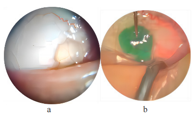
A50-year-old male reported one-year history with intermittent nasal bulbar redness and irritation in his left eye, and not in the right eye. The topical lubrications was used to relieve the symptomatic. No ocular surgery history was reported. The best corrected visual acuity in both eyes were 1.0/1.0. in the examination. With slit lamp exammination, a focal cystic area was found nasally within the bulbar conjunctiva in  the left eye(figure 1a). which was diagnosed as CL in clinical practice. The patient underwent outpatient surgery for CL excision  and detailed surgery procedureare described in the following section.  Subsequent post operative follow-up was uneventful.
20240425115035_6617.png
Figure 1  Conjunctival Lymphangiectasia before and after indocyanine green (ICG) gel injection
(A) Conjunctival Lymphangiectasia under slit lamp biomicroscopy;
 (B) Explicit range of Conjunctival Lymphangiectasia with ICG gel injected and subconjunctival anaesthesia was performed.

SURGICAL TECHNIQUE FOR CL EXCISION

After employing appropriate aseptic techniques and positioning the blepharostat, surface anesthesia was induced by instilling 1% lidocaine eye drops three times. A prepared formulation known as ICG gel, a homogeneous amalgamation consisting of 3 drops of ICG solution (5 mg/ml) and 1 ml of Viscoat (which contains sodium chondroitin sulfate 4% and sodium hyaluronate 1.65%, produced by Alcon), was utilized[4]. The ICG gel was then meticulously and gradually injected into the conjunctival cyst using a 1 ml syringe equipped with a 30-gauge needle, exerting appropriate pressure. Injection was discontinued once the cyst appeared sufficiently flled and the ICG gel was uniformly distributed, aiding in easier identification of the focal lesion (see Figure 1b). Subsequently, subconjunctival anesthesia was administered approximately 2 mm away from the cyst, employing a combination of 2% lidocaine and 0.75% bupivacaine. A small incision was made along the border of the focal lesion on the bulbar conjunctiva, followed by a delicate blunt dissection of the surrounding bulbar conjunctiva using a 0.4 mm inclined syringe needle. The distended lesion, along with the intact cyst wall, could be completely dissected away from adjacent tissues. Minimal bleeding was observed during the procedure, and no sutures were employed due to the small incision. To conclude the surgery, 0.3% tobramycin ointment was applied topically prior to patching. Apressure dressing was kept in place for 24 hours, and a course of antibiotics was prescribed for topical administration four times daily for a week.

DISCUSSION

The conjunctiva, the eye's most lymphatic-rich tissue, is characterized by subconjunctival lymphatics that develop from nasal lymphatics without involvement of conjunctival lymph nodes.[1] The dilation of lymphatic vessels within the bulbar conjunctiva arises from the obstruction of local lymphatic flow, likely triggered by trauma, radiotherapy, and various ocular surgeries.[1] Beyond the common causes, conjunctival lymphangiectasia (CL) is recognized as an ocular manifestation of several systemic diseases.[1, 5-6]

CL presents in two main forms: difusely enlarged lymphatics clinically referred to as conjunctivochalasis, and translucent, cystic lesions or a string of pearls appearance resulting from focal or beaded dilation of lymphatic vessels.[1] Due to its translucent nature and potential coexistence with conjunctivochalasis, CL can only be provisionally diagnosed through direct observation using a slit lamp or through conjunctival fluorescein staining to enhance visualization.[7] By identifying the precise location of the lesion, a preliminary differentiation can be made between CL and conjunctival epithelial cysts, as the latter primarily occur in the upper part of the conjunctiva.[1] However, a definitive diagnosis of CL necessitates pathological confrmation post-excision and, if necessary, immunohistochemical staining for D2-40 to diferentiate lymphatic endothelium from capillaries.[1]

Surgical intervention is warranted for individuals experiencing persistent and symptomatic conjunctival lymphangiectasia (CL). However, traditional methods such as cyst punctures have shown a high recurrence rate, while newer non-excision therapies also carry the risk of relapse.[1-3] Despite various surgical excision treatments available for CL, none have reported a method to accurately delineate the extent of the lesion during the procedure. The absence of clear demarcation poses challenges in determining the excision range, resulting in inevitable additional tissue damage. Although a prior study employed a mixture of healon and trypan blue solution to delineate conjunctival myxoma, this approach may not be suitable for CL considering its specific properties.[8]

Indocyanine green (ICG), an FDA-approved fuorescent dye known for its safety and native coloring, has recently found application in lymphography, yielding superior outcomes.[9-10] Previous research has demonstrated that subconjunctival injection of ICG effectively identifies subconjunctival lymphatics. Compared to trypan blue and fuorescein, ICG produces sharper and more defined images of lymphatic vessels without any leakage due to its larger molecular weight.[11-12] On the other hand, Viscoat, a viscoelastic substance, exhibits viscosity and cohesion, rendering it an ideal material for sealing puncture sites and maintaining lesion volume. Previous studies have described a combination of ICG and Viscoat as a fluorescent viscoelastic material that amalgamates the advantages of both approaches.[4] However, to the best of my knowledge, ICG gel has not been applied in CL surgery before.

We choose ICG gel as a staining and bulking agent for surgical excision of CL because of its fluorescent color, relatively lymphatic-specific, and viscoelastic properties. The lymphatic-specifc nature of ICG enables the distinction between dilated lymphatic vessels and capillaries  due to its greenish color. Besides, the viscoelastic nature of Viscoat allows for the long-standing distension of the cyst by sealing the puncture site and preventing leakage. By injecting ICG gel into the CL, it enhances lesion visualization and facilitates differentiation from the normal tenon's capsule during the procedure. This well-demarcated lesion enables safer and more precise excision, ensuring complete removal of cyst wall without residual lesions or additional tissue damage. Additionally, ICG gel naturally disappears upon CL removal, minimizing postoperative complications. 

In summary, this is the first report advocating for the application of ICG gel injection into conjunctival lymphangiectasia (CL) to streamline surgical procedures. This innovative technique holds the potential to decrease CL recurrence rates while minimizing the extent of additional tissue damage. There are some limitations in this case. To begin with, in this case, we solely rely on clinical diagnosis and lack a definitive pathological diagnosis. Additionally, this article exclusively presents our inventive technique based on a single case of conjunctival lymphangiectasia (CL) with a solitary cystic lesion, thereby limiting its applicability to cases characterized by difuse lymphatic enlargement. In order to assess and establish this novel technique, further research through larger-scale studies is required for evaluation and standardization.

Correction notice

None

Acknowledgement

None

Author Contributions

(I) Conception and design: Jingyao Chang, Chunyan Xue
(II)Administrative support: Chunyan Xue, Qian Cao
(III) Provision of study materials or patients: Chunyan Xue, Qian Cao(IV) Collection and assembly of data: Jingyao Chang, Xiaoran Zhang, Qian Cao
(V) Data analysis and interpretation: Jingyao Chang, Xiaoran Zhang
(VI) Manuscript writing:All authors
(VII) Final approval of manuscript:All authors

Funding

The funding organizations had no role in the following aspects: design and conduct of the study; the collection, management, analysis, and interpretation of the data; preparation, review, or approval of the manuscript; and the decision to submit the manuscript for publication.

Confict of Interests

None of the authors has any conflicts of interest to disclose. All authors have declared in the completed the ICMJE uniform disclosure form.

Patient consent for publication

None

Ethical Statement

This study does not contain any studies with human or animal subjects performed by any of the authors

Provenance and Peer Review

This article was a standard submission to our journal. The article has undergone peer review with ouranonymous review system

Data Sharing Statement

None

OpenAccess Statement

This is an Open Access article distributed in accordance with the Creative Commons Attribution-NonCommercial-NoDerivs 4.0 International License (CC BY-NC-ND 4.0), which permits the non-commercial replication and distribution of the article with the strict proviso that no changes or edits are made and the original work is properly cited (including links to both the formal publication through the relevant DOI and the license). See: https://creativecommons.org/licenses/by-nc-nd/4.0/.
1、Welch J, Srinivasan S, Lyall D, Roberts F. Conjunctival lymphangiectasia: a report of 11 cases and review of literature. Surv Ophthalmol 2012;57:136–48.Welch J, Srinivasan S, Lyall D, Roberts F. Conjunctival lymphangiectasia: a report of 11 cases and review of literature. Surv Ophthalmol 2012;57:136–48.
2、Choi%20SM%2C%20Jin%20KH%2C%20Kim%20TG.%20Successful%20treatment%20of%20conjunctival%20lymphangiectasia%20accompanied%20by%20corneal%20dellen%20using%20a%20high-frequency%20radiowave%20electrosurgical%20device.%C2%A0Indian%20J%20Ophthalmol.%202019%3B67(3)%3A409-411.Choi%20SM%2C%20Jin%20KH%2C%20Kim%20TG.%20Successful%20treatment%20of%20conjunctival%20lymphangiectasia%20accompanied%20by%20corneal%20dellen%20using%20a%20high-frequency%20radiowave%20electrosurgical%20device.%C2%A0Indian%20J%20Ophthalmol.%202019%3B67(3)%3A409-411.
3、Tan JC, Mann S, Coroneo MT. Successful Treatment of Conjunctival Lymphangiectasia With Subconjunctival Injection of Bevacizumab. Cornea. 2016;35(10):1375-1377.Tan JC, Mann S, Coroneo MT. Successful Treatment of Conjunctival Lymphangiectasia With Subconjunctival Injection of Bevacizumab. Cornea. 2016;35(10):1375-1377.
4、Park%20J%2C%20Lee%20J%2C%20Lee%20H%2C%20Baek%20S.%20Effectiveness%20of%20indocyanine%20green%20gel%20in%20the%20identification%20and%20complete%20removal%20of%20the%20medial%20wall%20of%20the%20lacrimal%20sac%20during%20endoscopic%20endonasal%20dacryocystorhinostomy.%C2%A0Can%20J%20Ophthalmol.%202017%3B52(5)%3A494-498.Park%20J%2C%20Lee%20J%2C%20Lee%20H%2C%20Baek%20S.%20Effectiveness%20of%20indocyanine%20green%20gel%20in%20the%20identification%20and%20complete%20removal%20of%20the%20medial%20wall%20of%20the%20lacrimal%20sac%20during%20endoscopic%20endonasal%20dacryocystorhinostomy.%C2%A0Can%20J%20Ophthalmol.%202017%3B52(5)%3A494-498.
5、Bunod%20R%2C%20Adams%20D%2C%20Cauquil%20C%2C%20Francou%20B%2C%20Labeyrie%20C%2C%20Bourenane%20H%2C%20et%20al.%20Conjunctival%20lymphangiectasia%20as%20a%20biomarker%20of%20severe%20systemic%20disease%20in%20Ser77Tyr%20hereditary%20transthyretin%20amyloidosis.%C2%A0Br%20J%20Ophthalmol.%202020%3B104(10)%3A1363-1367.Bunod%20R%2C%20Adams%20D%2C%20Cauquil%20C%2C%20Francou%20B%2C%20Labeyrie%20C%2C%20Bourenane%20H%2C%20et%20al.%20Conjunctival%20lymphangiectasia%20as%20a%20biomarker%20of%20severe%20systemic%20disease%20in%20Ser77Tyr%20hereditary%20transthyretin%20amyloidosis.%C2%A0Br%20J%20Ophthalmol.%202020%3B104(10)%3A1363-1367.
6、Hayek%20S%2C%20Adam%20C%2C%20Adams%20D%2C%20et%C2%A0al.%20Conjunctival%20lymphangiectasia%3A%20a%20novel%20ocular%20manifestation%20of%20hereditary%20transthyretin%20amyloidosis.%20Amyloid%202019%3B26%3A94%E2%80%935.Hayek%20S%2C%20Adam%20C%2C%20Adams%20D%2C%20et%C2%A0al.%20Conjunctival%20lymphangiectasia%3A%20a%20novel%20ocular%20manifestation%20of%20hereditary%20transthyretin%20amyloidosis.%20Amyloid%202019%3B26%3A94%E2%80%935.
7、%20Park%20ES%2C%20Kim%20MS%2C%20Jun%20I%2C%20et%20al.%20A%20Rare%20Case%20of%20Conjunctival%20Myxoma%20Initially%20Misdiagnosed%20as%20a%20Conjunctival%20Inclusion%20Cyst.%C2%A0Korean%20J%20Ophthalmol.%202021%3B35(5)%3A419-420.%20Park%20ES%2C%20Kim%20MS%2C%20Jun%20I%2C%20et%20al.%20A%20Rare%20Case%20of%20Conjunctival%20Myxoma%20Initially%20Misdiagnosed%20as%20a%20Conjunctival%20Inclusion%20Cyst.%C2%A0Korean%20J%20Ophthalmol.%202021%3B35(5)%3A419-420.
8、Al-Ghadeer H, Al-Assiri A, Al-Odhaib S, Alkatan H. Successful removal of a conjunctival myxoma. Middle East Afr J Ophthalmol. 2012 Jul-Sep;19(3):352-3. doi: 10.4103/0974-9233.97968. PMID: 22837636; PMCID: PMC3401812.Al-Ghadeer H, Al-Assiri A, Al-Odhaib S, Alkatan H. Successful removal of a conjunctival myxoma. Middle East Afr J Ophthalmol. 2012 Jul-Sep;19(3):352-3. doi: 10.4103/0974-9233.97968. PMID: 22837636; PMCID: PMC3401812.
9、Mihara M, Hara H, Araki J, Kikuchi K, Narushima M, Yamamoto T, et al. Indocyanine green (ICG) lymphography is superior to lymphoscintigraphy for diagnostic imaging of early lymphedema of the upper limbs. PLoS One. 2012;7(6):e38182. Mihara M, Hara H, Araki J, Kikuchi K, Narushima M, Yamamoto T, et al. Indocyanine green (ICG) lymphography is superior to lymphoscintigraphy for diagnostic imaging of early lymphedema of the upper limbs. PLoS One. 2012;7(6):e38182.
10、Koual%20M%2C%20Benoit%20L%2C%20Nguyen-Xuan%20HT%2C%20Bentivegna%20E%2C%20Aza%C3%AFs%20H%2C%20Bats%20AS.%20Diagnostic%20value%20of%20indocyanine%20green%20fluorescence%20guided%20sentinel%20lymph%20node%20biopsy%20in%20vulvar%20cancer%3A%20A%20systematic%20review.%20Gynecol%20Oncol.%202021%3B161(2)%3A436-441.Koual%20M%2C%20Benoit%20L%2C%20Nguyen-Xuan%20HT%2C%20Bentivegna%20E%2C%20Aza%C3%AFs%20H%2C%20Bats%20AS.%20Diagnostic%20value%20of%20indocyanine%20green%20fluorescence%20guided%20sentinel%20lymph%20node%20biopsy%20in%20vulvar%20cancer%3A%20A%20systematic%20review.%20Gynecol%20Oncol.%202021%3B161(2)%3A436-441.
11、Akiyama G, Saraswathy S, Bogarin T, Pan X, Barron E, Wong TT, et al. Functional, structural, and molecular identification of lymphatic outflow from subconjunctival blebs. Exp Eye Res. 2020;196:108049.Akiyama G, Saraswathy S, Bogarin T, Pan X, Barron E, Wong TT, et al. Functional, structural, and molecular identification of lymphatic outflow from subconjunctival blebs. Exp Eye Res. 2020;196:108049.
12、Freitas-Neto%20CA%2C%20Costa%20RA%2C%20Kombo%20N%2C%20Freitas%20T%2C%20Or%C3%A9fice%20JL%2C%20Or%C3%A9fice%20F%2C%20et%20al.%20Subconjunctival%20indocyanine%20green%20identifies%20lymphatic%20vessels.%C2%A0JAMA%20Ophthalmol.%202015%3B133(1)%3A102-104.Freitas-Neto%20CA%2C%20Costa%20RA%2C%20Kombo%20N%2C%20Freitas%20T%2C%20Or%C3%A9fice%20JL%2C%20Or%C3%A9fice%20F%2C%20et%20al.%20Subconjunctival%20indocyanine%20green%20identifies%20lymphatic%20vessels.%C2%A0JAMA%20Ophthalmol.%202015%3B133(1)%3A102-104.
上一篇
下一篇
出版者信息
目录