您的位置: 首页 > 2024年12月 第1卷 第4期 > 文字全文
2023年7月 第38卷 第7期11
目录

Surgical excision of subfoveal nodules and histopathological findings in pediatric patients with Coats’ disease

Surgical excision of subfoveal nodules and histopathological findings in pediatric patients with Coats’ disease

来源期刊: Eye Science | 2024年12月 第1卷 第4期 306-313 发布时间:2024-11-28 收稿时间:2024/12/11 14:52:55 阅读量:77
作者:
关键词:
Coats’ disease subfoveal nodule surgical excision pediatric
DOI:
10.12419/es24082401
Received date:
2024-08-24 
Accepted date:
2024-10-16 
Published online:
2024-11-28 
Purpose: To report on surgical outcomes of removing subfoveal nodules and to evaluate the histopathological findings of subfoveal nodules in pediatric patients with coats’ disease. Methods: This was a retrospective, interventional case series in which 6 pediatric patients had large (>1 disk diameter) subfoveal nodules. Vitrectomy and excision of subfoveal nodules with silicon oil tamponade were performed. Silicon oil was removed 3 months later. Results: This study was carried out in 6 patients with a mean follow-up of 9.2±1.5 months (range: 7-11 months), and the mean age was 5.2±2.4 years (range: 2-8 years). Preoperative visual acuity ranged from light perception (LP) to 20/250, and postoperative visual acuity ranged from LP to 20/200. Histopathology revealed nodules composed of proliferating fibrous tissue, hyaline degeneration with foamy histiocytes, focal myofibroblast hyperplasia, ossified tissue, and cholesterol fissures, with chronic cellular infiltration. No nodules regressed during the follow-up period. Conclusion: Certain eyes of pediatric patients with coats’ disease who underwent subfoveal nodule removal and no evidence of nodule regression may benefit from submacular surgery. Histopathological findings revealed that anti-proliferative and anti-fibrotic agents could be targets for treating coats disease.
Purpose: To report on surgical outcomes of removing subfoveal nodules and to evaluate the histopathological findings of subfoveal nodules in pediatric patients with coats’ disease. Methods: This was a retrospective, interventional case series in which 6 pediatric patients had large (>1 disk diameter) subfoveal nodules. Vitrectomy and excision of subfoveal nodules with silicon oil tamponade were performed. Silicon oil was removed 3 months later. Results: This study was carried out in 6 patients with a mean follow-up of 9.2±1.5 months (range: 7-11 months), and the mean age was 5.2±2.4 years (range: 2-8 years). Preoperative visual acuity ranged from light perception (LP) to 20/250, and postoperative visual acuity ranged from LP to 20/200. Histopathology revealed nodules composed of proliferating fibrous tissue, hyaline degeneration with foamy histiocytes, focal myofibroblast hyperplasia, ossified tissue, and cholesterol fissures, with chronic cellular infiltration. No nodules regressed during the follow-up period. Conclusion: Certain eyes of pediatric patients with coats’ disease who underwent subfoveal nodule removal and no evidence of nodule regression may benefit from submacular surgery. Histopathological findings revealed that anti-proliferative and anti-fibrotic agents could be targets for treating coats disease.

HIGHLIGHTS

• No effective treatment to subfoveal nodule in Coats’ disease was reported. We investigate the surgical outcome of excision of subfoveal nodules in pediatric patients with Coats’ disease and find Surgery to remove subfoveal fibrous nodules can be a safe and efficacious treatment option in pediatric patients with Coats’ disease.
• Nodule development is associated with reactive myofibroblast proliferation and collagen production, as some nodules generate hyalinized tissue and ossified tissue. These histopathological findings suggested that early application of antifibrosis therapy could be potentially beneficial when the macula was involved.
• Future large-scale studies with longer follow-up are needed to confirm the efficacy of surgical excision of subfoveal nodules and to provide additional insight into histopathological features.

INTRODUCTION

Coats’ disease is a sporadic, chronic retinal vascular condition characterized by telangiectatic and aneurysmal retinal vessels with intraretinal and subretinal lipid exudation and fluid. Georges Coats first described it in 1908 as a unilateral condition characterized by exudative retinal detachment and telangiectasia in a young man.[1] The symptoms of patients with Coats’ disease were decreased vision, strabismus, leukocoria and amblyogenic refractive error.[2-4] Lipid exudation and massive accumulation in or under the retina are the main causes of vision loss. Long-term accumulation of large amounts of lipids may lead to migration to the macula fovea, where they form subfoveal nodules. These nodules can progress to fibrous nodules over time, which are difficult to subside. Daruih et al. reported that 52.5% of Coats’ disease patients with stage 2B or 3A1 presented with a subfoveal nodule, which was associated with a worse visual outcome.[5]

The treatment modalities for Coats’ disease include ablative therapy with laser and cryotherapy, intravitreal anti-vascular endothelial growth factor (VEGF) therapy, and surgical options, including external subretinal drainage with or without scleral buckling and vitrectomy.[6] Many ophthalmologists have explored other approaches to treat the specific complications caused by Coats disease, includingsurgical drainage of large macular cystoid spaces,[7] perfluoro-octane-assisted external drainage of severe exudative retinal detachment.[8] But there are no good solutions reported for the massive subfoveal exudates and fibrosis, while thick foveal exudates and fibrosis usually predict a worse functional outcome. Surgical excision of subfoveal membranes and collaborative submacular surgery have been described for subfoveal neovascularization in children secondary to choroidal neovascularization.[9] The removal of subfoveal nodules and exudation has not been reported in children secondary to Coats’ disease. The purpose of the present study was to investigate the surgical outcome of excision of subfoveal nodules in pediatric patients with Coats’ disease and to report the histopathological findings of subfoveal nodules from these patients.

METHOD

We retrospectively reviewed a consecutive series of 6 pediatric patients with stage 3A or 3B Coats’ disease with subfoveal nodules in the macula who underwent submacular surgery. General ophthalmic examination, including best-corrected visual acuity (BCVA), slit lamp examination, and intraocular pressure (IOP), were performed preoperatively and postoperatively in all the patients. Young children who are unable to cooperate with visual acuity assessment was recorded not applicabele (N/A), and those who are unable to cooperate with non-contact tonometry was assessed withfinger method for intraocular pressure assessment. Fundus photography and fluorescein and angiography were performed using a RetCam II (Clarity Medical Systems, Pleasan-ton, CA, USA). The study was approved by the Institutional Review Board of West China Hospital and was performed in accordance with the Declaration of Helsinki. Written informed consent was obtained from the guardians of those patients.

A subfoveal nodule was defined on fundus examination as a yellow, exudative, protruding, spheroidal lesion. The nodule size was estimated as the papillary diameter. The lesion was confirmed by fundoscopy, fundus fluorescein angiography (FFA) and optical coherence tomography (OCT). On fundoscopy, the presence of an associated vascular component and the presence of macular exudation were also noted. Findings from fluorescein angiography on a RetCam II were recorded. Spectral-domain OCT (SD-OCT; Cirrus, Zeiss, CA) data were also recorded when available.

One experienced vitreoretinal surgeon (F.L.) performed all the surgeries. First, pars plana vitrectomy was performed after posterior vitreous detachment, and then the internal limiting membrane was removed. Second, a 27-gauge syringe needle was inserted into the subretina, and a balanced salt solution was injected to form a small bubble to elevate the neuroretina. Retinal scissors were used to perform a retinotomy, and retinal forceps were inserted beneath the neuroretina to grasp the nodule and remove it gently. The exudate was washed away by the balanced salt solution. After that, perfluorocarbons were used to flatten the retina, and fluid-air exchange was performed prior to laser photocoagulation of the retinal incision. Photocoagulation to the telangiectasias was usually selected. Cryotherapy was employed when the retinal detachment was shallow enough to allow approximation of the cryoprobe to the telangiectasias using the scleral technique. Finally, silicon oil was injected into the vitreous. The patient was instructed to keep a face-down position after surgery for 1 month. Silicon oil was removed 3 months later, and all the patients were followed up for at least 6 months.

All of the 6 eyes underwent nodule removal, pathological procedures were performed at our institution, and the nodules were available for the histopathological analysis.

RESULTS

The six patients were aged between 1 and 8 years, and four of the six patients were male. The course of disease ranged from 6 months to 24 months. Three patients in this series had unilateral 3A1, two had 3A2, and one had 3B Coats’ disease with total exudative retinal detachments. Three patients had previously received anti-VEGF therapy and laser therapy. Five patients had the right eye involved. On presentation, vision in the affected eye ranged from 0.08 to light perception. All of the six patients had foveal nodules. The average age of patients at the time of surgery was 5.17±2.40 years old. After surgical removal of the subfoveal nodules, the patients had an average follow-up of 9.2±1.5 months (range, 7-11 months), and postoperative visual acuity ranged from light perception to 0.15. For older children (patient #1,2,3) who can cooperate with non-invasive intraocular pressure measurement, we performed intraocular pressure testing, which were 13.1mmHg, 9.5mmHg and 17.3mmHg; For young children (Patient 4,5,6) who are unable to cooperate with non-invasive intraocular pressure measurement, we performed finger intraocular pressure measurement, which were Tn for these patient.These clinical findings are summarized in Table 1.

Table 1 Clinical characteristics of six patients with Coats’ disease

Case

Gender

Age(y)

Course of disease(m)

Pretreatment

Eye

Stage

Number of quadrants with telangiectasia

Size(mm)


Baseline VA


Post-surgery VA


Follow up(m)

1

M

8

14

Native

OD

3A1

2

3*2

CF

LP

10

2

M

5

12

Anti VEGFlaser

OD

3A1

4

4*2

CF

CF

9

3

F

8

13

Anti VEGFlaser

OS

3A2

3

3*4

0.08

0.1

10

4

M

2

15

Subretinal fluid drainage, Anti VEGF, laser

OD

3B

4

2*3

NA

NA

11

5

F

4

24

Native

OD

3A2

3

1*1

HM

0.15

8

6

M

4

6

Native

OD

3A1

4

3*2

CF

0.04

7

Abbreviations: OD: right eye, OS: left eye, VEGF: Vascular endothelial growth factor, VA: visual acuity, CF: counting figures, HM: hand movement, LP: light perception.

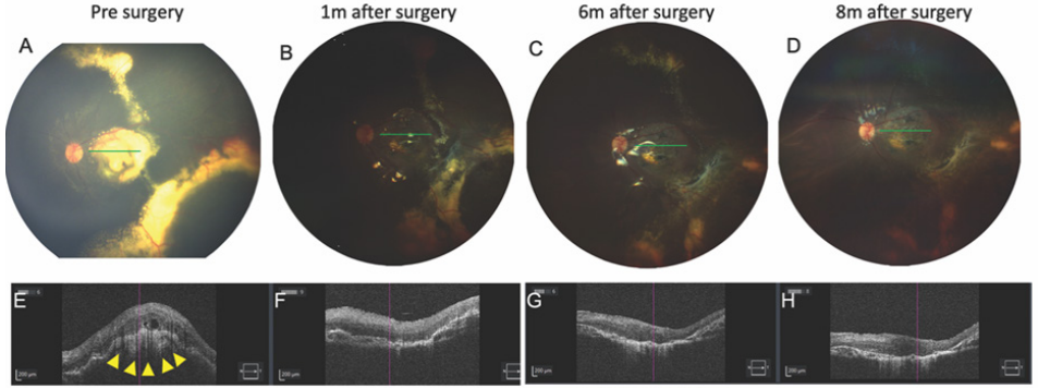
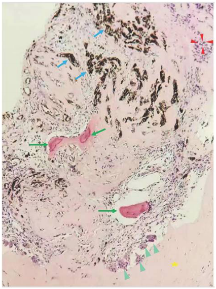
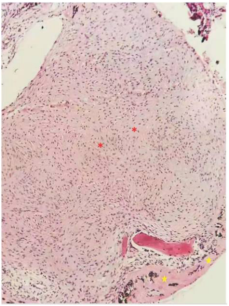
Figure 1 shows the presurgical fundus image and postoperative fundus images of patient 3. SD-OCT image at the level of the fibrotic nodule at the presentation showed a hyperreflective, sharply demarcated subfoveal lesion associated with a posterior shadowing effect, moderate retinal layer disorganization and retinal thickening, with abrupt elevation of retinal layers by the underlying hyperreflective nodule. Histopathological examination of the excised tissue revealed that the subfoveal nodules were fibrous tissue, which was composed mainly of fibroblasts and myofibroblasts. In these six nodules, 5 of them have cholesterol fissures, 4 have foreign body giant cells, all of them have chorionic cells and myofibroblastic hyperplasia. In addition, 3 of the 6 patients have ossified tissue in their subfoveal nodules.These features were associated with ossification and hyaline degeneration (Figures 2 and 3).
Figure 1 Color fundus and OCT images showing the retina of patient 3 before and after surgery at follow-up.
A-D, Color fundus photographs showing that the macula was stable with atrophy but without exudation again after subfoveal nodule removal.E-H, SD-OCT B-scan images showing macular subfoveal nodules before and after surgery corresponding to the location of green line in color fundus images of A-D. Yellow arrowheads indicate the nodules underneath macula.
Figure 2 Histopathological analysis of a subfoveal nodule removed from one eye (Patient 1) with Coats' disease.
H&E staining was performed with hematoxylin and eosin (X126). The red arrowheads indicate small blood vessel proliferation. The green arrows show the ossifications that formed. The blue arrows show hyperpigmentation. Cyan-blue arrowheads indicate foreign body giant cell reactions and histiocyte aggregation. The yellow pentagram shows fibroplasia with collagenization.
Figure 3 Histopathological analysis of a subfoveal nodule removed from another eye (Patient 3) with Coats' disease.
H&E staining was performed with hematoxylin and eosin (X126). The red asterisks denote myofibroblastic hyperplasia in the tissue. The yellow pentagrams denote hyalinization of the tissue.

DISCUSSION

Subfoveal nodules are ominous results of exudate accumulation with fibrosis in the macula of patients with Coats disease with or without treatment. This is one of the causes of vision loss in patients with Coats’ disease. In our study, 6 patients with subfoveal nodules presented with stage 3A1, 3A2 or 3B disease and had low vision at presentation (from LP to 0.08). After surgical removal of subfoveal nodules, 4/6 of the patients had a visual acuity better than that before surgery, and there was no evidence of spontaneous regression of fibrosis. No increase in intraocular pressure, cataracts, retinal detachment, vitreous hemorrhage, or endophthalmitis was observed during the follow-up.

Anti-VEGF therapy is a first-line treatment for choroidal neovascularization but has a limited effect on macular fibrosis. Surgical removal of choroidal neovascularization has been reported in children and adults, but there is no evidence of a significant improvement in visual acuity supports the concept that submacular surgery is safe in children with neovascular membranes.[9] Our study showed that surgical removal of submacular nodules was safe in children with Coats’ disease. Several factors may lead to favorable surgical outcomes in pediatric patients who undergo removal of subfoveal nodules compared with surgical outcomes inpediatricand adults patients with subfoveal membranes 
associated with choroidal neovascularization. First, the subfoveal nodule in Coats’ disease is composed of mature fibrotic tissue with less vascularization, and isolating the nodule from other tissues was less likely to be associated with massive bleeding. Second, subfoveal nodules have a solitary ingrowth site in the subretina, unlike choroidal neovascularization, for example, in age-related macular degeneration, which has multiple ingrowth sites in the subRPE.[10] Therefore, we speculate that the characteristics and location of lesion tissue may have contributed to a favorable result to some extent. However, during our long-term follow-up, vision did not improve significantly, and the RPE and neuroretinal layers exhibited some atrophy. These findings suggest that early treatment and minimizing the chance of nodule occurrence are needed in the future.

Cholesterol granuloma, calcium and osseous metaplasia in subfoveal nodule were reported in enucleated children’s eyes with Coats’ disease in 1984.[11]Daruich reported subfoveal nodule from two eyes with Coats’ disease including fibrin admixed with spindle cells, macrophages and pigmented cells.[5] In our study, histopathologic analysis revealed that these nodules were fibrous tissue, which was composed mainly of fibroblasts and myofibroblasts, cholesterol clefts, lipid-laden macrophages, multinucleated giant cells, ossified tissue, pigments and foam cells. Fibroblasts and myofibroblasts are essential for wound healing, and myofibroblasts are activated fibroblasts that participate in tissue remodeling following insult.[12] When a large amount of lipids are deposited in the macular fovea, injury to macular tissue causes the recruitment and activation of inflammatory cells and fibroblasts, the proliferation of activated fibroblasts (myofibroblasts), and the deposition of extracellular matrix (collagens, fibronectin, laminin and glycosaminoglycans); additionally, the extracellular matrix spreads to cover the damaged tissue.[13] The exudates from telangiectasia contain many lipids and attract macrophages to lesions to swallow lipids. A group of macrophages, called lipid-laden macrophages, localize to lipid deposits and become lipid-laden cells, giving them a foamy appearance under the microscope. Some cholesterol clefts could also be observed in the tissue of subofveal nodules. Ossified tissue and hyalinization were first observed in the margins of nodules in patients with Coats’ disease, and fibrous tissue collagen was speculated to be the origin of the hyalinized material, which turned into ossified tissue. Pigmentation was observed in the nodule, possibly because of the destruction and phagocytosis of RPE cells during nodular growth. These pathological changes suggested that pharmaceutical approach may have a limited effect on subfoveal nodule in late stage of the disease, which requires surgical treatment alternatively. However, more importantly, the ultimate goals for treatment should be preventing fibrosis in an ealier stage of the disease and preventing atrophy after the surgical removal of the nodules.

Limitations of the current study are the small number of pediatric patients included because of the low prevalence of Coats’ disease, and only six patients with subfoveal nodules were included. Future large-scale studies with longer follow-up periods areneeded to confirm the efficacy of surgical excision of subfoveal nodules and to provide additional insight into histopathological features.

In conclusion, subfoveal fibrous nodules are pathological structures that result in poor vision due to long-term chronic inflammation within the retina. Surgery to remove subfoveal fibrous nodules can be a safe and efficacious treatment option in pediatric patients with Coats’ disease. Nodule development is associated with reactive myofibroblast proliferation and collagen production, as some nodules generate hyalinized tissue and ossified tissue. These histopathological findings suggested that early application of antifibrosis therapy could be potentially beneficial when the macula was involved.

Correction notice

None

Acknowledgement

None

Author Contributions

(Ⅰ) Conception and design: Xiaoshuang Jiang, Fang Lu 
(Ⅱ) Administrative support: Fang Lu
(Ⅲ) Provision of study materials or patients: Xiaoshuang Jiang, You Li, Yiliu Yang
(Ⅳ) Collection and assembly of data: Xuejiao Yu, Licong Liang
(Ⅴ) Data analysis and interpretation: Mengxi Shen, Guina Liu
(Ⅵ) Manuscript writing: All authors
(Ⅶ) Final approval of manuscript: All authors

Funding

This work was supported by Natural Science Foundation of Sichuan Provincial Science and Technology Department (2023NSFSC1666).

Conflict of Interests

None of the authors has any conflicts of interest to disclose. All authors have declared in the completed the ICMJE uniform disclosure form.

Patient consent for publication

Written informed consent was obtained from the guardians of those patients.

Ethical Statement

The study was approved by the Institutional Review Board of West China Hospital.

Provenance and Peer Review

This article was a standard submission to our journal. The article has undergone peer review with our anonymous review system.

Data Sharing Statement

None

Open Access Statement

This is an Open Access article distributed in accordance with the Creative Commons AttributionNonCommercial-NoDerivs 4.0 International License (CC BY-NC-ND 4.0), which permits the non-commercial replication and distribution of the article with the strict proviso that no changes or edits are made and the original work is properly cited (including links to both the formal publication through the relevant DOI and the license).
1、C.G(Coats G).Forms of retinal disease with massive exudation. Royal London Ophthalmic Hospital Rep. 1908,17(3):440-525.C.G(Coats G).Forms of retinal disease with massive exudation. Royal London Ophthalmic Hospital Rep. 1908,17(3):440-525.
2、Shields JA, Shields CL, Honavar SG, et al. Classification and management of Coats disease: the 2000 Proctor Lecture. Am J Ophthalmol. 2001, 131(5): 572-583. DOI: 10.1016/s0002-9394(01)00896-0.Shields JA, Shields CL, Honavar SG, et al. Classification and management of Coats disease: the 2000 Proctor Lecture. Am J Ophthalmol. 2001, 131(5): 572-583. DOI: 10.1016/s0002-9394(01)00896-0.
3、Altamirano F, Gonzalez E, Shah AS, et al. Preventable vision loss in children with Coats disease. J AAPOS. 2024, 28(5): 104000. DOI: 10.1016/j.jaapos.2024.104000.Altamirano F, Gonzalez E, Shah AS, et al. Preventable vision loss in children with Coats disease. J AAPOS. 2024, 28(5): 104000. DOI: 10.1016/j.jaapos.2024.104000.
4、Kang HG, Kim JD, Choi EY, et al. Clinical features and prognostic factors in 71 eyes over 20years from patients with Coats' disease in Korea. Sci Rep. 2021, 11(1): 6124. DOI: 10.1038/s41598-021-85739-9.Kang HG, Kim JD, Choi EY, et al. Clinical features and prognostic factors in 71 eyes over 20years from patients with Coats' disease in Korea. Sci Rep. 2021, 11(1): 6124. DOI: 10.1038/s41598-021-85739-9.
5、Daruich AL, Moulin AP, Tran HV, et al. SUBFOVEAL NODULE IN COATS' DISEASE: toward an updated classification predicting visual prognosis. Retina. 2017, 37(8): 1591-1598. DOI: 10.1097/IAE.0000000000001399.Daruich AL, Moulin AP, Tran HV, et al. SUBFOVEAL NODULE IN COATS' DISEASE: toward an updated classification predicting visual prognosis. Retina. 2017, 37(8): 1591-1598. DOI: 10.1097/IAE.0000000000001399.
6、Sigler EJ, Randolph JC, Calzada JI, et al. Current management of Coats disease. Surv Ophthalmol. 2014, 59(1): 30-46. DOI: 10.1016/j.survophthal.2013.03.007.Sigler EJ, Randolph JC, Calzada JI, et al. Current management of Coats disease. Surv Ophthalmol. 2014, 59(1): 30-46. DOI: 10.1016/j.survophthal.2013.03.007.
7、Yiu G, Hang A, Fong R. Surgical drainage of large macular cystoid spaces in Coats disease. J Vitreoretin Dis. 2024, 8(3): 355-358. DOI: 10.1177/24741264241240320.Yiu G, Hang A, Fong R. Surgical drainage of large macular cystoid spaces in Coats disease. J Vitreoretin Dis. 2024, 8(3): 355-358. DOI: 10.1177/24741264241240320.
8、Sivalingam MD, Wakabayashi T, Kusaka S, et al. Lenssparing perfluoro-octane-assisted external drainage of total exudative retinal detachment in Coats' disease. Ophthalmic Surg Lasers Imaging Retina. 2024, 55(8): 430-432. DOI: 10.3928/23258160-20240704-01.Sivalingam MD, Wakabayashi T, Kusaka S, et al. Lenssparing perfluoro-octane-assisted external drainage of total exudative retinal detachment in Coats' disease. Ophthalmic Surg Lasers Imaging Retina. 2024, 55(8): 430-432. DOI: 10.3928/23258160-20240704-01.
9、Sears J, Capone A Jr, Sr TA, et al. Surgical management of subfoveal neovascularization in children. Ophthalmology. 1999, 106(5): 920-924. DOI: 10.1016/S0161- 6420(99)00510-2.Sears J, Capone A Jr, Sr TA, et al. Surgical management of subfoveal neovascularization in children. Ophthalmology. 1999, 106(5): 920-924. DOI: 10.1016/S0161- 6420(99)00510-2.
10、Melberg NS, Thomas MA, Burgess DB. The surgical removal of subfoveal choroidal neovascularization. Ingrowth site as a predictor of visual outcome. Retina. 1996, 16(3): 190-195. DOI: 10.1097/00006982- 199616030-00002.Melberg NS, Thomas MA, Burgess DB. The surgical removal of subfoveal choroidal neovascularization. Ingrowth site as a predictor of visual outcome. Retina. 1996, 16(3): 190-195. DOI: 10.1097/00006982- 199616030-00002.
11、Chang MM, McLean IW, Merritt JC. Coats' disease: a study of 62 histologically confirmed cases. J Pediatr Ophthalmol Strabismus. 1984, 21(5): 163-168. DOI: 10.3928/0191-3913-19840901-03.Chang MM, McLean IW, Merritt JC. Coats' disease: a study of 62 histologically confirmed cases. J Pediatr Ophthalmol Strabismus. 1984, 21(5): 163-168. DOI: 10.3928/0191-3913-19840901-03.
12、Hinz B. Myofibroblasts. Exp Eye Res. 2016,142:56-70. DOI: 10.1016/j.exer.2015.07.009.Hinz B. Myofibroblasts. Exp Eye Res. 2016,142:56-70. DOI: 10.1016/j.exer.2015.07.009.
13、Little K, Ma JH, Yang N, et al. Myofibroblasts in macular fibrosis secondary to neovascular age-related macular degeneration - the potential sources and molecular cues for their recruitment and activation. EBioMedicine. 2018, 38: 283-291. DOI: 10.1016/j.ebiom.2018.11.029.Little K, Ma JH, Yang N, et al. Myofibroblasts in macular fibrosis secondary to neovascular age-related macular degeneration - the potential sources and molecular cues for their recruitment and activation. EBioMedicine. 2018, 38: 283-291. DOI: 10.1016/j.ebiom.2018.11.029.
1、This work was supported by Natural Science Foundation of Sichuan Provincial Science and Technology Department (2023NSFSC1666).()
上一篇
下一篇
出版者信息
目录