您的位置: 首页 > 2024年12月 第1卷 第4期 > 文字全文
2023年7月 第38卷 第7期11
目录

A case of interface fluid syndrome following the enhancement surgery after small incision lenticule extraction

A case of interface fluid syndrome following the enhancement surgery after small incision lenticule extraction

来源期刊: Eye Science | 2024年12月 第1卷 第4期 443-452 发布时间:2024-12-20 收稿时间:2024/12/17 9:24:29 阅读量:12
作者:
关键词:
refractive enhancement surgery interface fluid syndrome intraocular pressure measurement
DOI:
10.12419/es24091001
Received date:
2024-09-10 
Accepted date:
2024-12-10 
Published online:
2024-12-20 
Purpose: To report a case of interface fluid syndrome following small incision lenticule extraction (SMILE) and subsequent CIRCLE enhancement. Case Presentation: A 30-year-old female experienced progressively worsening vision following refractive enhancement surgery. The patient had experienced a transient increase in intraocular pressure (IOP) after SMILE, normalized poststeroid cessation. Three months after the enhancement, her best-corrected visual acuity deteriorated from 20/20 in both eyes before the surgery to 20/300. IOP measured by non-contact tonometry was 25.3 mmHg in the right eye and 26.7 mmHg in the left eye, while the measurements off the flap using iCare were 55.3 mmHg and 47.8 mmHg, respectively. Examination revealed moderate corneal edema, interface fluid pockets, and haze, which were confirmed by anterior segment optical coherence tomography. Treatment involved the discontinuation of steroids and the introduction of hypotensive medication, leading to significant symptom relief. Conclusion: This case highlights the importance of cautious and conservative steroid use, particularly in steroid-responsive patients. When steroids are administered in cases potentially involving diffuse lamellar keratitis and haze, monitoring peripheral IOP is essential.
Purpose: To report a case of interface fluid syndrome following small incision lenticule extraction (SMILE) and subsequent CIRCLE enhancement. Case Presentation: A 30-year-old female experienced progressively worsening vision following refractive enhancement surgery. The patient had experienced a transient increase in intraocular pressure (IOP) after SMILE, normalized poststeroid cessation. Three months after the enhancement, her best-corrected visual acuity deteriorated from 20/20 in both eyes before the surgery to 20/300. IOP measured by non-contact tonometry was 25.3 mmHg in the right eye and 26.7 mmHg in the left eye, while the measurements off the flap using iCare were 55.3 mmHg and 47.8 mmHg, respectively. Examination revealed moderate corneal edema, interface fluid pockets, and haze, which were confirmed by anterior segment optical coherence tomography. Treatment involved the discontinuation of steroids and the introduction of hypotensive medication, leading to significant symptom relief. Conclusion: This case highlights the importance of cautious and conservative steroid use, particularly in steroid-responsive patients. When steroids are administered in cases potentially involving diffuse lamellar keratitis and haze, monitoring peripheral IOP is essential.

HIGHLIGHTS

• Differentiating between interface fluid syndrome and diffuse lamellar keratitis is essential in the early stage of interface haze.

• Clinicians should prioritize ruling out interface fluid syndrome before initiating steroid therapy.

• Peripheral intraocular pressure monitoring is critical when interface fluid syndrome is diagnosed or suspected.

INTRODUCTION

Ninety years ago, Prof. Frank B. Walsh and Prof. Eugene Chan documented a unique case of band-shaped keratitis with pearly, thickened conjunctiva extending onto the cornea.[1] Their insights into the etiology and pathophysiology of band-shaped keratitis, as well as their exploration of the potential pathological properties of conjunctival changes, have had lasting clinical significance. In homage to Professor Eugene Chan, we present a case of interface fluid syndrome following refractive enhancement surgery.

Interface fluid syndrome (IFS) is an uncommon complication of lamellar refractive surgeries that can result in irreversible vision loss if misdiagnosed or improperly treated. Recently, we have seen a case happened after CIRCLE enhancement following the small incision lenticule extraction (SMILE), and as a search of the literature has not resulted in our finding a similar one, we venture to present it.

CASE REPORT 

A Chinese female aged 30 years, Asian descent, clerk, came to Zhongshan Ophthalmic Center complaining of progressive decreased vision in both eyes since the CIRCLE enhancement 3 months previously. 

The patient underwent a CIRCLE enhancement (cap-to-flap conversion program in the VisuMax platform) surgery 3 months ago for the residual refractive error left by a SMILE surgery 11 months before. Before the CIRCLE enhancement, the refraction was -0.75 -0.50 x 30 in the right eye and -0.75 -0.50 x 165 in the left eye with best-corrected visual acuity (BCVA) of 20/20 in both eyes. The surgery was uneventful and the first day BCVA was 20/25 in both eyes. Tobradex eye drops (tobramycin 0.3%, dexamethasone 0.1%, Alcon) were started every two hours as part of the normal postoperative regimen. Three days later, the BCVA diminished to 20/60 in the right eye and 20/50 in the left eye, with the intraocular pressure (IOP) increased to 20.5 mmHg in the right eye and 21 mmHg in the left eye measured by a non-contact tonometer (NCT) (compared with 17 mmHg the right eye and 16.7 mmHg the left eye before the surgery). TobraDex was replaced by 0.1% fluorometholone, 4 times a day, and 2% carteolol, twice a day was added. At the 1-month postoperative visit, the BCVA remained 20/40 in both eyes and a mild haze could be seen in both eyes under a slit-lamp examination, with IOP of 18 mmHg and 19 mmHg. Brimonidine, 0.2%, and tacrolimus, 0.1%, twice a day were added to carteolol and fluorometholone. Two weeks later, the haze persisted, and the TobraDex ointment was given for presumptive diffuse lamellar keratitis. At the 2-month postoperative visit, the BCVA was decreased to 20/60 in both eyes with the IOP measured by NCT of 25.3 mmHg the right eye and 23 mmHg the left eye. The patient was referred for consultation.

Family history:

No similar disease history of immediate family members, including mother, father, and a brother.

Ophthalmic history:

Patient underwent an uneventful SMILE surgery 11 months ago. The preoperative refraction was -7.00 -0.75 x 91 in the right eye and -6.50 -0.50 x 94 in the left eye with BCVA of 20/20 in both eyes. After the surgery, a transient elevated IOP was observed after using Tobradex eye drops for one week with 30.3 mmHg the right eye and 25.7 mmHg the left eye measured by NCT, which decreased to normal after the discontinuation of steroids. 

Ophthalmological examination:

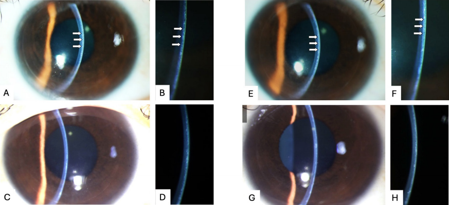
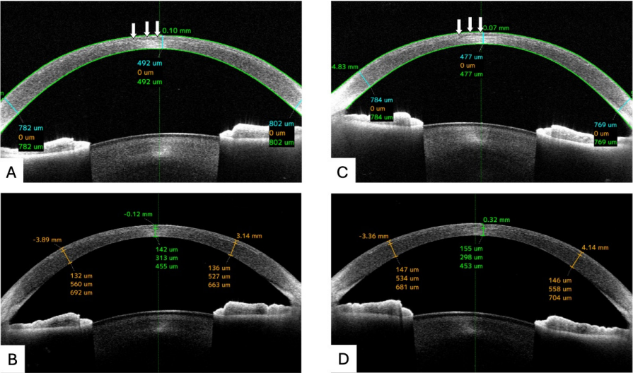
The visual acuity was 20/125 without improvement by correction in both eyes. IOP measured with NCT was 25.3 mmHg in the right eye and 26.7 mmHg in the left eye, while the pressure was 55.3 mmHg and 47.8 mmHg when measured off the flap using an iCare, nearly 2 mm away from nasal limbal. Slit-lamp examination revealed a shallow fluid interface under the epithelium in both eyes with brown irises (Figure 1). Anterior segment optical coherence tomographic (AS-OCT) image revealed the edematous cornea and a fluid interface between the stromal bed and the flap (Figure 2). The dilated fundoscopy examination was unremarkable. 
Figure 1 Ophthalmic examination at diagnosis and after 1-month treatment. 
The slit-lamp examination of the right eye (A-D) and the left eye (E-H).
A, B, E, F: 2 months after CIRCLE enhancement. Slit-lamp photograph showing a prominent flap. The beam focused on the cornea reveals a shallow fluid interface between the stromal bed and the flap (arrow). A & E. magnification 10x; B & F. magnification 45x. 
C, D, G, H: After the patient received hypotensive agents for 1 month. Slit-lamp photograph showing a significant resolution of fluid accumulation under the flap. C & G. magnification 10x; D & H. magnification 45x.

Figure 2 Anterior segment optical coherence tomographic (AS-OCT) image at diagnosis and after 1-month treatment.
The AS-OCT images of the right eye (A, B) and the left eye (C, D).
A & C. 2 months after CIRCLE enhancement: AS-OCT image revealing a hypo-reflective area (arrows) corresponding to the area of fluid accumulation at the interface and hyper-reflective stroma.
B & D. After the patient received hypotensive agents for 1 month: AS-OCT image revealing a resolution of interface fluid and decreased stromal hyper-reflectivity

Treatment and outcome:

The patient was diagnosed with interface fluid syndrome (IFS) and steroid-induced ocular hypertension in both eyes. Steroids were discontinued immediately. Mannitol, 20%, 250mL, was given intravenously. The patient started oral glycerin, 90mL, twice daily. Azarga (carbonic anhydrase inhibitor brinzolamide, 1%; timolol, 0.5%), brimonidine, 0.2%, bromfenac 0.1%, and tacrolimus, 0.1% were applied in both eyes twice a day. By the third day, BCVA had improved to 20/30 in both eyes. Intraocular pressure using the iCare was 6.0 mmHg centrally and 11.7 mmHg nasally in the right eye; 6.9 mmHg centrally and 15.4 mmHg nasally in the left eye. One month later, the interface fluid had nearly resolved (Figure 1 & 2). The BCVA was 20/30 in both eyes. Three months after treatment, the BCVA was 20/25 in both eyes.IFS is a LASIK flap-associated complication that was first reported by Lyle and Jin in 1999.[2] Awareness of this condition has increased with the growing number of reported cases.[3–5] Most previous reports have documented IFS occurring after LASIK surgery,[6] while a few cases have been reported following SMILE, [5,7,8] bioptics,[9] and surface ablation.[10] Re-treatment rates, or enhancement rates, after LASIK or SMILE range from 5% to 28%.[11–13] Three IFS cases have reported after LASIK enhancement.[14–16] To our knowledge, this is the first case of IFS observed after the CIRCLE enhancement following the SMILE surgery.

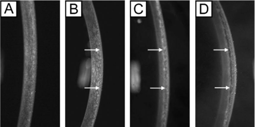
In our case, moderate edematous abnormalities along the interface wound causing a diffuse smudgy interface haze were observed, corresponding to stage 2 IFS as Dawson et al. classified (Supplemental Figure 1).[3] Clinically, stage 3 IFS can be detected with a careful slitlamp examination, appearing as an optically empty space between the flap and the residual stromal bed. However, stage 1 and 2 IFS could be easy to mistake for the diffuse granular appearance of diffuse lamellar keratitis (DLK), but the treatments of the two diseases are totally distinct. [17] Misdiagnosis of DLK may result in an increased dosage of topical steroids, potentially exacerbating the clinical condition.[16] Severe ocular hypertension, which can be obscured by interface fluid, may persist for an extended period, leading to optic nerve damage and irreversible visual field loss.[5] Our patient shares similar characteristics with previously reported IFS cases following SMILE, including Asian descent, myopia, and darkly pigmented irises. These factors place the patient at higher risk for developing ocular hypertension and IFS.[18]

Our patient experienced decreased vision the day after enhancement and steroid use, presenting earlier the typical onset for IFS. This early presentation is akin to that reported by Miya et al.,[14] where increased IOP and decreased BCAV were noted the day after steroid application (Table 1, Case 2). Typically, IFS manifests between 2 to 4 weeks post-operation, in contrast to the 2 to 5 days associated with DLK. Therefore, in both Case 2 and our case, considering a diagnosis of DLK is understandable. However, patients treated with topical steroids for DLK who do not see improvement within 10 days should be meticulously reassessed for interface fluid.[4]

Table 1. Previous cases of IFS following the enhancement.

Case

Author

Age(y)

Sex

Eye

The first LASIK

Cause of IFS

Symptoms onset

Treatment

Outcome

After surgery

After steroid used

Resolution period

Measures

Visual acuity

IOP

1

Russell et al. 2004 1

50

M

OD

UCVA 20/20

Steroid-induced elevated pressure

5 months

10 days

3 days

Steroid cessation + antihypertensive

1.0

18

OS

BCVA 20/20

Steroid-induced elevated pressure

5 months

10 days

3 days

1.0

18

2

Miya et al. 2007 2

53

M

OD

Uneventful

Steroid-induced elevated pressure

2 weeks

1 day

2 weeks

Steroid cessation + antihypertensive

UCVA 0.9

12

OS

Uneventful

Steroid-induced elevated pressure

2 weeks

1 day

2 weeks

UCVA 1.2

12

3

Hoffman et al. 2008 3

65

F

OD

-

Steroid-induced glaucoma, and endothelial dysfunction

4 years

Several months

1 day

Steroid cessation+ antihypertensive + DSEK + flap elevation

BCVA 1.0

-

 IFS = interface fluid syndrome, LASIK = laser in situ keratomileusis, UCVA = uncorrected visual acuity, BCVA = best-corrected visual acuity, IOP = intraocular pressure.
Reference:
1. Russell GE, Jafri B, Lichter H, Waring GO. Late Onset Decreased Vision in a Steroid Responder After LASIK Associated With Interface Fluid. J Refract Surg. 2004;20(1):91-92. doi:10.3928/1081-597X-20040101-21.
2. Miyai T, Yonemura T, Nejima R, Otani S, Miyata K, Amano S. Interlamellar Flap Edema Due to Steroid-Induced Ocular Hypertension After Laser In SituKeratomileusis. Jpn J Ophthalmol. 2007;51(3):228-230. doi:10.1007/s10384-006-0441-y.
3. Hoffman RS, Fine HI, Packer M. Persistent interface fluid syndrome. J Cataract Refract Surg. 2008;34(8):1405-1408. doi:10.1016/j.jcrs.2008.03.042.

The ocular hypertension and endothelial dysfunction are two mechanisms involved in IFS.[3,19] Experimental studies have confirmed that increased aqueous diffusion into the cornea, caused by higher endothelial stress caused by IOP and compromised endothelial barrier function, resulted in fluid accumulation in the interface.[3] At the mild stage, the fluid is taken up by the proteoglycans, leading to slight thickening of the interface with minimal to no haze. As the fluid accumulation progresses, the capacity of abnormally large proteoglycan–binding sites becomes overwhelmed, resulting in the formation of small fluid pockets. In cases of severe endothelial cell dysfunction or increased IOP, a significant amount of aqueous fluid can diffuse into the corneal stroma, creating large diffuse interface fluid pockets.[3]

Clinically, steroid-induced IOP elevation is the most common reason of IFS.[5] Other causes of IFS following the refractive surgeries include Posner Schlossman Syndrome,[8] anterior uveitis,[20] Fuchs endothelial dystrophy,[19] traumatic hyphema,[21]Amantadine,[22] and certain procedures such as cataract surgery,[23] vitreoretinal surgery,[24] trabeculectomy,[25]keratoplasty. [26] Our patient had a clear history of steroid use and elevated intraocular pressure, similar to the three previously documented cases of IFS after enhancement (Table 1). Case 3, with the longest duration of topical steroid use, was diagnosed with IFS secondary to steroidinduced glaucoma. IFS persisted due to concurrent endothelial failure. In Cases 1 and 2, symptoms emerged after 10 and 14 days of topical steroid use, respectively. Visual acuity improved significantly upon discontinuation of steroids and commencement of intraocular pressurelowering medications.After SMILE, the connection between the corneal cap and the stromal bed becomes loose, creating a potential space for fluid accumulation. Consequently, the incidence of IFS post-SMILE may be much higher than what is currently reported in the literature.[27] In patients with severe IFS and significant fluid accumulation, IOP tends to be low due to central corneal thinning and the buffering effect of the fluid.[28] Therefore, measuring IOP is critical for diagnosing IFS and differentiating diseases such as DLK, but IOP readings in IFS patients can be inaccurate and falsely low.[6] Ex vivo studies have shown that fluid beneath the corneal flap following keratotomy can significantly decrease IOP measurements, as applanation-based IOP measurement relies on both the thickness of the corneal stroma and the physical integrity of the cornea.[29]

One method to address inaccurate IOP readings is to employ alternative techniques less affected by corneal biomechanical properties and thickness,[30] such as the Tono-Pen and rebound tonometry (iCare).[30-31]Studies suggest that the Tono-Pen and iCare provide more accurate IOP values compared to the ocular response analyzer and Goldmann applanation tonometry, likely due to their smaller contact area with the cornea, which reduces dependency on corneal biomechanical properties. [32]

Another approach is to measure IOP outside the corneal flap. Previous research has shown that the discrepancy between central and peripheral IOP measurements with iCare ranges from -0.34 to 3.1 mmHg.[33-34] In the normal participants, iCare measurements of central IOP compared to nasal and temporal peripheral pressures show slight variations (central: 17.17 mmHg vs. nasal: 16.83 mmHg vs. temporal: 18.57 mmHg).[33] In participants who have undergone LASIK with corneal collagen crosslinking, the peripheral IOP measurements with iCare provide a good estimation of anterior chamber pressure.[34] In our case, we observed a 22 mmHg discrepancy between central and peripheral measurements, similar to previous findings.[2,4,35]

In IFS caused by ocular hypertension, IOP control is the main goal of treatment.[19] For hypertension induced by steroid, the cessation or taper of steroids is imperative. With adjunct topical and systemic hypotensive medication, the condition is usually reversed in 1-2 weeks.[2,19,36] In our case, after one month of treatment, vision acuity is yet fully recover, likely due to the prolonged course IFS. A systematic review indicated that a duration of IFS shorter than one month is associated with a significantly higher likelihood of achieving a final corrected distance visual acuity of 20/25 or better, with an adjusted odds ratio of 7.71 (P = 0.02).[6] At the early stage of our case and similar previous ones,[28,36-37]despite intensifying antihypertensive therapy, the trial use of steroids further exacerbated the symptoms. Additionally, systemic steroid use also worsened symptoms.[4] Therefore, clinicians should be wary or more conservative in reaching for steroids if IFS is possible, especially in those who are sensitive to steroids. Monitoring peripheral IOP is crucial when administering steroids under these conditions.

Correction notice

None

Acknowledgement

The authors thank all staff and subjects in the ZOC.

Author Contributions

(Ⅰ) Conception: Lingyi Liang 
(Ⅱ) Collection of case imaging and diagnostic data: Longyue Li
(Ⅲ) Manuscript drafting: Boyu Yang, Longyue Li.
(Ⅳ) Final approval of manuscript: All authors

Fundings

This work was supported by the National Natural Science Foundation of China General Program (82371021, 82070922). The Guangdong Natural Science Foundation of General Program (2023A1515012336, 2024A1515011384).

Conflict of Interests

None

Patient consent for publication

None

Ethical Statement

None

Provenance and Peer Review

This article was a standard submission to our journal. The article has undergone peer review with our anonymous review system.

Data Sharing Statement

None

Open Access Statement

This is an Open Access article distributed in accordance with the Creative Commons AttributionNonCommercial-NoDerivs 4.0 International License (CC BY-NC-ND 4.0), which permits the non-commercial replication and distribution of the article with the strict proviso that no changes or edits are made and the original work is properly cited (including links to both the formal publication through the relevant DOI and the license).

Supplemental Material

Supplemental Figure 1
Dawson et al.[3] classification of IFS: Representative slit-lamp clinical photographs (slit-beam illumination) of a (A) normal cornea, (B) normal laser in situ keratomileusis (LASIK) cornea, and (C, D) corneas with interface fluid syndrome (IFS). C, A LASIK cornea with moderate (stage 2) IFS that has moderate edematous abnormalities along the interface wound causing a diffuse smudgy interface haze. D, A LASIK cornea with severe (stage 3) IFS that has severe edematous abnormalities along the interface wound causing an optically empty space between the LASIK flap and the residual stromal bed because of interface fluid accumulation. White arrows, LASIK interface.
Supplemental Figure 2 Anterior segment optical coherence tomographic (AS-OCT) image after 3-month treatment.


1、Walsh FB, Chan E. A case of corneal calcification (bandshaped keratitis) with conjunctival changes. Am J Ophthalmol. 1934, 17(3): 238-241. DOI: 10.1016/s0002-9394(34)92587-3.Walsh FB, Chan E. A case of corneal calcification (bandshaped keratitis) with conjunctival changes. Am J Ophthalmol. 1934, 17(3): 238-241. DOI: 10.1016/s0002-9394(34)92587-3.
2、Lyle WA, Jin GJ. Interface fluid associated with diffuse lamellar keratitis and epithelial ingrowth after laser in situ keratomileusis. J Cataract Refract Surg. 1999, 25(7): 1009- 1012. DOI: 10.1016/s0886-3350(99)00083-8.Lyle WA, Jin GJ. Interface fluid associated with diffuse lamellar keratitis and epithelial ingrowth after laser in situ keratomileusis. J Cataract Refract Surg. 1999, 25(7): 1009- 1012. DOI: 10.1016/s0886-3350(99)00083-8.
3、Dawson DG, Schmack I, Holley GP, et al. Interface fluid syndrome in human eye bank corneas after LASIK: causes and pathogenesis. Ophthalmology. 2007, 114(10): 1848- 1859. DOI: 10.1016/j.ophtha.2007.01.029.Dawson DG, Schmack I, Holley GP, et al. Interface fluid syndrome in human eye bank corneas after LASIK: causes and pathogenesis. Ophthalmology. 2007, 114(10): 1848- 1859. DOI: 10.1016/j.ophtha.2007.01.029.
4、Hamilton DR, Manche EE, Rich LF, et al. Steroid-induced glaucoma after laser in situ keratomileusis associated with interface fluid. Ophthalmology. 2002, 109(4): 659-665. DOI: 10.1016/s0161-6420(01)01023-5.Hamilton DR, Manche EE, Rich LF, et al. Steroid-induced glaucoma after laser in situ keratomileusis associated with interface fluid. Ophthalmology. 2002, 109(4): 659-665. DOI: 10.1016/s0161-6420(01)01023-5.
5、Zheng K, Han T, Li M, et al. Corneal densitometry changes in a patient with interface fluid syndrome after small incision lenticule extraction. BMC Ophthalmol. 2017, 17(1): 34. DOI: 10.1186/s12886-017-0428-0.Zheng K, Han T, Li M, et al. Corneal densitometry changes in a patient with interface fluid syndrome after small incision lenticule extraction. BMC Ophthalmol. 2017, 17(1): 34. DOI: 10.1186/s12886-017-0428-0.
6、Ravipati A, Pradeep T, Donaldson KE. Interface fluid syndrome after LASIK surgery: retrospective pooled analysis and systematic review. J Cataract Refract Surg. 2023, 49(8): 885-889. DOI: 10.1097/ j.jcrs.0000000000001214.Ravipati A, Pradeep T, Donaldson KE. Interface fluid syndrome after LASIK surgery: retrospective pooled analysis and systematic review. J Cataract Refract Surg. 2023, 49(8): 885-889. DOI: 10.1097/ j.jcrs.0000000000001214.
7、Bansal AK, Murthy SI, Maaz SM, et al. Shifting “ectasia”: interface fluid collection after small incision lenticule extraction (SMILE). J Refract Surg. 2016, 32(11): 773- 775. DOI: 10.3928/1081597X-20160721-02.Bansal AK, Murthy SI, Maaz SM, et al. Shifting “ectasia”: interface fluid collection after small incision lenticule extraction (SMILE). J Refract Surg. 2016, 32(11): 773- 775. DOI: 10.3928/1081597X-20160721-02.
8、Mokumu D, Hu W, Damaola A, et al. Interface fluid syndrome after small incision lenticule extraction surgery secondary to posner schlossman syndrome - A case report. Heliyon. 2023, 9(11): e21863. DOI: 10.1016/ j.heliyon.2023.e21863.Mokumu D, Hu W, Damaola A, et al. Interface fluid syndrome after small incision lenticule extraction surgery secondary to posner schlossman syndrome - A case report. Heliyon. 2023, 9(11): e21863. DOI: 10.1016/ j.heliyon.2023.e21863.
9、Bardet E, Touboul D, Kerautret J, et al. Interface fluid syndrome after bioptics. J Refract Surg. 2011, 27(5): 383- 386. DOI: 10.3928/1081597X-20101111-01.Bardet E, Touboul D, Kerautret J, et al. Interface fluid syndrome after bioptics. J Refract Surg. 2011, 27(5): 383- 386. DOI: 10.3928/1081597X-20101111-01.
10、Guedes J, Vilares-Morgado R, Brazuna R, et al. Pressureinduced stromal keratopathy after surface ablation surgery. Case Rep Ophthalmol. 2024, 15(1): 532-541. DOI: 10.1159/000539701.Guedes J, Vilares-Morgado R, Brazuna R, et al. Pressureinduced stromal keratopathy after surface ablation surgery. Case Rep Ophthalmol. 2024, 15(1): 532-541. DOI: 10.1159/000539701.
11、Siedlecki J, Luft N, Priglinger SG, et al. Enhancement options after myopic small-incision lenticule extraction (SMILE): a review. Asia Pac J Ophthalmol. 2019, 8(5): 406-411. DOI: 10.1097/APO.0000000000000259.Siedlecki J, Luft N, Priglinger SG, et al. Enhancement options after myopic small-incision lenticule extraction (SMILE): a review. Asia Pac J Ophthalmol. 2019, 8(5): 406-411. DOI: 10.1097/APO.0000000000000259.
12、Netto MV, Wilson SE. Flap lift for LASIK retreatment in eyes with myopia. Ophthalmology. 2004, 111(7): 1362- 1367. DOI: 10.1016/j.ophtha.2003.11.009.Netto MV, Wilson SE. Flap lift for LASIK retreatment in eyes with myopia. Ophthalmology. 2004, 111(7): 1362- 1367. DOI: 10.1016/j.ophtha.2003.11.009.
13、Riau AK, Liu YC, Lim CHL, et al. Retreatment strategies following Small Incision Lenticule Extraction (SMILE): in vivo tissue responses. PLoS One. 2017, 12(7): e0180941. DOI: 10.1371/journal.pone.0180941.Riau AK, Liu YC, Lim CHL, et al. Retreatment strategies following Small Incision Lenticule Extraction (SMILE): in vivo tissue responses. PLoS One. 2017, 12(7): e0180941. DOI: 10.1371/journal.pone.0180941.
14、Miyai T, Yonemura T, Nejima R, et al. Interlamellar flap edema due to steroid-induced ocular hypertension after laser in situ keratomileusis. Jpn J Ophthalmol. 2007, 51(3): 228-230. DOI: 10.1007/s10384-006-0441-y.Miyai T, Yonemura T, Nejima R, et al. Interlamellar flap edema due to steroid-induced ocular hypertension after laser in situ keratomileusis. Jpn J Ophthalmol. 2007, 51(3): 228-230. DOI: 10.1007/s10384-006-0441-y.
15、Russell GE, Jafri B, Lichter H, et al. Late onset decreased vision in a steroid responder after LASIK associated with interface fluid. J Refract Surg. 2004, 20(1): 91-92. DOI: 10.3928/1081-597X-20040101-21.Russell GE, Jafri B, Lichter H, et al. Late onset decreased vision in a steroid responder after LASIK associated with interface fluid. J Refract Surg. 2004, 20(1): 91-92. DOI: 10.3928/1081-597X-20040101-21.
16、Hoffman RS, Fine IH, Packer M. Persistent interface fluid syndrome. J Cataract Refract Surg. 2008, 34(8): 1405- 1408. DOI: 10.1016/j.jcrs.2008.03.042.Hoffman RS, Fine IH, Packer M. Persistent interface fluid syndrome. J Cataract Refract Surg. 2008, 34(8): 1405- 1408. DOI: 10.1016/j.jcrs.2008.03.042.
17、Senthil S, Rathi V, Garudadri C. Misleading Goldmann applanation tonometry in a post-LASIK eye with interface fluid syndrome. Indian J Ophthalmol. 2010, 58(4): 333- 335. DOI: 10.4103/0301-4738.64133.Senthil S, Rathi V, Garudadri C. Misleading Goldmann applanation tonometry in a post-LASIK eye with interface fluid syndrome. Indian J Ophthalmol. 2010, 58(4): 333- 335. DOI: 10.4103/0301-4738.64133.
18、Asif MI, Bafna RK, Mehta JS, et al. Complications of small incision lenticule extraction. Indian J Ophthalmol. 2020, 68(12): 2711-2722. DOI: 10.4103/ijo.IJO_3258_20.Asif MI, Bafna RK, Mehta JS, et al. Complications of small incision lenticule extraction. Indian J Ophthalmol. 2020, 68(12): 2711-2722. DOI: 10.4103/ijo.IJO_3258_20.
19、Koronis S, Diafas A, Tzamalis A, et al. Late-onset interface fluid syndrome: a case report and literature review. Semin Ophthalmol. 2022, 37(7-8): 839-848. DOI: 10.1080/08820538.2022.2102928.Koronis S, Diafas A, Tzamalis A, et al. Late-onset interface fluid syndrome: a case report and literature review. Semin Ophthalmol. 2022, 37(7-8): 839-848. DOI: 10.1080/08820538.2022.2102928.
20、Goto S, Koh S, Toda R, et al. Interface fluid syndrome after laser in situ keratomileusis following herpetic keratouveitis. J Cataract Refract Surg. 2013, 39(8): 1267- 1270. DOI: 10.1016/j.jcrs.2013.04.026.Goto S, Koh S, Toda R, et al. Interface fluid syndrome after laser in situ keratomileusis following herpetic keratouveitis. J Cataract Refract Surg. 2013, 39(8): 1267- 1270. DOI: 10.1016/j.jcrs.2013.04.026.
21、Pham MT, Peck RE, Dobbins KRB. Nonarteritic ischemic optic neuropathy secondary to severe ocular hypertension masked by interface fluid in a post-LASIK eye. J Cataract Refract Surg. 2013, 39(6): 955-957. DOI: 10.1016/ j.jcrs.2013.04.027.Pham MT, Peck RE, Dobbins KRB. Nonarteritic ischemic optic neuropathy secondary to severe ocular hypertension masked by interface fluid in a post-LASIK eye. J Cataract Refract Surg. 2013, 39(6): 955-957. DOI: 10.1016/ j.jcrs.2013.04.027.
22、Altman A, Jaffry M, Dastjerdi MH. Amantadine induced interface fluid formation after LASIK. A case report. Am J Ophthalmol Case Rep. 2023, 32: 101895. DOI: 10.1016/ j.ajoc.2023.101895.Altman A, Jaffry M, Dastjerdi MH. Amantadine induced interface fluid formation after LASIK. A case report. Am J Ophthalmol Case Rep. 2023, 32: 101895. DOI: 10.1016/ j.ajoc.2023.101895.
23、Ortega-Usobiaga J, Martin-Reyes C, Llovet-Osuna F, et al. Interface fluid syndrome in routine cataract surgery 10 years after laser in situ keratomileusis. Cornea. 2012, 31(6): 706-707. DOI: 10.1097/ICO.0b013e3182254020.Ortega-Usobiaga J, Martin-Reyes C, Llovet-Osuna F, et al. Interface fluid syndrome in routine cataract surgery 10 years after laser in situ keratomileusis. Cornea. 2012, 31(6): 706-707. DOI: 10.1097/ICO.0b013e3182254020.
24、Wirbelauer C, Pham DT. Imaging interface fluid after laser in situ keratomileusis with corneal optical coherence tomography. J Cataract Refract Surg. 2005, 31(4): 853- 856. DOI: 10.1016/j.jcrs.2004.08.045.Wirbelauer C, Pham DT. Imaging interface fluid after laser in situ keratomileusis with corneal optical coherence tomography. J Cataract Refract Surg. 2005, 31(4): 853- 856. DOI: 10.1016/j.jcrs.2004.08.045.
25、Kang SJ, Dawson DG, Hopp LM, et al. Interface fluid syndrome in laser in situ keratomileusis after complicated trabeculectomy. J Cataract Refract Surg. 2006, 32(9): 1560-1562. DOI: 10.1016/j.jcrs.2006.03.040.Kang SJ, Dawson DG, Hopp LM, et al. Interface fluid syndrome in laser in situ keratomileusis after complicated trabeculectomy. J Cataract Refract Surg. 2006, 32(9): 1560-1562. DOI: 10.1016/j.jcrs.2006.03.040.
26、Villarrubia A, Cano-Ortiz A. Delayed-onset interface fluid syndrome after laser-assisted in situ keratomileusis secondary to descemet stripping automated endothelial keratoplasty. JAMA Ophthalmol. 2014, 132(8): 1028- 1029. DOI: 10.1001/jamaophthalmol.2014.960.Villarrubia A, Cano-Ortiz A. Delayed-onset interface fluid syndrome after laser-assisted in situ keratomileusis secondary to descemet stripping automated endothelial keratoplasty. JAMA Ophthalmol. 2014, 132(8): 1028- 1029. DOI: 10.1001/jamaophthalmol.2014.960.
27、Xu J, Li C, Zhang X, et al. Interface fluid syndrome caused by the corneal perforation injury after small incision lenticule extraction: a case report. BMC Ophthalmol. 2024, 24(1): 117. DOI: 10.1186/s12886-024-03339-3.Xu J, Li C, Zhang X, et al. Interface fluid syndrome caused by the corneal perforation injury after small incision lenticule extraction: a case report. BMC Ophthalmol. 2024, 24(1): 117. DOI: 10.1186/s12886-024-03339-3.
28、Najman-Vainer J, Smith RJ, Maloney RK. Interface fluid after LASIK: misleading tonometry can lead to end-stage glaucoma. J Cataract Refract Surg. 2000, 26(4): 471-472. DOI: 10.1016/s0886-3350(00)00382-5.Najman-Vainer J, Smith RJ, Maloney RK. Interface fluid after LASIK: misleading tonometry can lead to end-stage glaucoma. J Cataract Refract Surg. 2000, 26(4): 471-472. DOI: 10.1016/s0886-3350(00)00382-5.
29、Fillmore P, Gerding P, Sayegh S. Effect of an intra– corneal fluid interface following keratotomy on intraocular pressure measurement by applanation. Investig Ophthalmol Vis Sci, 2004, 45: 197.Fillmore P, Gerding P, Sayegh S. Effect of an intra– corneal fluid interface following keratotomy on intraocular pressure measurement by applanation. Investig Ophthalmol Vis Sci, 2004, 45: 197.
30、Bamashmus MA, Saleh MF. Post-LASIK interface fluid syndrome caused by steroid drops. Saudi J Ophthalmol, 2013, 27(2): 125-128. DOI: 10.1016/j.sjopt.2013.03.003.Bamashmus MA, Saleh MF. Post-LASIK interface fluid syndrome caused by steroid drops. Saudi J Ophthalmol, 2013, 27(2): 125-128. DOI: 10.1016/j.sjopt.2013.03.003.
31、Chui WS, Lam A, Chen D, et al. The influence of corneal properties on rebound tonometry. Ophthalmology, 2008, 115(1): 80-84. DOI: 10.1016/j.ophtha.2007.03.061.Chui WS, Lam A, Chen D, et al. The influence of corneal properties on rebound tonometry. Ophthalmology, 2008, 115(1): 80-84. DOI: 10.1016/j.ophtha.2007.03.061.
32、Neuburger%20M%2C%20Maier%20P%2C%20B%C3%B6hringer%20D%2C%20et%20al.%20The%20impact%20of%20%0Acorneal%20edema%20on%20intraocular%20pressure%20measurements%20using%20%0Agoldmann%20applanation%20tonometry%2C%20Tono-Pen%20XL%2C%20iCare%2C%20%0Aand%20ORA%3A%20an%20in%20vitro%20model.%20J%20Glaucoma%2C%202013%2C%2022(7)%3A%20%0A584-590.%20DOI%3A%2010.1097%2FIJG.0b013e31824cef11.Neuburger%20M%2C%20Maier%20P%2C%20B%C3%B6hringer%20D%2C%20et%20al.%20The%20impact%20of%20%0Acorneal%20edema%20on%20intraocular%20pressure%20measurements%20using%20%0Agoldmann%20applanation%20tonometry%2C%20Tono-Pen%20XL%2C%20iCare%2C%20%0Aand%20ORA%3A%20an%20in%20vitro%20model.%20J%20Glaucoma%2C%202013%2C%2022(7)%3A%20%0A584-590.%20DOI%3A%2010.1097%2FIJG.0b013e31824cef11.
33、Jorge JMM, González-Méijome JM, Queirós A, et al. Correlations between corneal biomechanical properties measured with the ocular response analyzer and ICare rebound tonometry. J Glaucoma, 2008, 17(6): 442-448. DOI: 10.1097/IJG.0b013e31815f52b8.Jorge JMM, González-Méijome JM, Queirós A, et al. Correlations between corneal biomechanical properties measured with the ocular response analyzer and ICare rebound tonometry. J Glaucoma, 2008, 17(6): 442-448. DOI: 10.1097/IJG.0b013e31815f52b8.
34、Lam AK, Tam K, Kwok P, et al. Central and peripheral rebound tonometry in myopic LASIK without and with corneal collagen crosslinking. Invest Ophthalmol Vis Sci. 2015,56(7):2026.Lam AK, Tam K, Kwok P, et al. Central and peripheral rebound tonometry in myopic LASIK without and with corneal collagen crosslinking. Invest Ophthalmol Vis Sci. 2015,56(7):2026.
35、Ramos JLB, Zhou S, Yo C, et al. High-resolution Imaging of Complicated LASIK Flap Interface Fluid Syndrome. Ophthalmic Surg Lasers Imaging. 2008,39(4 Suppl):S80-S82. DOI:10.3928/15428877-20080715-04.Ramos JLB, Zhou S, Yo C, et al. High-resolution Imaging of Complicated LASIK Flap Interface Fluid Syndrome. Ophthalmic Surg Lasers Imaging. 2008,39(4 Suppl):S80-S82. DOI:10.3928/15428877-20080715-04.
36、Wheeldon CE, Hadden BO, Niederer RL, et al. Presumed late diffuse lamellar keratitis progressing to interface fluid syndrome. J Cataract Refract Surg. 2008,34(2):322-326. DOI:10.1016/j.jcrs.2007.09.025.Wheeldon CE, Hadden BO, Niederer RL, et al. Presumed late diffuse lamellar keratitis progressing to interface fluid syndrome. J Cataract Refract Surg. 2008,34(2):322-326. DOI:10.1016/j.jcrs.2007.09.025.
37、Rehany U, Bersudsky V, Rumelt S. Paradoxical hypotony after laser in situ keratomileusis. J Cataract Refract Surg. 2000,26(12):1823-1826. DOI:10.1016/s0886- 3350(00)00763-x.Rehany U, Bersudsky V, Rumelt S. Paradoxical hypotony after laser in situ keratomileusis. J Cataract Refract Surg. 2000,26(12):1823-1826. DOI:10.1016/s0886- 3350(00)00763-x.
1、This work was supported by the National Natural Science Foundation of China General Program (82371021, 82070922). The Guangdong Natural Science Foundation of General Program (2023A1515012336, 2024A1515011384).()
下一篇
出版者信息
目录