Location: Home > March 2025 Vol.2 No.1 > Details
March 2025 Vol.2 No.1
Contents

Role of omics approaches in the study of pediatric cataract

Role of omics approaches in the study of pediatric cataract

Source: Eye Science | March 2025 Vol.2 No.1 1-7 Posted: 2025-03-10 Received:2025/3/13 16:59:23 Hits: 168
Authors:
 
1.State Key Laboratory of Ophthalmology, Zhongshan Ophthalmic Center, Sun Yat-sen University, Guangdong Provincial Key Laboratory of Ophthalmology and Visual Science, Guangzhou 510060, China.
Keywords:
pediatric cataract omics pathogenesisbiomarkers
DOI:
10.12419/es24071605
Received date:
2024-08-02 
Revised date:
2024-10-11 
Accepted date:
2024-11-04 
Published online:
2025-03-10 
Pediatric cataract, a leading cause of blindness in children globally, imposing a significant financial burden on both families and society. The extensive phenotypic heterogeneity of this condition means that the underlying mechanisms remain poorly understood, limiting the development of precise and effective treatments. The advent of omics technologies has provided potent tools for unraveling the pathogenesis of pediatric cataract. By mapping expression profiles across various molecular levels, these omics approaches enhance our understanding of the disease’s etiological mechanisms, aid in the identification of novel biomarkers and key pathways, and offer researchers new insights for the innovative strategies in disease diagnosis and targeted therapies. In this review, we summarize the application of omics approaches in clinical and basic research on pediatric cataract over the past decade, encompassing genomics, transcriptomics, proteomics, and metabolomics. Furthermore, we discuss the current challenges and future prospects of omics analyses in pediatric cataract studies.

Highlights

1.Critical Discoveries and Outcomes

This review summarizes the application of omics approaches in the study of pediatric cataract over the past decade

2.Methodological Innovations

This paper employs a comprehensive review methodology to integrate the knowledge of multiple omics in the field of pediatric cataract, discussing the advantages, challenges, and future prospects of omics concerning this disease.

3. Prospective Applications and Future Directions

The review depicts a future landscape of multi-omics in pediatric cataract:
Mechanism Research: Systematically surveying the molecular expression profiles of pediatric cataract at “gene-mRNA-protein-metabolite” levels to understand its etiological mechanisms.
Therapeutic Improvements: Exploration of potential biomarkers and key pathways to improve personalized diagnosis and treatment.

Introduction

Pediatric cataract, characterized by lens opacity present at birth or developing during childhood, is a major cause of visual impairment and blindness in children globally,[1] affecting 1 to 15 per 10,000 newborns.[2] This condition severely impacts the quality of visual information during the critical period of children's visual system development. If left uncorrected, it can lead to irreversible visual impairment or even permanent blindness, imposing a substantial economic burden on families and societies. Congenital cataract, the most prevalent form of pediatric cataract, incurs annual global costs 3.1 to 5.2 billion dollars.[3] Surgical removal of the form deprivation is the recommended treatment to facilitate visual rehabilitation. However, the surgical management of pediatric cataract is considerably more challenging than that of adult-onset cataract, with risks of postoperative complications including glaucoma, posterior capsule opacification, and retinal detachment.[4] Consequently, researchers are committed to investigating regulatory mechanisms and key pathway targets underlying the pathogenesis of pediatric cataract , which may pave the way for exploring non-surgical therapies to prevent or reverse lens opacification, thereby improving the prevention, treatment and visual prognosis in children. The pathogenesis of pediatric cataract is diverse. While clear causes such as trauma, complications, drug-induced effects, and metabolic disorders are identified in some cases, the pathogenesis remains unclear in over 50% of instances.[5] Furthermore, given the high degree of heterogeneity in pediatric cataract, comprehensive and in-depth investigations at the molecular level are essential to accurately elucidate the pathogenesis, enhancing the prospects for accurate diagnosis, precise treatment, and prognosis assessment.

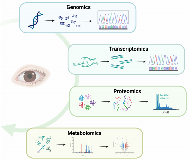
Advances in high-throughput sequencing technology and bioinformatics methods have introduced omics as a novel tool to facilitate our understanding of the complex pathogenesis and specific biomolecular processes underlying pediatric cataract. Omics refers to a set of disciplines that aim to measure and characterize biomolecules in a system using high-throughput approaches. Genomics focuses on identifying genetic variants implicated in disease, providing valuable insights into the discovery of pathogenic variations or mutations.[6] Transcriptomics serves as a powerful tool for the qualitative and quantitatively detection of the genome-wide RNA expression patterns, offering crucial information on gene expression, structure and regulation.[6] High-throughput proteomics enables the acquisition of information on peptide abundance, post-translational modifications, and protein-protein interactions across thousands of proteins, reflecting global protein expression.[7] As the omics field most intimately linked to the phenotype, metabolomics can quantify various small molecules, such as amino acids, fatty acids, carbohydrates and other products of cellular metabolism, providing metabolic profiles and identifying potential biomarkers and pathological metabolic pathways.[7] Based on the genetic central dogma, multi-omics approaches, including genomics, transcriptomics, proteomics, and metabolomics, offer a systematic and comprehensive understanding of disease states and biological processes at biomolecular levels of "DNA-mRNA-proteome-metabolite". Omics technology has been widely applied in ophthalmology to enhance our understanding of eye diseases at the biomolecular level, aiding in diagnosis and the identification of new therapeutic targets.

This study aims to provide a comprehensively review of the application of omics in the study of pediatric cataract over the past decade, encompassing genomics, transcriptomics, proteomics, and metabolomics (Figure 1), it will discuss the advantages and value of these applications in both clinical and laboratory setting, and outline the current challenges as well as future development directions of omics in the field of pediatric cataract.

Figure1. Omics approaches for the study of pediatric cataract

20250318153951_6873.png

Genomics of Pediatric Cataract

Overview of Genomics

Genomics identifies genetic variants and key genes based on DNA. Genomics can comprehensively analyze the organism’s genetic information by studying the structure and function of DNA sequences, including all coding and non-coding regions of a genome, as well as regions between genes.[8] The main genomics technologies include microarrays, whole exome sequencing(WES), whole genome sequencing (WGS), etc.[9] Microarrays can be used to identify loci of preset disease variants.[9] WES primarily sequences the coding regions of a genome, making it relatively efficient and cost-effective, while WGS obtains all genes of an individual, including both coding and non-coding regions, providing a more comprehensive but more expensive approach.[9] Genomics compares the sequenced genomes to reference genomes to identify genetic variants, such as DNA insertions or deletions, single nucleotide variants, copy number variants, and structural variants. Further annotation of their functionality can reveal possible causative gene mutations and provide references for disease diagnosis and genotype-phenotype associations.[10,11]

Applications of Genomics in Pediatric Cataract

Genetic factors contribute to 8 to 29% of pediatric cataract cases.[13] Pediatric cataract demonstrates significant genetic and phenotypic heterogeneity, and genotype-phenotype associations remain limited, posing challenges for clinical detection and diagnosis. With the advancement of high-throughput sequencing technology, genetic testing is increasingly utilized for clinical diagnosis, guiding clinical decision-making, predicting prognosis, providing reproductive guidance, and combining with phenotypic data to enhance diagnostic capabilities and explore new therapeutic options.
Utilizing genomics technology, a substantial number of causative genes have been implicated in pediatric cataract. Over 50 genes related to pediatric cataract have been identified, including crystallin genes (CRYAA, CRYAB, CRYBB1, CRYBB2, CRYBB3, CRYBA1, CRYBA3, CRYBA2, CRYBA4, CRYGC, CRYGD, CRYGS),[12-18] membrane protein genes (GJA3, GJA8, MIP, LIM2),[19,20] cytoskeletal protein genes (VIM, BFSP1, BFSP2),[21] transcription factor genes (PAX6, MAF, PITX3),[22-24] signal molecule genes (EPHA2),[25] syndrome-related genes (NHS, C10orf71, OCRL, SMARCA1, HMX1, RGS6, STX3, GALK1),[26-33] and other genes (PIKFYVE, PGRMC1).[34,35]

Next-generation genome sequencing methods can achieve diagnostic rates ranging from 37.5% to 77% in pediatric cataract cohorts, aiding in the expansion of the gene mutation spectrum associated with pediatric cataract. In 2017, Javadiyan et al.[36] utilized high-throughput gene sequencing technology to sequence 51 previously reported pediatric cataract genes in 33 Australian pediatric cataract children with a family history. Their study identified potential disease-causing variants in 42% of patients and uncovered 11 potentially novel mutations. In the same year, Patel et al.[37] applied multiple-gene panel sequencing and exome sequencing to 166 pediatric cataract patients from 74 families in Saudi Arabia, targeting 322 previously reported genes mutation linked to inherited eye diseases. The detected mutation rate among these patients was 58%, and 15 novel gene mutations were identified. In 2021, Ma et al. [38] applied genome sequencing techniques, including WES and WGS, to 52 Australian children with congenital cataracts, achieving an overall diagnosis rate of 77%. Recently, in 2023, Rossen et al.[39] assessed diagnostic rates of different genetic testing approaches in a 20-year US cohort of congenital cataracts, with WES demonstrating the highest diagnostic rate at 46.2%.

Diagnostic efficacy varies among different genetic testing approaches, with the WGS exhibiting the highest diagnostic rate, followed by WES. In contrast, panel-based sequencing and microarrays targeting preset cataract-related genes demonstrate lower diagnostic rates. Previously, a cohort study of congenital cataract  evaluated the diagnostic efficacy of various genetic testing methods, and found the WES group had the highest diagnostic rate (46.2%), followed by the group undergoing targeted sequencing of congenital cataract-associated genes (37.5%), with the lowest rate observed in the microarrays group (11%).[39] Another cohort study involving 52 congenital cataract patients revealed that the genome sequencing could improve diagnostic yield by an additional 10% (up to 77%), although exome sequencing detected variants in 67% of cases.[38] Panel-based or targeted sequencing of cataract-associated genes is limited by the originally designed gene set, which may explain the correspondingly  low overall diagnostic rate. WES analysis encompasses all regions of the exome and covers more than 20,000 genes, enabling comprehensive screening to identify potential disease-causing mutation in over 85% of human genetic diseases. Furthermore, WGS has a broader scope of detection, including coding and non-coding regions, and can detect small copy number variants, DNA indels in repetitive regions, or single nucleotide variants in GC-enriched regions that are not detectable by microarrays, panel-based assays, or WES.[38]

Transcriptomics of Pediatric Cataract

3.1 Overview of Transcriptomics

Transcriptomics identifies the expressions of key genes in organisms based on RNA. It detects the presence of transcripts, novel splice sites, RNA editing sites, and the expression levels of individual transcripts through quantitative and qualitative examination of RNA within genomes of a biological sample.[38] Although only about 3% of genomes encode proteins, up to 80% of genomes is transcribed.[40] In addition to the protein-coding transcriptome, thousands of long-stranded non-coding RNAs transcribed in cells play crucial roles in physiological processes, and dysregulation of short non-coding RNAs and circular RNAs is potentially relevant to various diseases.[6]

Current transcriptome technologies encompass microarrays, bulk RNA sequencing, single-cell/single-nucleus RNA sequencing, and spatial transcriptomics, etc.[41] Microarrays quantify the expression levels of a predetermined set of transcript sequences, whereas high-throughput bulk RNA sequencing can capture the sequences of all transcript. Single-cell/single-nucleus RNA sequencing represents transcriptomics at the  single-cell level, utilizing high-throughput techniques to sequence all RNAs from a single cell/nucleus. Spatial transcriptomics techniques enable the measurements of cellular-level expression activity within a morphological context. Applications of transcriptomics facilitate the comparison of gene expression differences between pathological and normal biological samples, identifying significantly differentially expressed transcripts that can be enriched in key functional pathways. This contributes to a deeper understanding of the disease pathogenesis and reveals important biomarkers and potential pathways.[10]

3.2 Application of Transcriptomics in Pediatric Cataract

Studies investigated the transcriptome of pediatric cataract patients are limited. Lin et al.[42] performed mRNA sequencing of the lenses from six patients with posterior subcapsular congenital cataracts and eight normal children (lenses obtained from an eye bank). The study revealed that, compared with controls, down-regulated genes in the lens epithelium of posterior subcapsular congenital cataracts were significantly enriched in processes related to the structural constituent, lens development, and lens fibroblast differentiation. In the lens cortex and nucleus, down-regulated genes were enriched in wound healing and acid metabolic processes. Furthermore, priority candidate genes associated with posterior subcapsular congenital cataracts were screened using databases, including GRIFIN, HTRA1, and DAPL1. [42]

Several studies have utilized transcriptomics to exploring the transcriptional regulatory networks and molecular mechanisms underlying congenital cataract-associated genes, as well as to elucidate the specific roles of downstream molecular changes and functional effects following mutations in specific genes in the pathogenesis and progression of congenital cataract. To investigate the regulation of lens mRNAs by the congenital cataract-linked tudor domain-containing protein gene TDRD7, Anand et al.[43] conducted a transcriptome analysis of Tdrd7-targeted knockout mice at postnatal days 4 and 20. They identified 18 preferentially reduced target mRNAs compared to lenses from normal mice. Gene ontology enrichment analysis and miRNA-RNA networks revealed several new mRNA targets and associated pathways related to lens biology.[43] Additionally, Tu et al.[44] performed a transcriptome analysis from Tdrd7-targeted knockout mice and found that the mRNA expression of Tbc1d20, a key regulator of autophagosomes maturation, was significantly down-regulated. Further molecular experiments revealed that TDRD7-mediated autophagosome maturation plays a role in lens development. Using transcriptomics, Zhang et al.[45] discovered that differentially expressed genes in lenses of HSF4 mutant mice, which developed congenital cataract,  included a group of autophagy-related genes. Subsequent molecular experiments led to the identification of the core autophagy molecule ATG9a, suggesting that autophagy activation may be a potential therapeutic strategy for HSF4-associated congenital cataracts. To investigate the pathogenesis of congenital cataract caused by βB2-crystallin mutations, Xiao et al.[46] analyzed the transcriptome of lenses from βB2-crystallin mutant mice and found that significantly up-regulated genes were enriched in endoplasmic reticulum stress, lysosomal pathways, apoptosis, cell migration and fibrosis, which may contribute to the development of congenital cataract. To explore the role of miR-184 mutations in hereditary eye diseases, including congenital cataract, Luo et al.[47] performed transcriptome sequencing in human lens epithelial cells transfected with mutant miR-184. They found that several genes and circular RNAs were differentially regulated by mutant miR-184, providing insights into the pathogenic role of mutant miR-184 in hereditary eye diseases.

4 Proteomics of Pediatric Cataract

4.1 Overview of Proteomics

Proteomics examines alterations in protein expression and post-translational modifications under both physiological and pathological conditions. By comparing the diversity and abundance of proteins (encompassing isoforms and post-translational modifications) in healthy individuals or those undergoing natural aging with those in diseased states or under treatments  [48,49] ,proteomics can offer novel insights into the  pathogenesis of eye disease The application of proteomics can facilitate the identification of disease-associated biomarkers, which can help identify individuals at risk of developing diseases, unravel disease-specific regulatory networks and signaling pathways, and monitor individual responses to therapeutic interventions.[50]

The main techniques in proteomics encompass non-targeted and targeted analyses. Non-targeted proteomics involves the qualitative analysis and relative quantification of all proteins present in one or more samples. Targeted proteomics, on the other hand, focuses on the identification and absolute quantification of specific proteins or peptides within a complex protein mixture. Following mass spectrometry of protein peptides in biological samples and subsequent protein identification and quantification, proteomics can further analyze the data from multiple perspectives through bioinformatics analyses, such as identifying differentially expressed proteins, conducting pathway enrichments, and constructing protein-protein interaction networks.

4.2 Proteomics in Pediatric Cataract

Studies on proteomics of pediatric cataract patients remain relatively scarce. In 2017, Wu et al.[51] quantified proteomic data from human lens in congenital cataract, secondary cataract, and age-related cataracts, revealing that congenital cataracts are associated with gene expression and the vascular endothelial growth factor signaling pathway. Their study confirmed the involvement of genetic predisposition and developmental disorders in congenital cataract, thereby enhancing our molecular understanding of this disease.[51] To gain insights into protein composition of posterior capsular plaque (PCP) in congenital unilateral cataract with anterior vitreolenticular interface dysgenesis, Looveren et al.[52] collected PCP during surgery, performed proteomic analysis, and found that the protein composition of the PCP was similar to that known to be present in lens epithelial cells and fibres. In 2023, Theophanous et al.[50] conducted the first proteomics analysis of aqueous humor in pediatric cataract patients and discovered that, compared to adult controls, crystallin proteins were significantly up-regulated in patients with congenital cataract or traumatic cataract. They also revealed that inflammatory and oxidative stress pathways were up-regulated in pediatric cataract, potentially contributing to its formation. Despite the age difference and relatively small sample size, this study provides new insights into the pathological mechanisms of pediatric cataract.

Proteomics has proven to be a powerful tool for elucidating underlying mechanisms of congenital cataract in various animal models. In a proteomics analysis of lenses from congenital cataract-associated Ubiquitin-mutant (K6w-Ub) mice, Shang et al.[53] observed down-regulation of γ-crystallin , which is maintaining  the optical properties of lens. Using mass spectrometry-based tandem mass tagging, Bejarano et al.[54] conducted a quantitative proteomics study employing K6w-Ub model of congenital cataract and indicated that an unbalanced redox state, resulting from an imbalance of taurine and glutathione, may be a major determinant of lens opacity that occurs early in life. Their finding confirm that amino acid metabolism-regulating proteins are potential therapeutic targets for congenital cataract. As the application of proteomics in animal models of congenital or early-onset cataract-related phenotypes remains relatively rare, it is anticipated that proteomics will be more widely utilized in the future to explore pediatric cataract biomarkers and potential pathways involved in pediatric cataract.

5 Metabolomics of Pediatric Cataract

5.1 Overview of Metabolomics

Metabolomics is gaining increasing popularity as a means of investigating disease mechanisms. It is a discipline that involves the separation, qualitatively and quantitatively analysis of all small molecule metabolites in organisms, tissues or cells, with the aim of identifying metabolite changes or differences that are related to physiological and phenotypic changes, thereby addressing biological questions. Metabolites are highly diverse and include compounds such as sugars, amino acids, lipids, and nucleotides.[55] A key objective of metabolomics is to discover potential biomarkers and facilitate their translation into clinical practice, enabling personalized diagnostics and a deeper understanding of disease pathogenesis.

Mass spectrometry-based metabolomics analysis can be categorized into two types: untargeted and targeted metabolomics.[56] Untargeted metabolomics detects all metabolites in biological samples without bias, screens for differential metabolites between control and experimental groups, and further explains biological roles played by these differential metabolites in metabolic pathways. This approach offers the advantages of unbiasedness, broad metabolite coverage, high throughput, and high resolution. However, it also has limitations, including poor reproducibility and complex data processing.[57] In contrast, targeted metabolomics utilizes metabolite standards as references to specifically detect and analyze particular metabolites. The advantages of targeted metabolomics include high sensitivity, qualitative and quantitative accuracy. Its limitations lie in the reliance on standards and limited metabolite coverage (low throughput).[58] Compared to genomics and proteomics, metabolomics is a relatively simpler technology, as metabolites can be easily detected, validated, and analyzed. The metabolic level is closer to the phenotypic level of organisms, and thereby changes in metabolite levels can reveal physiological and pathological states of the organism in real time, establishing a direct correlation with phenotype changes.[59]

5.2 Metabolomics in Pediatric Cataract

There are limited metabolomics studies on pediatric cataract. In 2024, Chen et al.[60] compared the metabolomic profiles of aqueous humor between pediatric cataract and age-related cataract patients using untargeted metabolomics analysis. They found that differential metabolites were mainly enriched in histidine metabolism and tryptophan metabolism pathways, and identified 11 metabolites that could serve as potential biomarkers for pediatric cataract patients. Despite the age differences between groups, this study offers new insights into potential metabolic mechanisms underlying the pathogenesis of pediatric cataract .

6 Summary and future prospects

In this review, we summarize the current applications of omics approaches, including genomics, transcriptomics, proteomics, and metabolomics, in the study of pediatric cataracts (Table 1). Although the application of omics in pediatric cataract research is still in its infancy, it has increasingly become an important tool for researchers to explore the pathogenesis of pediatric cataracts over the past decade. Multi-omics data can provide a systematic overview of molecular expression profiles in pediatric cataract at “gene-mRNA-protein-metabolite” levels, enabling a more comprehensive and in-depth understanding of etiological mechanisms. Additionally, this approach allows for the exploration of potential biomarkers and key pathways, which may offer great opportunities for personalized and precise diagnosis, treatment, and prognosis improvements for pediatric cataract patients in the future.

Despite the significant advances in high-throughput sequencing and mass spectrometry chromatography, challenges exit in the effective integration of omics applications into clinical research. Firstly, since pediatric cataract is a relatively rare disease, it may be challenging to conduct large-scale studies of different subtypes or to collect sufficient biological samples. Secondly, it is difficult to find age-matched pediatric controls with other pathology to make the identification of pediatric cataract-specific proteins and metabolites more rigorous. Furthermore, the collation of large-scale, complex omics data collation requires advanced bioinformatics technology combined with a solid foundation of clinical, genetic, and biological knowledge to identify key molecules and pathways from a vast amount of information. Additionally, most current omics studies of pediatric cataract are based on a single omics approach, and each study is relatively fragmented and independent. Due to high heterogeneity of pediatric cataract, it is difficult to integrate and complement for multi-omics studies from different sample sources.

In addition, the limitations of different omics approaches should be considered when applied individually to explore the mechanisms underlying pediatric cataracts. The development of pediatric cataract is influenced by multiple factors, including genetics and environment, whereas a single omics can only provide information from a single perspective. As genomics focuses on gene mutation sites, it is challenging to elucidate downstream molecular expressions and the regulation of biological processes such as metabolism, leading to limitations in exploring gene-environment interactions and explaining the mechanisms behind different pediatric cataract phenotypes resulting from the same gene mutation. Although transcriptomics can reveal changes in gene expression, it cannot directly unveil structural variants, mutations, or copy number changes in genes, which may play a crucial role in the pathogenesis of pediatric cataract. Proteomics and metabolomics are susceptible to environmental influences, such as interference from drugs and food. Relying only on proteomics or metabolomics, it is difficult to discern which protein/metabolite changes are truly disease-related or indirectly caused by other factors, making it hard to rigorously screen for potential disease-related markers. Moreover, protein expression and metabolism are downstream of biological processes and may only reflect common pathogenic processes, it is difficult to delineate the differences in the pathogenesis of various pediatric cataract subtypes. In short, each single omics approach can only provide information at single molecule level, which limits our comprehensive understanding of the complex mechanisms of pediatric cataracts and the screening for intervention targets.

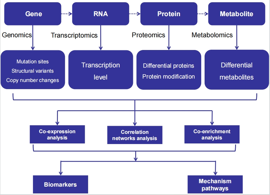
Multi-omic integration is anticipated to offer a comprehensive insight into pediatric cataracts, as it offers a glimpse into the intricate web of molecular interactions and systems biology regulations (Figure 2). By adhering to the central tenet, multi-omic approaches can unveil genotyping traits, RNA transcription, differences in protein expression, and systemic metabolic shifts in pediatric cataract patients. Through the integration of multidimensional omics data, encompassing genetic variants and expression disparities in RNA, proteins, and metabolite during disease states, along with the conduct of correlation networks analyses, we can identify reliable pathogenesis-related pathways and disease-related biomarkers, including specific proteins and metabolites. This holistic approach, characterized by mutual validation and complementation between the upstream and downstream molecular levels, facilitates the identification of the key pathways in pathogenesis-related disorder and enhances our understanding of the biological regulatory network underlying the disease. This, in turn, paves the way for the development of novel therapeutic targets and more efficacious therapeutic strategies. Furthermore, multi-omics data can be further analyzed in conjunction with the phenotypes manifestations of pediatric cataracts to identify the specific molecular pathogenic mechanisms of particular subtypes, potentially aiding in the formulation of individualized clinical diagnosis and treatment strategies.

Figure2. Combined analyses of multi-omics data to explore the molecular pathogenesis

20250318154331_5001.png
In the future, it is recommended to conduct multi-omics analysis on various biological materials, including peripheral blood, aqueous humor, and lens epithelium, obtained from pediatric cataract patients, particularly from the same cohort of patients. The combined analysis of genomics, proteomics, and metabolomics data from the same patient may have additional clinical value, enabling us to integrate multiple biological processes, systematically and reliably investigate the molecular mechanisms underlying pediatric cataract, and rigorously identify candidate biomarkers and key pathways. Furthermore, multi-omics can establish molecular profiles at different stages of the disease, which, when combine with clinical phenotype data, can achieve the link of between "genotype-protein-metabolite-phenotype". This integration can provide researchers with new perspectives for developing new diagnostic and targeted therapeutic strategies for pediatric cataract.

Table 1 Main technologies and biological samples of omics approaches that have been used in studies of pediatric cataracts

Omics layer

Biological samples

Main technologies

Genomics

Peripheral blood

Whole genome sequencing


Whole exome sequencing


Panel-based sequencing


Microarray analysis


Transcriptomics

Peripheral blood

Whole RNA sequencing


Lens or lens contents

mRNA sequencing


Human lens epithelial cells

Microarray analysis


Proteomics

Aqueous humor

Non-targeted proteomics


Lens or lens contents

Targeted proteomics


Metabolomics

Aqueous humor

Non-targeted metabolomics

Correction notice

None

Acknowledgements

None

Author Contributions

(I) Conception and design: Zhenzhen Liu
(II) Administrative support: Zhenzhen Liu
(III) Provision of study materials or patients: Not applicable  
(IV) Collection and assembly of data: Yinglin Yu, Shuo Gao
(V) Data analysis and interpretation: Yinglin Yu, Shuo Gao
(VI) Manuscript writing: All authors
(VII) Final approval of manuscript: All authors

Funding

This study was supported by the General Program of Natural Science Foundation of Guangdong Province (2023A1515011102).

Conflict of Interests

None of the authors has any conflicts of interest to disclose. All authors have declared in the completed the ICMJE uniform disclosure form.

Patient consent for publication

None

Ethics approval and consent to participate

None

Data availability statement

None

Open access

This is an Open Access article distributed in accordance with the Creative Commons AttributionNonCommercial-NoDerivs 4.0 International License (CC BY-NC-ND 4.0), which permits the non-commercial replication and distribution of the article with the strict proviso that no changes or edits are made and the original work is properly cited (including links to both the formal publication through the relevant DOI and the license).

Figure1. Omics approaches for the study of pediatric cataract

20250318153951_6873.png

Figure2. Combined analyses of multi-omics data to explore the molecular pathogenesis

20250318154331_5001.png

Table 1 Main technologies and biological samples of omics approaches that have been used in studies of pediatric cataracts

Omics layer

Biological samples

Main technologies

Genomics

Peripheral blood

Whole genome sequencing


Whole exome sequencing


Panel-based sequencing


Microarray analysis


Transcriptomics

Peripheral blood

Whole RNA sequencing


Lens or lens contents

mRNA sequencing


Human lens epithelial cells

Microarray analysis


Proteomics

Aqueous humor

Non-targeted proteomics


Lens or lens contents

Targeted proteomics


Metabolomics

Aqueous humor

Non-targeted metabolomics

1、Zhao QH, Zhao YE. Commentary review: challenges of intraocular lens implantation for congenital cataract infants. Int J Ophthalmol. 2021, 14(6): 923-930. DOI: 10.18240/ijo.2021.06.19. Zhao QH, Zhao YE. Commentary review: challenges of intraocular lens implantation for congenital cataract infants. Int J Ophthalmol. 2021, 14(6): 923-930. DOI: 10.18240/ijo.2021.06.19.
2、Trumler AA. Evaluation of pediatric cataracts and systemic disorders. Curr Opin Ophthalmol. 2011, 22(5): 365-379. DOI: 10.1097/ICU.0b013e32834994dc. Trumler AA. Evaluation of pediatric cataracts and systemic disorders. Curr Opin Ophthalmol. 2011, 22(5): 365-379. DOI: 10.1097/ICU.0b013e32834994dc.
3、Rahi JS, Dezateux C, British Congenital Cataract Interest Group. Measuring and interpreting the incidence of congenital ocular anomalies: lessons from a national study of congenital cataract in the UK. Invest Ophthalmol Vis Sci. 2001, 42(7): 1444-1448. Rahi JS, Dezateux C, British Congenital Cataract Interest Group. Measuring and interpreting the incidence of congenital ocular anomalies: lessons from a national study of congenital cataract in the UK. Invest Ophthalmol Vis Sci. 2001, 42(7): 1444-1448.
4、Singh R, Barker L, Chen SI, et al. Surgical interventions for bilateral congenital cataract in children aged two years and under. Cochrane Database Syst Rev. 2022, 9(9): CD003171. DOI: 10.1002/14651858.CD003171.pub3. Singh R, Barker L, Chen SI, et al. Surgical interventions for bilateral congenital cataract in children aged two years and under. Cochrane Database Syst Rev. 2022, 9(9): CD003171. DOI: 10.1002/14651858.CD003171.pub3.
5、SanGiovanni JP, Chew EY, Reed GF, et al. Infantile cataract in the collaborative perinatal project: prevalence and risk factors. Arch Ophthalmol. 2002, 120(11): 1559-1565. DOI: 10.1001/archopht.120.11.1559.SanGiovanni JP, Chew EY, Reed GF, et al. Infantile cataract in the collaborative perinatal project: prevalence and risk factors. Arch Ophthalmol. 2002, 120(11): 1559-1565. DOI: 10.1001/archopht.120.11.1559.
6、Hasin Y, Seldin M, Lusis A. Multi-omics approaches to disease. Genome Biol. 2017, 18(1): 83. DOI: 10.1186/s13059-017-1215-1. Hasin Y, Seldin M, Lusis A. Multi-omics approaches to disease. Genome Biol. 2017, 18(1): 83. DOI: 10.1186/s13059-017-1215-1.
7、Kowalczyk T, Ciborowski M, Kisluk J, et al. Mass spectrometry based proteomics and metabolomics in personalized oncology. Biochim Biophys Acta Mol Basis Dis. 2020, 1866(5): 165690. DOI: 10.1016/j.bbadis.2020.165690.Kowalczyk T, Ciborowski M, Kisluk J, et al. Mass spectrometry based proteomics and metabolomics in personalized oncology. Biochim Biophys Acta Mol Basis Dis. 2020, 1866(5): 165690. DOI: 10.1016/j.bbadis.2020.165690.
8、Ahmad Dar M, Arafah A, Ahmad Bhat K, et al. Multiomics technologies: role in disease biomarker discoveries and therapeutics. Brief Funct Genom. 2023, 22(2): 76-96. DOI: 10.1093/bfgp/elac017. Ahmad Dar M, Arafah A, Ahmad Bhat K, et al. Multiomics technologies: role in disease biomarker discoveries and therapeutics. Brief Funct Genom. 2023, 22(2): 76-96. DOI: 10.1093/bfgp/elac017.
9、Wang X, Fan D, Yang Y, et al. Integrative multi-omics approaches to explore immune cell functions: Challenges and opportunities. iScience. 2023, 26(4): 106359. DOI: 10.1016/j.isci.2023.106359.Wang X, Fan D, Yang Y, et al. Integrative multi-omics approaches to explore immune cell functions: Challenges and opportunities. iScience. 2023, 26(4): 106359. DOI: 10.1016/j.isci.2023.106359.
10、Lancaster SM, Sanghi A, Wu S, et al. A customizable analysis flow in integrative multi-omics. Biomolecules. 2020, 10(12): 1606. DOI: 10.3390/biom10121606. Lancaster SM, Sanghi A, Wu S, et al. A customizable analysis flow in integrative multi-omics. Biomolecules. 2020, 10(12): 1606. DOI: 10.3390/biom10121606.
11、Pettini F, Visibelli A, Cicaloni V, et al. Multi-omics model applied to cancer genetics. Int J Mol Sci. 2021, 22(11): 5751. DOI: 10.3390/ijms22115751. Pettini F, Visibelli A, Cicaloni V, et al. Multi-omics model applied to cancer genetics. Int J Mol Sci. 2021, 22(11): 5751. DOI: 10.3390/ijms22115751.
12、Delas F, Koller S, Feil S, et al. Novel CRYGC mutation in conserved ultraviolet-protective tryptophan (p.Trp131Arg) is linked to autosomal dominant congenital cataract. Int J Mol Sci. 2023, 24(23): 16594. DOI: 10.3390/ijms242316594. Delas F, Koller S, Feil S, et al. Novel CRYGC mutation in conserved ultraviolet-protective tryptophan (p.Trp131Arg) is linked to autosomal dominant congenital cataract. Int J Mol Sci. 2023, 24(23): 16594. DOI: 10.3390/ijms242316594.
13、Zhou Z, Zhao L, Guo Y, et al. A novel mutation in CRYGC mutation associated with autosomal dominant congenital cataracts and microcornea. Ophthalmol Sci. 2022, 2(1): 100093. DOI: 10.1016/j.xops.2021.100093.Zhou Z, Zhao L, Guo Y, et al. A novel mutation in CRYGC mutation associated with autosomal dominant congenital cataracts and microcornea. Ophthalmol Sci. 2022, 2(1): 100093. DOI: 10.1016/j.xops.2021.100093.
14、Peng Y, Zheng Y, Deng Z, et al. Case report: a de novo variant of CRYGC gene associated with congenital cataract and microphthalmia. Front Genet. 2022, 13: 866246. DOI: 10.3389/fgene.2022.866246. Peng Y, Zheng Y, Deng Z, et al. Case report: a de novo variant of CRYGC gene associated with congenital cataract and microphthalmia. Front Genet. 2022, 13: 866246. DOI: 10.3389/fgene.2022.866246.
15、Reis LM, Tyler RC, Muheisen S, et al. Whole exome sequencing in dominant cataract identifies a new causative factor, CRYBA2, and a variety of novel alleles in known genes. Hum Genet. 2013, 132(7): 761-770. DOI: 10.1007/s00439-013-1289-0. Reis LM, Tyler RC, Muheisen S, et al. Whole exome sequencing in dominant cataract identifies a new causative factor, CRYBA2, and a variety of novel alleles in known genes. Hum Genet. 2013, 132(7): 761-770. DOI: 10.1007/s00439-013-1289-0.
16、Chen D, Yang T, Zhu S. Novel likely pathogenic variants identified by panel-based exome sequencing in congenital cataract patients. J Ophthalmol. 2021, 2021: 3847409. DOI: 10.1155/2021/3847409. Chen D, Yang T, Zhu S. Novel likely pathogenic variants identified by panel-based exome sequencing in congenital cataract patients. J Ophthalmol. 2021, 2021: 3847409. DOI: 10.1155/2021/3847409.
17、Ha C, Kim D, Bak M, et al. CRYAB stop-loss variant causes rare syndromic dilated cardiomyopathy with congenital cataract: expanding the phenotypic and mutational spectrum of alpha-B crystallinopathy. J Hum Genet. 2024, 69(3-4): 159-162. DOI: 10.1038/s10038-023-01218-1. Ha C, Kim D, Bak M, et al. CRYAB stop-loss variant causes rare syndromic dilated cardiomyopathy with congenital cataract: expanding the phenotypic and mutational spectrum of alpha-B crystallinopathy. J Hum Genet. 2024, 69(3-4): 159-162. DOI: 10.1038/s10038-023-01218-1.
18、Javadiyan S, Craig JE, Souzeau E, et al. Recurrent mutation in the crystallin alpha A gene associated with inherited paediatric cataract. BMC Res Notes. 2016, 9: 83. DOI: 10.1186/s13104-016-1890-0. Javadiyan S, Craig JE, Souzeau E, et al. Recurrent mutation in the crystallin alpha A gene associated with inherited paediatric cataract. BMC Res Notes. 2016, 9: 83. DOI: 10.1186/s13104-016-1890-0.
19、Li R, Xu P, Zou Y, Li J, Gao Y. Analysis of novel compound heterozygous variants of the GJA8 gene in a child with congenital cataract..Zhonghua Yi Xue Yi Chuan Xue Za Zhi. 2022, 39(11): 1262-1265. DOI:10.3760/cma.j.cn511374-20211218-01005.Li R, Xu P, Zou Y, Li J, Gao Y. Analysis of novel compound heterozygous variants of the GJA8 gene in a child with congenital cataract..Zhonghua Yi Xue Yi Chuan Xue Za Zhi. 2022, 39(11): 1262-1265. DOI:10.3760/cma.j.cn511374-20211218-01005.
20、Berry V, Ionides ACW, Pontikos N, et al. Whole-genome sequencing reveals a recurrent missense mutation in the Connexin 46 (GJA3) gene causing autosomal-dominant lamellar cataract. Eye (Lond). 2018, 32(10): 1661-1668. DOI: 10.1038/s41433-018-0154-8. Berry V, Ionides ACW, Pontikos N, et al. Whole-genome sequencing reveals a recurrent missense mutation in the Connexin 46 (GJA3) gene causing autosomal-dominant lamellar cataract. Eye (Lond). 2018, 32(10): 1661-1668. DOI: 10.1038/s41433-018-0154-8.
21、Song S, Landsbury A, Dahm R, et al. Functions of the intermediate filament cytoskeleton in the eye lens. J Clin Invest. 2009, 119(7): 1837-1848. DOI: 10.1172/JCI38277. Song S, Landsbury A, Dahm R, et al. Functions of the intermediate filament cytoskeleton in the eye lens. J Clin Invest. 2009, 119(7): 1837-1848. DOI: 10.1172/JCI38277.
22、Goolam S, Carstens N, Ross M, et al. Familial congenital cataract, coloboma, and nystagmus phenotype with variable expression caused by mutation in PAX6 in a South African family. Mol Vis. 2018, 24: 407-413. Goolam S, Carstens N, Ross M, et al. Familial congenital cataract, coloboma, and nystagmus phenotype with variable expression caused by mutation in PAX6 in a South African family. Mol Vis. 2018, 24: 407-413.
23、Javadiyan S, Craig JE, Sharma S, et al. Novel missense mutation in the bZIP transcription factor, MAF, associated with congenital cataract, developmental delay, seizures and hearing loss (Aymé-Gripp syndrome). BMC Med Genet. 2017, 18(1): 52. DOI: 10.1186/s12881-017-0414-7. Javadiyan S, Craig JE, Sharma S, et al. Novel missense mutation in the bZIP transcription factor, MAF, associated with congenital cataract, developmental delay, seizures and hearing loss (Aymé-Gripp syndrome). BMC Med Genet. 2017, 18(1): 52. DOI: 10.1186/s12881-017-0414-7.
24、Ye X, Zhang G, Dong N, et al. Human pituitary homeobox-3 gene in congenital cataract in a Chinese family. Int J Clin Exp Med. 2015, 8(12): 22435-22439.Ye X, Zhang G, Dong N, et al. Human pituitary homeobox-3 gene in congenital cataract in a Chinese family. Int J Clin Exp Med. 2015, 8(12): 22435-22439.
25、Berry V, Pontikos N, Albarca-Aguilera M, et al. A recurrent splice-site mutation in EPHA2 causing congenital posterior nuclear cataract. Ophthalmic Genet. 2018, 39(2): 236-241. DOI: 10.1080/13816810.2017.1381977.Berry V, Pontikos N, Albarca-Aguilera M, et al. A recurrent splice-site mutation in EPHA2 causing congenital posterior nuclear cataract. Ophthalmic Genet. 2018, 39(2): 236-241. DOI: 10.1080/13816810.2017.1381977.
26、Chen X, Xu P, Li J, Niu Y, Kang R, Gao Y. Identification of a novel variant of NHS gene underlying Nance-Horan syndrome.. Zhonghua Yi Xue Yi Chuan Xue Za Zhi. 2021,,38(11): 1077-1080. doi:10.3760/cma.j.cn511374-20200817-00603.Chen X, Xu P, Li J, Niu Y, Kang R, Gao Y. Identification of a novel variant of NHS gene underlying Nance-Horan syndrome.. Zhonghua Yi Xue Yi Chuan Xue Za Zhi. 2021,,38(11): 1077-1080. doi:10.3760/cma.j.cn511374-20200817-00603.
27、Chograni M, Alahdal HM, Rejili M. Autosomal recessive congenital cataract is associated with a novel 4-bp splicing deletion mutation in a novel C10orf71 human gene. Hum Genomics. 2023, 17(1): 41. DOI: 10.1186/s40246-023-00492-6. Chograni M, Alahdal HM, Rejili M. Autosomal recessive congenital cataract is associated with a novel 4-bp splicing deletion mutation in a novel C10orf71 human gene. Hum Genomics. 2023, 17(1): 41. DOI: 10.1186/s40246-023-00492-6.
28、Damián A, Ionescu RO, Rodríguez de Alba M, et al. Fine breakpoint mapping by genome sequencing reveals the first large X inversion disrupting the NHS gene in a patient with syndromic cataracts. Int J Mol Sci. 2021, 22(23): 12713. DOI: 10.3390/ijms222312713. Damián A, Ionescu RO, Rodríguez de Alba M, et al. Fine breakpoint mapping by genome sequencing reveals the first large X inversion disrupting the NHS gene in a patient with syndromic cataracts. Int J Mol Sci. 2021, 22(23): 12713. DOI: 10.3390/ijms222312713.
29、Zheng B, Chen Q, Wang C, et al. Whole-genome sequencing revealed an interstitial deletion encompassing OCRL and SMARCA1 gene in a patient with Lowe syndrome. Mol Genet Genomic Med. 2019, 7(9): e876. DOI: 10.1002/mgg3.876. Zheng B, Chen Q, Wang C, et al. Whole-genome sequencing revealed an interstitial deletion encompassing OCRL and SMARCA1 gene in a patient with Lowe syndrome. Mol Genet Genomic Med. 2019, 7(9): e876. DOI: 10.1002/mgg3.876.
30、Gillespie RL, Urquhart J, Lovell SC, et al. Abrogation of HMX1 function causes rare oculoauricular syndrome associated with congenital cataract, anterior segment dysgenesis, and retinal dystrophy. Investig Ophthalmol Vis Sci. 2015, 56(2): 883-891. DOI: 10.1167/iovs.14-15861.Gillespie RL, Urquhart J, Lovell SC, et al. Abrogation of HMX1 function causes rare oculoauricular syndrome associated with congenital cataract, anterior segment dysgenesis, and retinal dystrophy. Investig Ophthalmol Vis Sci. 2015, 56(2): 883-891. DOI: 10.1167/iovs.14-15861.
31、Chograni M, Alkuraya FS, Maazoul F, et al. RGS6: a novel gene associated with congenital cataract, mental retardation, and microcephaly in a Tunisian family. Invest Ophthalmol Vis Sci. 2014, 56(2): 1261-1266. DOI: 10.1167/iovs.14-15198.Chograni M, Alkuraya FS, Maazoul F, et al. RGS6: a novel gene associated with congenital cataract, mental retardation, and microcephaly in a Tunisian family. Invest Ophthalmol Vis Sci. 2014, 56(2): 1261-1266. DOI: 10.1167/iovs.14-15198.
32、Chograni M, Alkuraya FS, Ourteni I, et al. Autosomal recessive congenital cataract, intellectual disability phenotype linked to STX3 in a consanguineous Tunisian family. Clin Genet. 2015, 88(3): 283-287. DOI: 10.1111/cge.12489.Chograni M, Alkuraya FS, Ourteni I, et al. Autosomal recessive congenital cataract, intellectual disability phenotype linked to STX3 in a consanguineous Tunisian family. Clin Genet. 2015, 88(3): 283-287. DOI: 10.1111/cge.12489.
33、Chacon-Camacho OF, Buentello-Volante B, Velázquez-Montoya R, et al. Homozygosity mapping identifies a GALK1 mutation as the cause of autosomal recessive congenital cataracts in 4 adult siblings. Gene. 2014, 534(2): 218-221. DOI: 10.1016/j.gene.2013.10.057. Chacon-Camacho OF, Buentello-Volante B, Velázquez-Montoya R, et al. Homozygosity mapping identifies a GALK1 mutation as the cause of autosomal recessive congenital cataracts in 4 adult siblings. Gene. 2014, 534(2): 218-221. DOI: 10.1016/j.gene.2013.10.057.
34、Mei S, Wu Y, Wang Y, et al. Disruption of PIKFYVE causes congenital cataract in human and zebrafish. eLife. 2022, 11: e71256. DOI: 10.7554/elife.71256.Mei S, Wu Y, Wang Y, et al. Disruption of PIKFYVE causes congenital cataract in human and zebrafish. eLife. 2022, 11: e71256. DOI: 10.7554/elife.71256.
35、Jones JL, Corbett MA, Yeaman E, et al. A 127 kb truncating deletion of PGRMC1 is a novel cause of X-linked isolated paediatric cataract. Eur J Hum Genet. 2021, 29(8): 1206-1215. DOI: 10.1038/s41431-021-00889-8.Jones JL, Corbett MA, Yeaman E, et al. A 127 kb truncating deletion of PGRMC1 is a novel cause of X-linked isolated paediatric cataract. Eur J Hum Genet. 2021, 29(8): 1206-1215. DOI: 10.1038/s41431-021-00889-8.
36、Javadiyan S, Craig JE, Souzeau E, et al. High-throughput genetic screening of 51 pediatric cataract genes identifies causative mutations in inherited pediatric cataract in south eastern Australia. G3 (Bethesda). 2017, 7(10): 3257-3268. DOI: 10.1534/g3.117.300109. Javadiyan S, Craig JE, Souzeau E, et al. High-throughput genetic screening of 51 pediatric cataract genes identifies causative mutations in inherited pediatric cataract in south eastern Australia. G3 (Bethesda). 2017, 7(10): 3257-3268. DOI: 10.1534/g3.117.300109.
37、Patel N, Anand D, Monies D, et al. Novel phenotypes and loci identified through clinical genomics approaches to pediatric cataract. Hum Genet. 2017, 136(2): 205-225. DOI: 10.1007/s00439-016-1747-6. Patel N, Anand D, Monies D, et al. Novel phenotypes and loci identified through clinical genomics approaches to pediatric cataract. Hum Genet. 2017, 136(2): 205-225. DOI: 10.1007/s00439-016-1747-6.
38、Ma A, Grigg JR, Flaherty M, et al. Genome sequencing in congenital cataracts improves diagnostic yield. Hum Mutat. 2021, 42(9): 1173-1183. DOI: 10.1002/humu.24240. Ma A, Grigg JR, Flaherty M, et al. Genome sequencing in congenital cataracts improves diagnostic yield. Hum Mutat. 2021, 42(9): 1173-1183. DOI: 10.1002/humu.24240.
39、Rossen JL, Bohnsack BL, Zhang KX, et al. Evaluation of genetic testing in a cohort of diverse pediatric patients in the United States with congenital cataracts. Genes (Basel). 2023, 14(3): 608. DOI: 10.3390/genes14030608.Rossen JL, Bohnsack BL, Zhang KX, et al. Evaluation of genetic testing in a cohort of diverse pediatric patients in the United States with congenital cataracts. Genes (Basel). 2023, 14(3): 608. DOI: 10.3390/genes14030608.
40、Consortium EP. An integrated encyclopedia of DNA elements in the human genome. Nature. 2012, 489(7414): 57-74. DOI: 10.1038/nature11247. Consortium EP. An integrated encyclopedia of DNA elements in the human genome. Nature. 2012, 489(7414): 57-74. DOI: 10.1038/nature11247.
41、Zhao N, Quicksall Z, Asmann YW, et al. Network approaches for omics studies of neurodegenerative diseases. Front Genet. 2022, 13: 984338. DOI: 10.3389/fgene.2022.984338. Zhao N, Quicksall Z, Asmann YW, et al. Network approaches for omics studies of neurodegenerative diseases. Front Genet. 2022, 13: 984338. DOI: 10.3389/fgene.2022.984338.
42、Lin X, Li H, Yang T, et al. Transcriptomics analysis of lens from patients with posterior subcapsular congenital cataract. Genes. 2021, 12(12): 1904. DOI: 10.3390/genes12121904. Lin X, Li H, Yang T, et al. Transcriptomics analysis of lens from patients with posterior subcapsular congenital cataract. Genes. 2021, 12(12): 1904. DOI: 10.3390/genes12121904.
43、Anand D, Al Saai S, Shrestha SK, et al. Genome-wide analysis of differentially expressed miRNAs and their associated regulatory networks in lenses deficient for the congenital cataract-linked Tudor domain containing protein TDRD7. Front Cell Dev Biol. 2021, 9: 615761. DOI: 10.3389/fcell.2021.615761. Anand D, Al Saai S, Shrestha SK, et al. Genome-wide analysis of differentially expressed miRNAs and their associated regulatory networks in lenses deficient for the congenital cataract-linked Tudor domain containing protein TDRD7. Front Cell Dev Biol. 2021, 9: 615761. DOI: 10.3389/fcell.2021.615761.
44、Tu C, Li H, Liu X, et al. TDRD7 participates in lens development and spermiogenesis by mediating autophagosome maturation. Autophagy. 2021, 17(11): 3848-3864. DOI: 10.1080/15548627.2021.1894058. Tu C, Li H, Liu X, et al. TDRD7 participates in lens development and spermiogenesis by mediating autophagosome maturation. Autophagy. 2021, 17(11): 3848-3864. DOI: 10.1080/15548627.2021.1894058.
45、Zhang J, Jiang N, Du C, et al. HSF4 transcriptionally activates autophagy by regulating ATG9a during lens terminal differentiation. Invest Ophthalmol Vis Sci. 2023, 64(7): 5. DOI: 10.1167/iovs.64.7.5. Zhang J, Jiang N, Du C, et al. HSF4 transcriptionally activates autophagy by regulating ATG9a during lens terminal differentiation. Invest Ophthalmol Vis Sci. 2023, 64(7): 5. DOI: 10.1167/iovs.64.7.5.
46、Xiao W, Yang W, Zhang X, et al. Endoplasmic reticulum stress and the lysosomal pathway play crucial roles in the progression of βB2-crystallin mutation-induced congenital cataracts in mice. Invest Ophthalmol Vis Sci. 2023, 64(4): 34. DOI: 10.1167/iovs.64.4.34. Xiao W, Yang W, Zhang X, et al. Endoplasmic reticulum stress and the lysosomal pathway play crucial roles in the progression of βB2-crystallin mutation-induced congenital cataracts in mice. Invest Ophthalmol Vis Sci. 2023, 64(4): 34. DOI: 10.1167/iovs.64.4.34.
47、Luo Y, Liu S, Yao K. Transcriptome-wide Investigation of mRNA/circRNA in miR-184 and Its r.57c > u Mutant Type Treatment of Human Lens Epithelial Cells. Mol Ther Nucleic Acids. 2017, 7: 71-80. DOI: 10.1016/j.omtn.2017.02.008. Luo Y, Liu S, Yao K. Transcriptome-wide Investigation of mRNA/circRNA in miR-184 and Its r.57c > u Mutant Type Treatment of Human Lens Epithelial Cells. Mol Ther Nucleic Acids. 2017, 7: 71-80. DOI: 10.1016/j.omtn.2017.02.008.
48、Torres-Arroyo A, Ruiz-Lara A, Castillo-Villanueva A, et al. Proteomics: a tool to develop novel diagnostic methods and unravel molecular mechanisms of pediatric diseases. Bol Med Hosp Infant Mex. 2017, 74(3): 233-240. DOI: 10.1016/j.bmhimx.2017.03.009.Torres-Arroyo A, Ruiz-Lara A, Castillo-Villanueva A, et al. Proteomics: a tool to develop novel diagnostic methods and unravel molecular mechanisms of pediatric diseases. Bol Med Hosp Infant Mex. 2017, 74(3): 233-240. DOI: 10.1016/j.bmhimx.2017.03.009.
49、Semba RD, Enghild JJ, Venkatraman V, et al. The human eye proteome project: perspectives on an emerging proteome. Proteomics. 2013, 13(16): 2500-2511. DOI: 10.1002/pmic.201300075. Semba RD, Enghild JJ, Venkatraman V, et al. The human eye proteome project: perspectives on an emerging proteome. Proteomics. 2013, 13(16): 2500-2511. DOI: 10.1002/pmic.201300075.
50、Theophanous CN, Wolfgeher DJ, Farooq AV, et al. Biomarkers of pediatric cataracts: a proteomics analysis of aqueous fluid. Int J Mol Sci. 2023, 24(10): 9040. DOI: 10.3390/ijms24109040. Theophanous CN, Wolfgeher DJ, Farooq AV, et al. Biomarkers of pediatric cataracts: a proteomics analysis of aqueous fluid. Int J Mol Sci. 2023, 24(10): 9040. DOI: 10.3390/ijms24109040.
51、Wu X, Liu Z, Zhang X, et al. Proteomics analysis and proteogenomic characterization of different physiopathological human lenses. BMC Ophthalmol. 2017, 17(1): 253. DOI: 10.1186/s12886-017-0642-9. Wu X, Liu Z, Zhang X, et al. Proteomics analysis and proteogenomic characterization of different physiopathological human lenses. BMC Ophthalmol. 2017, 17(1): 253. DOI: 10.1186/s12886-017-0642-9.
52、Van Looveren J, Van Gerwen V, Schildermans K, et al. Proteomic analysis of posterior capsular plaques in congenital unilateral cataract. Acta Ophthalmol. 2018, 96(8): e963-e969. DOI: 10.1111/aos.13782. Van Looveren J, Van Gerwen V, Schildermans K, et al. Proteomic analysis of posterior capsular plaques in congenital unilateral cataract. Acta Ophthalmol. 2018, 96(8): e963-e969. DOI: 10.1111/aos.13782.
53、Shang F, Wilmarth PA, Chang ML, et al. Newborn mouse lens proteome and its alteration by lysine 6 mutant ubiquitin. J Proteome Res. 2014, 13(3): 1177-1189. DOI: 10.1021/pr400801v. Shang F, Wilmarth PA, Chang ML, et al. Newborn mouse lens proteome and its alteration by lysine 6 mutant ubiquitin. J Proteome Res. 2014, 13(3): 1177-1189. DOI: 10.1021/pr400801v.
54、Bejarano E, Whitcomb EA, Pfeiffer RL, et al. Unbalanced redox status network as an early pathological event in congenital cataracts. Redox Biol. 2023, 66: 102869. DOI: 10.1016/j.redox.2023.102869. Bejarano E, Whitcomb EA, Pfeiffer RL, et al. Unbalanced redox status network as an early pathological event in congenital cataracts. Redox Biol. 2023, 66: 102869. DOI: 10.1016/j.redox.2023.102869.
55、Zhang A, Sun H, Wang P, et al. Modern analytical techniques in metabolomics analysis. Analyst. 2012, 137(2): 293-300. DOI: 10.1039/c1an15605e.Zhang A, Sun H, Wang P, et al. Modern analytical techniques in metabolomics analysis. Analyst. 2012, 137(2): 293-300. DOI: 10.1039/c1an15605e.
56、Grochowski ET, Pietrowska K, Kowalczyk T, et al. Omics in myopia. J Clin Med. 2020, 9(11): 3464. DOI: 10.3390/jcm9113464. Grochowski ET, Pietrowska K, Kowalczyk T, et al. Omics in myopia. J Clin Med. 2020, 9(11): 3464. DOI: 10.3390/jcm9113464.
57、Schrimpe-Rutledge AC, Codreanu SG, Sherrod SD, et al. Untargeted metabolomics strategies: challenges and emerging directions. J Am Soc Mass Spectrom. 2016, 27(12): 1897-1905. DOI: 10.1007/s13361-016-1469-y. Schrimpe-Rutledge AC, Codreanu SG, Sherrod SD, et al. Untargeted metabolomics strategies: challenges and emerging directions. J Am Soc Mass Spectrom. 2016, 27(12): 1897-1905. DOI: 10.1007/s13361-016-1469-y.
58、Roberts LD, Souza AL, Gerszten RE, et al. Targeted metabolomics. Curr Protoc Mol Biol. 2012, Chapter 30: Unit30.2.1-Unit30.224. DOI: 10.1002/0471142727.mb3002s98. Roberts LD, Souza AL, Gerszten RE, et al. Targeted metabolomics. Curr Protoc Mol Biol. 2012, Chapter 30: Unit30.2.1-Unit30.224. DOI: 10.1002/0471142727.mb3002s98.
59、Cajka T, Fiehn O. Toward merging untargeted and targeted methods in mass spectrometry-based metabolomics and lipidomics. Anal Chem. 2016, 88(1): 524-545. DOI: 10.1021/acs.analchem.5b04491. Cajka T, Fiehn O. Toward merging untargeted and targeted methods in mass spectrometry-based metabolomics and lipidomics. Anal Chem. 2016, 88(1): 524-545. DOI: 10.1021/acs.analchem.5b04491.
60、Chen H, Yu Y, Hu L, et al. Metabolomic profiling of the aqueous humor in patients with pediatric cataract. Exp Eye Res. 2024, 243: 109906. DOI: 10.1016/j.exer.2024.109906. Chen H, Yu Y, Hu L, et al. Metabolomic profiling of the aqueous humor in patients with pediatric cataract. Exp Eye Res. 2024, 243: 109906. DOI: 10.1016/j.exer.2024.109906.
1、This study was supported by the General Program of Natural Science Foundation of Guangdong Province (2023A1515011102).
Previous
Next
  1. 1
/ 1
  1. 1
/ 1
  1. 1
/ 1
  1. 1
/ 1