您的位置: 首页 > 2025年3月 第2卷 第1期 > 文字全文
2023年7月 第38卷 第7期11
目录

Prevalence of idiopathic thoracic scoliosis in children and adolescents with superior oblique palsy: a cross‑sectional study

Prevalence of idiopathic thoracic scoliosis in children and adolescents with superior oblique palsy: a cross‑sectional study

来源期刊: Eye Science | 2025年3月 第2卷 第1期 1-8 发布时间:2025-03-10 收稿时间:2025/3/14 14:11:11 阅读量:147
作者:
关键词:
Idiopathic Thoracic Scoliosis Superior Oblique Palsy Children and Adolescents Binocular function
DOI:
10.12419/es24121601
Received date:
2024-12-16 
Revised date:
2025-01-08 
Accepted date:
2025-02-20 
Published online:
2025-03-10 

Purpose: Strabismus is associated with a higher prevalence of idiopathic thoracic scoliosis (ITS) in children. This study aims to investigate whether superior oblique palsy (SOP) poses a higher risk for developing ITS among children and adolescents.

Methods: A cross-sectional study. The study group comprised 347 consecutive candidates for SOP surgery aged 4 to 18 yearsPatients within the same age range with ocular trauma were enrolled as the control group. Preoperative chest plain radiographs were used to measure the Cobb angle. Demographic information and clinical data, including diopter, best corrected visual acuity, deviation degree, and binocular functionwere analyzed.

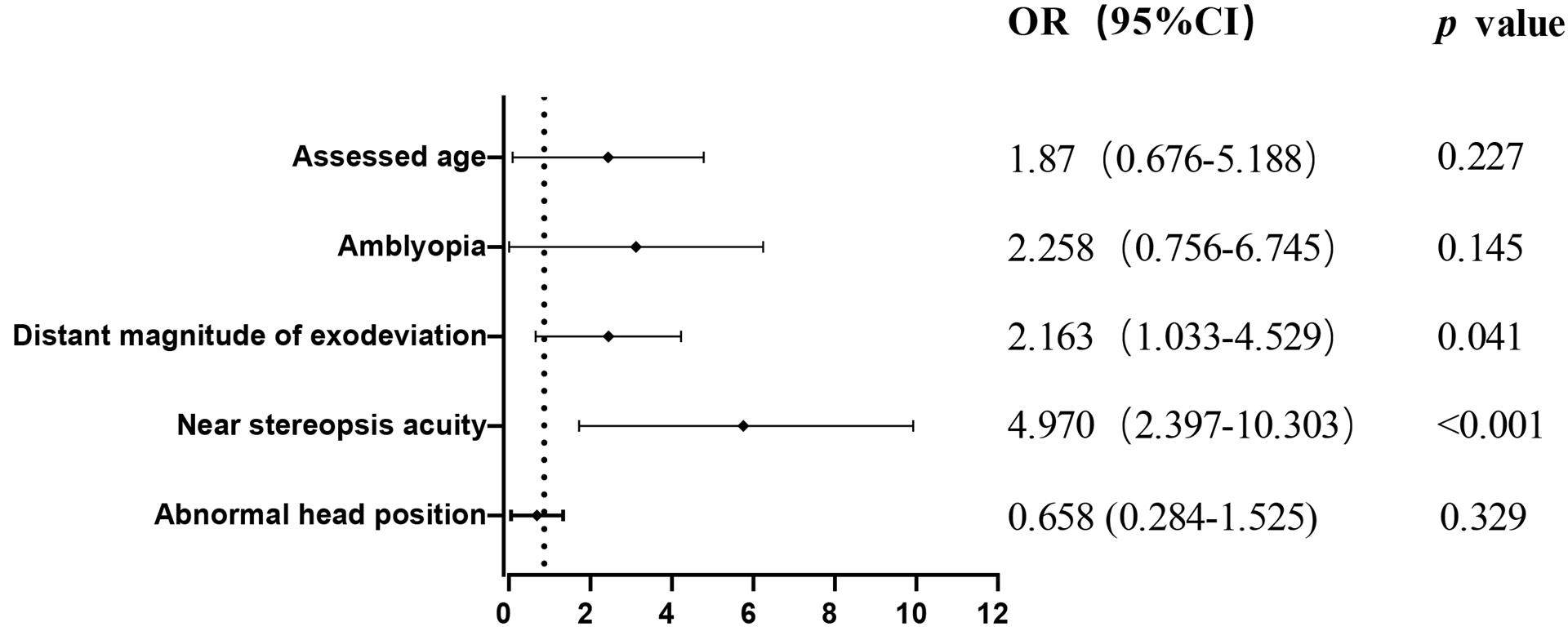
Results: A significantly higher prevalence of ITS was found in study group compared with control group (12.68% vs 4.18%p< 0.001) . Additionally, the mean Cobb angle was lagrger in SOP group than that in control group (5.02°±3.87° vs 3.84°±3.09°, < 0.001). Males in SOP group showed a higher prevalence of ITS  (12.9% vs 2.87%, p = 0.007), but there was no significant difference in females between two groups (12.3% vs 7.69%, p = 0.295). Good near stereopsis acuity was  significantly associated with high prevalence of thoracic scoliosis (p<0.001). Multivariate logistic regression analysis revealed that a large distant magnitude of deviation (>20PD) and near stereoacuity were significantly associated with ITS.

Conclusions: Patients with SOP have a significantly  higher risk of developing idiopathic thoracic scoliosis, especially those with good near stereoscopic and large distant magnitude of deviation. 

Highlights

1.Critical Discoveries and Outcomes

Patients with SOP have a significantly higher risk of developing idiopathic thoracic scoliosis, especially those with good near stereoscopic and large distant magnitude of deviation.

2.Methodological Innovations

X-ray is not only used for preoperative examination to exclude pulmonary diseases but can also be utilized for the early screening of scoliosis in children, particularly in children with SOP.

3. Prospective Applications and Future Directions

Earlier detection of potential scoliosis in children with SOP can prevent the progression of scoliosis and improve the quality of life for the children.

Introduction

Superior oblique palsy (SOP) represents one of the most prevalent forms of vertical strabismus, with origins that can be either congenital or acquired etiology[1]. Tollefson reported an annual incidence rate of 3.4 per 100,000 among children under 19 years of age in 2006, accounting for 31% of all paitents wih hypertropic[2]. Individuals with unilateral or asymmetric SOP exhibit hypertropia primary gaze and fundus extorsion. Consequently, to achieve normal binocular fusion, these patients often adopt an abnormal head posture (AHP) , such as tilting the head and turning towards the contralateral side of the palsy with the chin depressed. While the AHP aids patients with SOP in maintaining fusion, it also gives to additional complications, including facial asymmetry, deviation of the nasal tip, malpositioning of the teeth and jaws, and asymmetry of the sternocleidomastoid muscle due to the AHP[3-5]. Furthermore,, prolonged maintenane of an abnormal posture may contribute to a high prevalence of idiopathic thoracic scoliosis (ITS).

Scoliosis is defined as a lateral curvature of the spine greater than 10°, as assessed using the Cobb method on standing radiographic images. Previous studies have demonstrated that patients with idiopathic scoliosis often report back pain, sleep disturbance, and even depression, all of which can significantly impaire their quality of life[6, 7]. Patients with strabismus, particularly those with concomitant exotropia, have been shown to exhibit a markedly increased risk of developing idiopathic scoliosis[8]. However, it remains unclear whether congenital or early-onset scoliosis, which frequently presents with AHP, is associated with a higher prevalence of idiopathic scoliosis.

To date, X-ray images continue to serve as the gold standard for imaging in the evaluation of scoliosis. Consequently, we collected preoperative chest radiographs from SOP and children with ocular trauma, with the latter serving as a control group matched for age. The objective of this study was to identify the prevalence of ITS in children with SOP and to elucidate the relationship between ITS and demographic data, clinical variables, and factors related to scoliosis.

Materials and methods

Ethical Approval Statement

This study involves human participants and ethical approval (2023KYPJ271) was obtained from the Institutional Review Board of Zhongshan Ophthalmic Center, Sun Yat-sen University. This cross-sectional study was conducted in accordance with the tenets of Declaration of Helsinki. Informed written consent was obtained from guardians of all included participants, as they were all children under the age of 18.

Patient and Public Involvement statement

Patients or the public were not involved in the design, conduct, or reporting, or dissemination plans of our research.

Study design

This study included 347 consecutive patients who met the inclusion criteria (age range 4–18 years) and were diagnosed with unilateral or bilateral SOP and underwent operational treatment from January 2018 to December 2022. The control group comprised patients of the same age range (4–18 years) with ocular trauma who denied a history of eye disease during the same timeframe. Patients were excluded from this study if they had a history of strabismus correction, congenital ptosis, congenital craniofacial dysplasia, severe premature birth, or systemic disorders with musculoskeletal dysplasia. All strabismus and eye-traumatic surgeries were performed at the Zhongshan Ophthalmic Center. For each strabismus patient, the following data were recorded : sex, age of onset, disease duration, assessment age, degree of strabismus, best-corrected visual acuity, refractive diopter, vertical deviation diopter, stereoscopic vision, fundus torsion, AHP, (assumed to be a specific measurement or parameter relevant to the study), and chest X-ray images. In the traumatic group, we recorded the following data: sex, age of surgery, and chest X-ray images.

Cobb's angle measurement

Radiographic assessments were conducted using a standing anteroposterior plain chest X-ray (Digital Radiography System, Siemens Ysio Max), which is a routine preparatory procedure for general anesthesia at our institution. The coronal plane of the chest radiograph was selected for the measurement of Cobb's angle  using the integrated measuring tool, following adjustments to the grayscale and image enhancement to optimize visualization of the spine. Utilizing the Cobb method to measure the degree of scoliosis, we selected the most tilted vertebrae above and below the apex of the curve[9]. The angle between intersecting lines drawn perpendicular to the top of the top of the uppermost vertebra and the bottom of the lowermost vertebra is the Cobb angle. A Cobb’s angle greater than 10° was defined as scoliosis, which was independently measured by two of the authors[10]. Based on the thoracic scoliosis (TS) level, TS is classified as follows: proximal thoracic (T2–T5, PT), main thoracic (T6–T10, MT), and thoracolumbar (T11–T12, TL).

Statistical analysis

All continuous variables that followed a normal distribution were analyzed using independent-sample t-tests and are presented as mean ± standard deviation. Categorical variables are reported as frequency (percentage) and were analyzed using the chi-square test or Fisher’s exact test ( for small sample size). Based on the comparison of characteristics between ITS and non-ITS within the SOP groups,  variables with < 0.2 were included in the multivariate logistic regression analysis. A P< 0.05 was considered statistically significant, and data analysis was using SPSS Version 24 (SPSS Inc., Chicago, IL, USA).

Results

An higher prevalence of intraocular trauma with significant ocular patholoy (ITS) was observed in the SOP group compared to the control group (12.68% vs. 4.18%, χ 2=14.09, P < 0.001) (figure 1A). The demographic data of the included patients are presented in table 1. In the SOP group, we examined 130 male and 217 female patients with a mean age of 6.93 ± 3.16 years. In the control group, 287 patients with ocular trauma were selected for the study, comprising 209 males and 78 females. The average age of the SOP group (6.93, 95% CI 6.59 to 7.26) did not differ significantly  from that of the control group (7.23, 95%CI 6.88 to 7.59; P = 0.219). However, the proportion of males in the control group was significantly higher than that in the SOP group (χ 2 = 78.93; P < 0.001).

The mean Cobb angle was significantly larger in the SOP group (5.02, 95%CI 4.61 to 5.43 degree) compared to the control group (3.84, 95%CI 3.48 to 4.20); P <0.001), Furthermore, there were gender-specific The prevalence of ITS in males with SOP was significantly higher than that in the control group (12.9% vs. 2.87%; P = 0.007), whereas no significant difference was found between the two groups in females (12.3% vs 7.69%; P = 0.30. Table 1). Among the 44 patients with ITS in the SOP group, 43 revealed curvatures greater than or equal to 10° but less than 20°. Only one  patient exhibited a curvature exceeding 20°. Of these, 28 (63.6%) were located on the right, and 16 (36.4%) were on the left (Figure 1B). There was also no significant correlation between the direction of the ITS and the paralytic eye (χ 2 = 0.04; P = 0.84). ITS was distributed in the proximal thoracic (PT) in 10 patients (22.73%), the main thoracic (MT) region in 25 patients (56.82%), and the thoracolumbar (TL) region in 9 patients (20.45%) (Figure 1C). In the control group, 12 patients were found to have scoliosis of less than 20°, with a mean curvature of the scoliosis was 13.93 ± 2.60°. Of these, 10 (83.3%) were bent to the right and 2 (16.7%) to the left (Figure 1B). Scoliosis was distributed in the main thoracic spine in 10 patients (83.3%) and thoracolumbar spine in 2 (16.7%) (Figure 1C).

Table 1. Demographic data of the two groups

Characteristics


Groups


P value

SOP

Ocular trauma

Number of patients

347

287


Age (years)a

6.93 ± 3.16

7.23 ± 3.06

0.219

Gender (ratio)

Male

Female


130/347 (37.46%)

217/347 (52.54%)


209/287 (72.82%)

78/287 (27.18%)


<0.001

Mean Cobb anglea

5.02° ± 3.87°

3.84° ± 3.09°

<0.001

Prevalence of ITS

Male

Female

Overall


28/217(12.90%)

16/130(12.30%)

44/347(12.68%


6/209(2.87%)

6/78(7.69%)

12/287(4.18%)


0.007

0.295

P< 0.001

aMean ± standard deviation, P value <0.05
In the SOP group, the proportion of individuals with near stereopsis in the ITS group was significantly higher than that in the non-ITS (χ 2 =20.42; P < 0.001) (Table 2). Additionally, within the SOP group,comparisons of characteristics revealed that age, amblyopia, distance degree of strabismus deviation, and abnormal head position were no significantly associated with ITS, although p-values were <0.2, These variables,  along with near stereoacuity, were included in a multivariate Logistic regression analysis. However, the results of the multivariate logistic regression analysis indicated that near stereoacuity (≤400 acs) and large distant magnitude of deviation (> 20PD) were significantly associated with  ITS (P < 0.001 and P = 0.041, respectively)(Table 3).

Table 2. Characteristics of SOP Children with ITS vs SOP Children without ITS

Clinical features

Children, No.


P value

With ITS

(n=44)

Without ITS (n=303)

Sex

Male

Femal

assessed age (year)


28(63.6%)

16(36.4%)


18962.4%)

114(37.6%


1.00

<12

37 (84.1%)

282 (93.1%)


≥12

Onset time (year)

≤1

>1

Duration (year)

≤5

>5

Amblyopia

yes

no

Anisometropia

yes

no

Near magnitude of deviation, PD

≤20

>20

7 (15.9%)


20 (45.5%)

24 (54.5%)


30 (68.2%)

14 (31.8%)


5 (11.4%)

39 (88.6%)


2 (4.5%)

42 (95.5%)


36 (81.8%)

8 (18.2%)

21 (6.9%)


160 (52.8%)

143 (47.2%)


215 (71.0%)

88 (29.0%)


17 (77.3%)

286 (94.4%)


17 (5.6%)

281 (94.4%)


250 (82.5%)

53 (17.5%)

0.067


0.42


0.725


0.176


1.00


1.00

Distant magnitude of deviation, PD


≤20

30 (68.2%)

244 (80.5%)


>20

14 (31.8%)

59 (19.5%)

0.074

Near stereoacuity (acs)


≤400

27 (61.4%)

37 (12.2%)


Nil

17 (38.6%)

266 (87.8%)

<0.001

Distant stereoacuity


≤400

2 (4.5%)

9 (3.0%)


Nil

42 (95.5%)

294 (97.0%)        

0.637

Torsion degree

20.59±15.17)

19.60±11.07

0.678

Abnormal head position


yes

34 (77.3%)

258 (85.1%)


no

10 (22.7%)

45 (14.9%)

0.188

 N: Near (33cm), D: Distance (6m), P value <0.05

Figure 1. The overall prevalence of thoracic scoliosis of the two groups

(A). It was significantly higher in the SOP group (12.68%) than in the trauma group (4.18%) (χ 2=14.09, p < 0.001). Most direction of ITS distributed in the right (63.6%, 28/44 and 83.3%, 10/12) and a small amount distributed in the left (36.4%, 16/44 and 16.7%, 2/12) (B). The two groups’ patients mainly distributed in main thoracic (T6–T10, MT) (56.82%, 25/44; 83.3%, 10/12) (C). Position of Cobb angle was divided into four sections as follows: proximal thoracic (T2–T5, PT), main thoracic (T6–T10, MT), thoracolumbar (T11–T12, TL) and lumbar (L1–L5, L).

Table 3. Multivariate logistic regression analysis of variables among SOP Children with ITS 


P value <0.05

Discussion

In the SOP group, the prevalence of ITS was three times higher than that in the control group. The prevalence of ITS was associated with the magnitude of distant deviation and near stereo function; specifically, a higher likelihood of ITS was observed in SOP patients with good near stereo acuity. The incidence of ITS was not correlated with the age of onset, disease duration, amblyopia, anisometropia, near-vertical deviation, distance stereoacuity, AHP, or fundus torsion.

The incidence of ITS in SOP is significantly higher than that in the general population. Previous studies from China, Korea, and Japan have suggested that the overall incidence of ITS in school-aged children is approximatedly 3.85% (ranging from 0.70% to 5.14%)[11-17] (Table 4). A meta-analysis reported by Zhang et al. indicated that the prevalence of scoliosis among primary and middle school students in Mainland China was 1.02%[18]. Consistent with earlier reports, most patients with scoliosis in SOP exhibited mild ITS (10° to 19°)[19], with a smaller proportion falling between 20° and  29°. Previous studies have also noted that ITS typically presents with a convex curvature of the right thoracic region, primarily located in the main thoracic vertebrae[20, 21]. Similarly, our study demonstrated that ITS was more prevalent in the main thoracic vertebrae, and most curves were observed on the right side. The age range of the subjects in our study was comparable to that of previous studies, and the prevalence of ITS in the control group was similar to that reported in the existing literature. Furthermore, the incidence rate of ITS in SOP patients was three times higher than that of the control group, indicating that SOP patients may indeed have a higher incidence of ITS. Additionally, we found a higher  proportion of male with ITS in the SOP group compared to control group, which contradicts the conclusions of previous studies suggesting that ITS is more common in female population[15,19].  

Table 4. Comparison of prevalence rates by age group and sex based on data from various studies

Author(s)

Year

Region

Study population

Number of IS patients

Age (years)

Prevalence (%)

Screening methods

Zou Y et al.

2022

Zhejiang, Hangzhou

45547

1766

6-17

3.87%

Adam’s forward bending test 

Hu M et al.

2022

Shanghai

10731

213

11-16

1.98%

Adam’s forward bending test


Scoliometer measurements

Whole-spine X-ray examination

Zheng et al.

2016

Wuxi, Jiangsu

11024

282

6-13

2.56%

Adam’s forward bending test


Scoliometer measurements

Whole-spine X-ray examination

Fan H et al.

2016

Guangdong

99695

5125

10-19

5.14%

Adam’s forward bending test


Scoliometer measurements

Whole-spine X-ray examination

Huang N et al.

2011

Guangzhou, Guangdong

30142

211

7-20

0.70%

Adam’s forward bending test 

Whole-spine X-ray examination

Lee JY et al.

2014

Korean

37856

71

11

0.19%

Moiré topography

Whole-spine X-ray examination

Suh S et al.

2011

Korean

1134890

37339

10-14

3.26%

Adam’s forward bending test


Scoliometer measurements

Whole-spine X-ray examination

Ueno M et al.

2011

Tokyo

255875

2225

11-14

0.87%

Moiré topography

Whole-spine X-ray examination

We observed a higher incidence of ITS in subjects exhibiting near-stereoscopic functioning compared to those without. Impaired posture control has been implicated as the primary factor contributing to the onset and progression of scoliosis in individuals with strabismus, due to abnormal binocular vision and musculoskeletal deformities[22, 23]. Postural control is a complex multisensory function that requires the collaboration of visual, proprioceptive, vestibular, and somatosensory functions. Children and adolescents predomiantly rely on their visual perception to maintain postural control[24]. Evidence suggests that individuals with binocular vision  exhibit superior posture control compared to those relying on monocular viewing[25, 26]. Children with strabismus had near-stereoscopic functioning may better maintain stable postural control, inadvertently prolonging abnormal posture, which can lead to the development of ITS as they age. Furthermore, patients with SOP need AHP to maintain their binocular function, resulting in secondary facial and sternocleidomastoid muscle asymmetry. This asymmetry can lead to an imbalance of the back muscles, subsequently altering the thoracic spine position and ultimately causing scoliosis. No  significant difference in ITS prevalence was observed between patients with and without AHP in our study. However, the retrospective nature of our study may have the limited the detail and accuracy of AHO records. Future studies will utilize more comprehensive and accurate AHP data to assess postural control and the incidence of ITS in patients with strabismus.

Our data also indicate the possibility of residual AHP after strabismus surgery in children with strabismus should be considered preoperatively. A previous study reported a 68% success rate in eliminating significant abnormal head posture with strabismus surgery in children with SOP, while about 32% of children continued to exhibit noticeable AHP despite successful strabismus correction. Older age at surgery and tight sternocleidomastoid muscle were identified as factors associated with the residual AHP[27]. Our study supports the notion that a higher incidence of ITS is present in children with SOP, especially in older patients. Those with evident ITS have a high likelihood of retaining some degree of abnormal posture, including abnormal head posture, even after successful strabismus surgery.  

The current study has several limitations. Firstly, we only analyzed only patients with strabismus who underwent surgical treatment, excluding those treated with prism. Patients treated with prism may have experienced smaller deviations and shorter AHP durations of AHP due to earlier treatment. Secondly, as a retrospective study, our ability to assess whether scoliosis in strabismus can gradually improve with the correction of deviation is limited. We did not symmetrical and functional analysis of the back muscles in scoliosis patients, nor did we perform a detailed evaluation of  AHP due to the studys the retrospective design. Therefore, a prospective clinical study is necessary to obtain solid regarding the evidence of idiopathic scoliosis  and the risk factors associated with SOP patients. Previous studies have shown a significantly higher prevalence of ITS in girls compared to boys[16,19]; however,  achieving gender matching is challenging due to the nature of eye injuries.

Conclusion

Patients with SOP exhibit a significantly higher risk of developing idiopathic thoracic scoliosis, particularly those with near stereoacuity and a large magnitude of distant deviation. Early intervention is likely to decrease the incidence and progression of ITS. Nveertheless, the role of AHP in the onset and progression of ITS still warrants a prospective study.

Correction notice

None

Acknowledgements

None

Author Contributions

(I)Conception and design:  Xinping Yu, Shihuai Nie

(II) Administrative support: Xinping Yu,  Shihuai Nie

(III) Provision of study materials or patients: Xinping Yu,  Shihuai Nie

(IV) Collection and assembly of data: Yongguang Yuan,  Ming Zhou

(V) Data analysis and interpretation: Yongguang Yuan,  Ming Zhou

(VI) Manuscript writing: Yongguang Yuan,  Xinping Yu

(VII) Final approval of manuscript: All authors

Funding

This work is supported grants from the National Natural Science Foundation of China grant NSFC (82070995).

Conflict of Interests

None of the authors has any conflicts of interest to disclose.All authors have declared in the completed the ICMJE uniform disclosure form.

Patient consent for publication

None

Ethics approval and consent to participate

This study involves human participants and ethical approval (2023KYPJ271) was obtained from the Institutional Review Board of Zhongshan Ophthalmic Center, Sun Yat-sen University. Informed written consent was obtained from guardians of all included participantsas they were all children under the age of 18..

Data availability statement

None

Open access

This is an Open Access article distributed in accordance with the Creative Commons AttributionNonCommercial-NoDerivs 4.0 International License (CC BY-NC-ND 4.0), which permits the non-commercial replication and distribution of the article with the strict proviso that no changes or edits are made and the original work is properly cited (including links to both the formal publication through the relevant DOI and the license).

1、Khorrami-Nejad M, Akbari MR, Kangari H, et al. Abnormal head posture in unilateral superior oblique palsy. J Binocul Vis Ocul Motil. 2021, 71(1): 16-23. DOI: 10.1080/2576117x.2020.1845561.Khorrami-Nejad M, Akbari MR, Kangari H, et al. Abnormal head posture in unilateral superior oblique palsy. J Binocul Vis Ocul Motil. 2021, 71(1): 16-23. DOI: 10.1080/2576117x.2020.1845561.
2、Tollefson MM, Mohney BG, Diehl NN, et al. Incidence and types of childhood hypertropia: a population-based study. Ophthalmology. 2006, 113(7): 1142-1145. DOI: 10.1016/j.ophtha.2006.01.038. Tollefson MM, Mohney BG, Diehl NN, et al. Incidence and types of childhood hypertropia: a population-based study. Ophthalmology. 2006, 113(7): 1142-1145. DOI: 10.1016/j.ophtha.2006.01.038.
3、Yang HK, Jung S, WhangBo TK, et al. Characteristics of facial asymmetry in congenital superior oblique palsy according to trochlear nerve absence. J Ophthalmol. 2020, 2020: 9476749. DOI: 10.1155/2020/9476749. Yang HK, Jung S, WhangBo TK, et al. Characteristics of facial asymmetry in congenital superior oblique palsy according to trochlear nerve absence. J Ophthalmol. 2020, 2020: 9476749. DOI: 10.1155/2020/9476749.
4、Greenberg MF, Pollard ZF. Ocular plagiocephaly: ocular torticollis with skull and facial asymmetry. Ophthalmology. 2000, 107(1): 173-178;discussion178-9. DOI: 10.1016/s0161-6420(99)00004-4. Greenberg MF, Pollard ZF. Ocular plagiocephaly: ocular torticollis with skull and facial asymmetry. Ophthalmology. 2000, 107(1): 173-178;discussion178-9. DOI: 10.1016/s0161-6420(99)00004-4.
5、Sallmann R, Jaggi GP, Enz T, et al. Malposition of Teeth and Jaws in Patients with Congenital Superior Oblique Palsy. Klin Monbl Augenheilkd. 2016, 233(4): 424-428. DOI: 10.1055/s-0042-102616.Sallmann R, Jaggi GP, Enz T, et al. Malposition of Teeth and Jaws in Patients with Congenital Superior Oblique Palsy. Klin Monbl Augenheilkd. 2016, 233(4): 424-428. DOI: 10.1055/s-0042-102616.
6、Teles AR, St-Georges M, Abduljabbar F, et al. Back pain in adolescents with idiopathic scoliosis: the contribution of morphological and psychological factors. Eur Spine J. 2020, 29(8): 1959-1971. DOI: 10.1007/s00586-020-06489-2.Teles AR, St-Georges M, Abduljabbar F, et al. Back pain in adolescents with idiopathic scoliosis: the contribution of morphological and psychological factors. Eur Spine J. 2020, 29(8): 1959-1971. DOI: 10.1007/s00586-020-06489-2.
7、Wong%20AYL%2C%20Samartzis%20D%2C%20Cheung%20PWH%2C%20et%20al.%20How%20common%20is%20back%20pain%20and%20what%20biopsychosocial%20factors%20are%20associated%20with%20back%20pain%20in%20patients%20with%20adolescent%20idiopathic%20scoliosis%3F%20Clin%20Orthop%20Relat%20Res.%202019%2C%20477(4)%3A%20676-686.%20DOI%3A%2010.1097%2Fcorr.0000000000000569.%20Wong%20AYL%2C%20Samartzis%20D%2C%20Cheung%20PWH%2C%20et%20al.%20How%20common%20is%20back%20pain%20and%20what%20biopsychosocial%20factors%20are%20associated%20with%20back%20pain%20in%20patients%20with%20adolescent%20idiopathic%20scoliosis%3F%20Clin%20Orthop%20Relat%20Res.%202019%2C%20477(4)%3A%20676-686.%20DOI%3A%2010.1097%2Fcorr.0000000000000569.%20
8、Pan XX, Huang CA, Lin JL, et al. Prevalence of the thoracic scoliosis in children and adolescents candidates for strabismus surgery: results from a 1935-patient cross-sectional study in China. Eur Spine J. 2020, 29(4): 786-793. DOI: 10.1007/s00586-020-06341-7.Pan XX, Huang CA, Lin JL, et al. Prevalence of the thoracic scoliosis in children and adolescents candidates for strabismus surgery: results from a 1935-patient cross-sectional study in China. Eur Spine J. 2020, 29(4): 786-793. DOI: 10.1007/s00586-020-06341-7.
9、McCann KS, Kelham SA. Scoliosis. JAAPA. 2022, 35(6): 57-58. DOI: 10.1097/01.JAA.0000830204.55401.96.McCann KS, Kelham SA. Scoliosis. JAAPA. 2022, 35(6): 57-58. DOI: 10.1097/01.JAA.0000830204.55401.96.
10、Negrini S, Aulisa AG, Aulisa L, et al. 2011 SOSORT guidelines: Orthopaedic and Rehabilitation treatment of idiopathic scoliosis during growth. Scoliosis. 2012, 7(1): 3. DOI: 10.1186/1748-7161-7-3.Negrini S, Aulisa AG, Aulisa L, et al. 2011 SOSORT guidelines: Orthopaedic and Rehabilitation treatment of idiopathic scoliosis during growth. Scoliosis. 2012, 7(1): 3. DOI: 10.1186/1748-7161-7-3.
11、Zheng Y, Dang Y, Wu X, et al. Epidemiological study of adolescent idiopathic scoliosis in Eastern China. J Rehabil Med. 2017, 49(6): 512-519. DOI: 10.2340/16501977-2240. Zheng Y, Dang Y, Wu X, et al. Epidemiological study of adolescent idiopathic scoliosis in Eastern China. J Rehabil Med. 2017, 49(6): 512-519. DOI: 10.2340/16501977-2240.
12、Suh%20SW%2C%20Modi%20HN%2C%20Yang%20JH%2C%20et%20al.%20Idiopathic%20scoliosis%20in%20Korean%20schoolchildren%3A%20a%20prospective%20screening%20study%20of%20over%201%20million%20children.%20Eur%20Spine%20J.%202011%2C%2020(7)%3A%201087-1094.%20DOI%3A%2010.1007%2Fs00586-011-1695-8.%C2%A0Suh%20SW%2C%20Modi%20HN%2C%20Yang%20JH%2C%20et%20al.%20Idiopathic%20scoliosis%20in%20Korean%20schoolchildren%3A%20a%20prospective%20screening%20study%20of%20over%201%20million%20children.%20Eur%20Spine%20J.%202011%2C%2020(7)%3A%201087-1094.%20DOI%3A%2010.1007%2Fs00586-011-1695-8.%C2%A0
13、Ueno M, Takaso M, Nakazawa T, et al. A 5-year epidemiological study on the prevalence rate of idiopathic scoliosis in Tokyo: school screening of more than 250,000 children. J Orthop Sci. 2011, 16(1): 1-6. DOI: 10.1007/s00776-010-0009-z.Ueno M, Takaso M, Nakazawa T, et al. A 5-year epidemiological study on the prevalence rate of idiopathic scoliosis in Tokyo: school screening of more than 250,000 children. J Orthop Sci. 2011, 16(1): 1-6. DOI: 10.1007/s00776-010-0009-z.
14、Hu M, Zhang Z, Zhou X, et al. Prevalence and determinants of adolescent idiopathic scoliosis from school screening in Huangpu district, Shanghai, China. Am J Transl Res. 2022, 14(6): 4132-4138.Hu M, Zhang Z, Zhou X, et al. Prevalence and determinants of adolescent idiopathic scoliosis from school screening in Huangpu district, Shanghai, China. Am J Transl Res. 2022, 14(6): 4132-4138.
15、Zou Y, Lin Y, Meng J, et al. The prevalence of scoliosis screening positive and its influencing factors: a school-based cross-sectional study in Zhejiang Province, China. Front Public Health. 2022, 10: 773594. DOI: 10.3389/fpubh.2022.773594. Zou Y, Lin Y, Meng J, et al. The prevalence of scoliosis screening positive and its influencing factors: a school-based cross-sectional study in Zhejiang Province, China. Front Public Health. 2022, 10: 773594. DOI: 10.3389/fpubh.2022.773594.
16、Hengwei F, Zifang H, Qifei W, et al. Prevalence of idiopathic scoliosis in Chinese schoolchildren: a large, population-based study. Spine (Phila Pa 1976). 2016, 41(3): 259-264. DOI: 10.1097/brs.0000000000001197. Hengwei F, Zifang H, Qifei W, et al. Prevalence of idiopathic scoliosis in Chinese schoolchildren: a large, population-based study. Spine (Phila Pa 1976). 2016, 41(3): 259-264. DOI: 10.1097/brs.0000000000001197.
17、Zheng Y, Wu X, Dang Y, et al. Prevalence and determinants of idiopathic scoliosis in primary school children in Beitang district, Wuxi, China. J Rehabil Med. 2016, 48(6): 547-553. DOI: 10.2340/16501977-2098. Zheng Y, Wu X, Dang Y, et al. Prevalence and determinants of idiopathic scoliosis in primary school children in Beitang district, Wuxi, China. J Rehabil Med. 2016, 48(6): 547-553. DOI: 10.2340/16501977-2098.
18、Zhang H, Guo C, Tang M, et al. Prevalence of scoliosis among primary and middle school students in Mainland China: a systematic review and meta-analysis. Spine (Phila Pa 1976). 2015, 40(1): 41-49. DOI: 10.1097/brs.0000000000000664. Zhang H, Guo C, Tang M, et al. Prevalence of scoliosis among primary and middle school students in Mainland China: a systematic review and meta-analysis. Spine (Phila Pa 1976). 2015, 40(1): 41-49. DOI: 10.1097/brs.0000000000000664.
19、Du Q, Zhou X, Negrini S, et al. Scoliosis epidemiology is not similar all over the world: a study from a scoliosis school screening on Chongming Island (China). BMC Musculoskelet Disord. 2016, 17: 303. DOI: 10.1186/s12891-016-1140-6.Du Q, Zhou X, Negrini S, et al. Scoliosis epidemiology is not similar all over the world: a study from a scoliosis school screening on Chongming Island (China). BMC Musculoskelet Disord. 2016, 17: 303. DOI: 10.1186/s12891-016-1140-6.
20、Cotter SA, Tarczy-Hornoch K, Wang Y, et al. Visual acuity testability in African-American and hispanic children: the multi-ethnic pediatric eye disease study. Am J Ophthalmol. 2007, 144(5): 663-667. DOI: 10.1016/j.ajo.2007.07.014.Cotter SA, Tarczy-Hornoch K, Wang Y, et al. Visual acuity testability in African-American and hispanic children: the multi-ethnic pediatric eye disease study. Am J Ophthalmol. 2007, 144(5): 663-667. DOI: 10.1016/j.ajo.2007.07.014.
21、Pan Y, Tarczy-Hornoch K, Cotter SA, et al. Multi-Ethnic Pediatric Eye Disease Study Group. Visual acuity norms in pre-school children: the Multi-Ethnic Pediatric Eye Disease Study. Optom Vis Sci. 2009, 86(6): 607-612. DOI: 10.1097/OPX.0b013e3181a76e55.Pan Y, Tarczy-Hornoch K, Cotter SA, et al. Multi-Ethnic Pediatric Eye Disease Study Group. Visual acuity norms in pre-school children: the Multi-Ethnic Pediatric Eye Disease Study. Optom Vis Sci. 2009, 86(6): 607-612. DOI: 10.1097/OPX.0b013e3181a76e55.
22、Teodorescu L. Anomalous head postures in strabismus and nystagmus diagnosis and management. Rom J Ophthalmol. 2015, 59(3): 137-140. Teodorescu L. Anomalous head postures in strabismus and nystagmus diagnosis and management. Rom J Ophthalmol. 2015, 59(3): 137-140.
23、Nucci P, Kushner BJ, Serafino M, et al. A multi-disciplinary study of the ocular, orthopedic, and neurologic causes of abnormal head postures in children. Am J Ophthalmol. 2005, 140(1): 65-68. DOI: 10.1016/j.ajo.2005.01.037. Nucci P, Kushner BJ, Serafino M, et al. A multi-disciplinary study of the ocular, orthopedic, and neurologic causes of abnormal head postures in children. Am J Ophthalmol. 2005, 140(1): 65-68. DOI: 10.1016/j.ajo.2005.01.037.
24、Zanini S, Cordaro C, Martucci L, et al. Visual and vestibular functioning, and age and surgery effects on postural control in healthy children with vertical strabismus. Ther Adv Ophthalmol. 2018, 10: 2515841418788005. DOI: 10.1177/2515841418788005. Zanini S, Cordaro C, Martucci L, et al. Visual and vestibular functioning, and age and surgery effects on postural control in healthy children with vertical strabismus. Ther Adv Ophthalmol. 2018, 10: 2515841418788005. DOI: 10.1177/2515841418788005.
25、Isotalo E, Kapoula Z, Feret PH, et al. Monocular versus binocular vision in postural control. Auris Nasus Larynx. 2004, 31(1): 11-17. DOI: 10.1016/j.anl.2003.10.001. Isotalo E, Kapoula Z, Feret PH, et al. Monocular versus binocular vision in postural control. Auris Nasus Larynx. 2004, 31(1): 11-17. DOI: 10.1016/j.anl.2003.10.001.
26、Gaertner C, Creux C, Espinasse-Berrod MA, et al. Benefit of bi-ocular visual stimulation for postural control in children with strabismus. PLoS One. 2013, 8(4): e60341. DOI: 10.1371/journal.pone.0060341.Gaertner C, Creux C, Espinasse-Berrod MA, et al. Benefit of bi-ocular visual stimulation for postural control in children with strabismus. PLoS One. 2013, 8(4): e60341. DOI: 10.1371/journal.pone.0060341.
27、Lau FH, Fan DS, Sun KK, et al. Residual torticollis in patients after strabismus surgery for congenital superior oblique palsy. Br J Ophthalmol. 2009, 93(12): 1616-1619. DOI: 10.1136/bjo.2008.156687. Lau FH, Fan DS, Sun KK, et al. Residual torticollis in patients after strabismus surgery for congenital superior oblique palsy. Br J Ophthalmol. 2009, 93(12): 1616-1619. DOI: 10.1136/bjo.2008.156687.
1、This work is supported grants from the National Natural Science Foundation of China grant NSFC (82070995).()
下一篇
出版者信息
目录