您的位置: 首页 > 2025年6月 第2卷 第2期 > 文字全文
2023年7月 第38卷 第7期11
目录

A decade of progress in artificial intelligence for fundus image-based diabetic retinopathy screening (2014–2024): a bibliometric analysis

A decade of progress in artificial intelligence for fundus image-based diabetic retinopathy screening (2014–2024): a bibliometric analysis

来源期刊: Eye Science | 2025年6月 第2卷 第2期 1-14 发布时间:2025-06-23 收稿时间:2025/6/23 17:37:07 阅读量:120
作者:
关键词:
artificial intelligence fundus image diabetic retinopathy bibliometric
DOI:
10.12419/es24122605
Received date:
2024-12-26 
Revised date:
2025-03-17 
Accepted date:
2025-04-14 
Published online:
2025-06-23 

Background: Diabetic retinopathy (DR) screening using artificial intelligence (AI) has evolved significantly over the past decade. This study aimed to analyze research trends, developments, and patterns in AI-based fundus image DR screening from 2014 to 2024 through bibliometric analysis.Methods: The study used CiteSpace and Microsoft Excel to analyze 1,172 publications from the Web of Science Core Collection database. The analysis included publication trends over time, citation patterns, institutional collaborations, and the emergence of keywords. Results: From 2014-2022, there was a steady increase in the number of publications, reaching a peak in 2021. India (26%), China (20.05%), and the USA (9.98%) were the major contributors to research output in this field. Among the publication venues, IEEE ACCESS stood out as the leading one, with 44 articles published. The research landscape has evolved from traditional image processing techniques to deep learning approaches. In recent years, there has been a growing emphasis on multimodal AI models. The analysis identified three distinct phases in the development of AI-based DR screening: CNN-based systems (2014-2020), Vision Transformers and innovative learning paradigms (2020-2022), and large foundation models (2022-2024)Conclusions: The field has demonstrated a mature development in traditional AI approaches and is currently in the process of transitioning toward multimodal learning technologies. Future directions suggest an increased focus on the integration of telemedicine, innovative AI algorithms, and real-world implementation of these technologies in real-world settings.

Background: Diabetic retinopathy (DR) screening using artificial intelligence (AI) has evolved significantly over the past decade. This study aimed to analyze research trends, developments, and patterns in AI-based fundus image DR screening from 2014 to 2024 through bibliometric analysis. Methods: The study used CiteSpace and Microsoft Excel to analyze 1,172 publications from the Web of Science Core Collection database. The analysis included publication trends over time, citation patterns, institutional collaborations, and the emergence of keywords. Results: From 2014-2022, there was a steady increase in the number of publications, reaching a peak in 2021. India (26%), China (20.05%), and the USA (9.98%) were the major contributors to research output in this field. Among the publication venues, IEEE ACCESS stood out as the leading one, with 44 articles published. The research landscape has evolved from traditional image processing techniques to deep learning approaches. In recent years, there has been a growing emphasis on multimodal AI models. The analysis identified three distinct phases in the development of AI-based DR screening: CNN-based systems (2014-2020), Vision Transformers and innovative learning paradigms (2020-2022), and large foundation models (2022-2024). Conclusions: The field has demonstrated a mature development in traditional AI approaches and is currently in the process of transitioning toward multimodal learning technologies. Future directions suggest an increased focus on the integration of telemedicine, innovative AI algorithms, and real-world implementation of these technologies in real-world settings.

HIGHTLIGHTS

1.Critical Discoveries and Outcomes

    · Comprehensive mapping of research evolution in AI-based DR screening over the past decade.
    · Identification of research concentration in specific geographical areas and emerging trends in multimodal AI approaches.

2.Methodological Innovations

    · The study uses CiteSpace and Microsoft Excel to analyze publications from the Web of Science Core Collection database.
    · The analysis includes publication trends, citation patterns, institutional collaborations, and the emergence of keywords.
   · CiteSpace is used to analyze collaborative networks, research hotspots, and keyword emergence, while Microsoft Excel is employed for statistical analysis of publication distribution, venues, and citations.

3. Prospective Applications and Future Directions

    · Highlights the need for increased international collaboration and technology sharing.
    · Suggests focus areas for future research, including multimodal learning and real-world implementation.
    · Provides direction for healthcare organizations and researchers in adopting and developing AI-based DR screening technologies.

INTRODUCTION

Diabetic retinopathy (DR) represents one of the most prevalent chronic complications among diabetic patients and is a leading cause of blindness [1]. With the global prevalence of diabetes on the rise, DR has become as a substantial threat to the quality of life of those living wiht the disease [2]. Early screening for diabetic retinopathy is of paramount importance. It plays a crucial role in preventing vision loss by allowing for timely and cost-effective treatment. By detecting and managing the condicion at an early stage, before it advacnes to severe forms, it not only enhances the quality of life for patients but also reduces the overall burden on healthcare systems [3], [4].

Fundus imaging is a widely used approach for diabetic retinopathy (DR) screening due to its clear biomarkers, cost-effectiveness, and non-invasive nature [5]. However, traditional manual analysis of fundus images is typically a slow process, susceptible to errors, and highly labor-intensive. These drawbacks restrict its application in large-scale and rapid DR screening programs [6]. To tackle these challenges, there has been a growing trend towards adoption of artificial intelligence (AI)-based automation techniques for screening [7], [8].

The integration of artificial intelligence (AI) with fundus imaging has a research history that dates back to the 1960s [9]. From that time onward, for almost half a century until 2014, AI techniques applied to fundus images mainly depended on image processing algorithms to measure and assess the pathological biomarkers present within the images [9], [10] . A notable shift took place around 2012 with the advent of Deep Neural Networks (DNN), which marked the beginning  of the deep learning era [11]. In contrast to conventional image processing methods, deep learning exhibits superior capabilities in feature representation and pattern recognition. This has resulted in significant enhancements in both the accuracy and efficiency of DR screening. Consequently, since 2014, this technology has been rapidly and extensively adopted in the filed, as documented by the literature [12].

Looking back over the past decade since 2014, the field of fundus image-based diabetic retinopathy (DR) screening has witnessed revolutionary changes [13], [14]. From a technical standpoint, this evolution can be roughly categorized into three stages. During the first six years until 2020, research was predominantly centered around convolutional neural network (CNN)-based deep learning systems and their real-world deployment [15], [16]. From 2020 to 2022, new deep learning models, such as Vision Transformers and their variants, emerged. Simultaneously, there was a wider adoption of innovative learning paradigms, including self-supervised, unsupervised, and federated learning [17]. In the most recent two years, large foundation models, encompassing large language and vision models, have driven significant advancements. These advancements are further augmented by techniques like multi-modal learning and generative AI [18]. In summary, the past decade has seen a remarkable evolution of AI in fundus image-based DR screening, and this trend shows no signs of slowing down.

The driving force behind this unstoppable trend lies in the substantial efforts of countless researchers who have dedicated themselves to integrating novel AI techniques with fundus image-based DR screening. These efforts have yielded a substantial volume of academic output. The objective of this paper is to conduct a comprehensive and up-to-date review of the  research progress in this field over the past dcaded, from 2014 to 2024. This will be accomplished by employing bibliometric methods to visualize and analyze the relevant literature. The bibliometric analysis will utilized a variety of metrics, such as keywords, the number of publications, highly cited works, and contributions from research organizations and countries. This approach will enable us to identify research dynamics, future trends, and potential barriers in this field. We hope that this study will provide valuable references and insights for researchers and clinicians in related disciplines.

DATA AND METHODOLOGY

Initial data collection

A search for research literature on fundus image-based AI for diabetic retinopathy (DR) screening was carried out in the Web of Science (WOS) All Collection database. The  following search formula was used: “(Fundus OR Retinal) AND (Image OR Imaging OR photo OR photography) AND (Learning OR Network OR model OR intelligence OR CNN OR AI OR Language) AND (Screening OR Diagnosis OR Grading OR Classification) AND (DR OR Diabetic Retinopathy).” The literature types considered for inclusion were Article, Proceeding Paper, Review Article, and Early Access publications. The  publication indexing period was set from January 1, 2014, to July 14, 2024, which was the date this study was conducted. In the initial collection of literature, a total of 2,914 documents were retrieved.

The 2nd round inclusion and exclusion criteria

The inclusion criteria for this study were as follows: we considered studies that were spcifically designed to screen for unique features associated with DR. Theses features included microaneurysms, spot and imprint hemorrhages, hard exudates and/or cotton wool spots, vein beading, intraretinal microvascular anomalies (IRMA), and diabetic macular oedema. Importantly, studies were included even if the selected search terms did not explicitly appear in their titles or abstracts. The exclusion criteria for this study were: (1) Duplicate publications; (2) Informationally Incomplete Literature; (3) Research of Fundus Image-based AI applied to other diseases. The entire data collection workflow is illustrated in Figure 1. After applying the inclusion and exclusion criteria, a total of 1,172 documents were ultimately selected for bibliometric analysis.

Figure 1 The data collection workflow of this study

Figure 1. The data collection workflow of this study

Literature analysis methods

CiteSpace software (6.4.R1 64-bit Advanced) [19] was utilized to analyze the collaborative networks among countries and regions in the selected papers. Additionally, it was used to conduct hotspot analysis of research organizations, and perform emergence analysis of keywords. The emergence analysis provides insights into the frequency of keywords in articles published within a relatively short time frame. Longer emergence durations suggest that the keyword remain relevant over an extended period and are indicative of more  robust  research frontiers. Microsoft Excel (Version 2409 Build 16.0.18025.20030) [20] was employed for statistical analysis. This analysis encompassed the publication distribution of publications in the searched literature, the venues of publication, the ranking of highly-cited works, as well as  the analysis of organizations and countries.

Results

Annual distribution of publications

Figure 2 illustrates a consistent upward trend in the annual number of articles published in the field of fundus image-based AI for diabetic retinopathy (DR) screening. From 2014 to 2016, the number of publications was relatively ,modest, with with an average annual growth rate of 10.19%. This period can be characterized as the initial stage, during which  most studies conducted pilot studies to preliminarily validations of deep learning algorithms. Between 2017 to 2022, the average annual growth rate skyrocketed to 30.58%. The year 2021 marked a peak, with 42 more articles published compared to the previous year. However, since 2023, the growth rate has started to slow down. Data from the first half of 2024 shows that 105 articles have been published. If this trend continues, it is highly probable that the growth momentum will keep diminshing, suggesting that this research area may be approaching a state of saturated equilibrium.

Figure 2 Annual distribution of papers published from 2014 to 2024 on artificial intelligence for fundus image-based DR screening

Figure 2. Annual distribution of papers published from 2014 to 2024 on Artificial Intelligence for fundus image-based DR screening

Publication source analysis

Table 1 presents the analysis of  the top 20 journals with the highest number of publications within the reviewed literature. IEEE ACCESS emerges as the leading source, with 44 articles, followed closedly by MULTIMEDIA TOOLS AND APPLICATIONS with 42 articles, and BIOMEDICAL SIGNAL PROCESSING AND CONTROL with 35 articles. A significant proportion of these journals are classified in the Q1 division of the Journal Citation Reports (JCR) by Clarivate, which underscores their high academic standard in their respective fields. In terms of the Journal Impact Factor (JIF), the majority of these journals have a JIF above 2.5. Notably, COMPUTERS IN BIOLOGY AND MEDICINE boasts the highest JIF of 7, indicating that its articles are frequently cited and well-recognized within the academic community.

Regarding the JCR discipline categories, among these 20 journals, nine fall under computer science, six are interdisciplinary, two are in the engineering, two are in the ophthalmology, and one is in the medicine. This distribution is a testament to the inherently interdisciplinary nature of research in this field. Studies in this area not only focus on methodological innovations of AI techniques in medical practice but also emphasize on clinical translation within ophthalmology. The integration of ophthalmic medicine and computer science holds substantial promise for advancing this research domain.

At the same time, we note that the majority of the primary sources with the highest number of publications are from computer science. It is speculated that this could be because many computer science journals are open access, making them more available and, reaching a broader audience. However, as shown in Table 2, although the number of articles in ophthalmology or medical journals is relatively smaller, they generally receive higher citations. This suggests that papers published in medical journals are more representative of this field and tend to have a more profound social impact and practical significance.

Table 1 The top 20 sources with the most publications on artificial intelligence for fundus image-based DR screening. (COMPUTER SCIENCE=CS,MEDICINE =M,ENGINEERING-E,OPHTHALMOLOGY=O,INTERDISCIPLINARY=I)

Sources

Number of articles

JCR discipline

JCR

JIF (2023-2024)

IEEE ACCESS

44

CS

Q2

3.4

MULTIMEDIA TOOLS AND APPLICATIONS

42

CS

Q2

3

BIOMEDICAL SIGNAL PROCESSING AND CONTROL

35

I

Q1

4.9

DIAGNOSTICS

21

M

Q1

3

COMPUTERS IN BIOLOGY AND MEDICINE

19

I

Q1

7

SCIENTIFIC REPORTS

19

I

Q1

3.8

CMC-COMPUTERS MATERIALS & CONTINUA

14

CS

Q3

2

SENSORS

14

I

Q2

3.4

INTERNATIONAL JOURNAL OF IMAGING SYSTEMS AND TECHNOLOGY

13

E

Q2

3

APPLIED SCIENCES-BASEL

12

I

Q1

2.5

COMPUTER METHODS AND PROGRAMS IN BIOMEDICINE

12

CS

Q1

4.9

COMPUTER METHODS IN BIOMECHANICS AND BIOMEDICAL ENGINEERING-IMAGING AND VISUALIZATION

12

E

Q4

1.3

TRANSLATIONAL VISION SCIENCE & TECHNOLOGY

12

O

Q2

2.6

EYE

10

O

Q1

2.8

IEEE JOURNAL OF BIOMEDICAL AND HEALTH INFORMATICS

10

CS

Q1

6.7

PLOS ONE

10

I

Q1

2.9

JOURNAL OF INTELLIGENT & FUZZY SYSTEMS

9

CS

Q3

1.7

MEDICAL & BIOLOGICAL ENGINEERING & COMPUTING

9

CS

Q2

2.6

ARTIFICIAL INTELLIGENCE IN MEDICINE

8

CS

Q1

6.1

ELECTRONICS

8

CS

Q2

2.6

Highly cited papers

Table 2 presents the top 10 highly cited papers in this field from 2014 to 2024. Citation counts serve as a key metric for illustrating research trends during this period, as well as the subsequent influence and contributions of the cited work to the academic community. For example, Varun Gulshan et al. authors published their article ‘Development and Validation of a Deep Learning Algorithm for Detection of Diabetic’ in 2016 in JAMA [21], a high-impact journal of integrative clinical medicine. The study developed a deep learning-based algorithm capable of efficiently and accurately automating the detection of diabetic retinopathy and diabetic macular oedema, wiht the potential to enhance eye care for diabetic patients. As a result, the paper has garnered widespread attention as a result and has been cited 3,845 times. Moreover, the article "Screening for diabetic retinopathy: new perspectives and challenges," published in the study by Vujosevic, Stela et al. (2020) [22], is also highly regarded. It provides global epidemiological data on DR, demonstrating how the prevalence of DR has  changed in different populations since 1980. Furthermore, the article underscores the impact of novel technologies, including artificial intelligence, telemedicine, and user-friendly imaging devices, on the efficacy, precision, and cost-effectiveness of screening. These developments have facilitated the broader accessibility of screening in resource-limited settings. Additionally, the paper introduces a personalized screening interval model, which could enable the adaptation of screening programs to specific risk factors. This personalized approach may enhance the efficacy of screening and contribute to a more optimal allocation of limited healthcare resources.


By analyzing these highly cited articles, we can identify that most of them were published before 2018. Analyzing early highly cited articles is crucial for identifying emerging trends, understanding their impact on subsequent research, and guiding future studies. These articles often introduce foundational concepts and methodologies that shape this research field, establishing benchmarks for quality and credibility. Moreover, they offer historical context, enabling researchers to trace the evolution of ideas and practices over time.

Table 2 The top 10 articles with the most citations on artificial intelligence for fundus image-based DR screening

Article Title

Year

Authors

Source Title

Times Cited, WoS Core

Development and Validation of a Deep Learning Algorithm for Detection of Diabetic Retinopathy in Retinal Fundus Photographs

2016

Gulshan, V; Peng, L; Coram, M;et al

JAMA-JOURNAL OF THE AMERICAN MEDICAL ASSOCIATION

3845

Development and Validation of a Deep Learning System for Diabetic Retinopathy and Related Eye Diseases Using Retinal Images From Multiethnic Populations With Diabetes

2017

Ting, DSW; Cheung, CYL; Lim, G;et al

JAMA-JOURNAL OF THE AMERICAN MEDICAL ASSOCIATION

1239

Automated Identification of Diabetic Retinopathy Using Deep Learning

2017

Gargeya, R; Leng, T

OPHTHALMOLOGY

737

Improved Automated Detection of Diabetic Retinopathy on a Publicly Available Dataset Through Integration of Deep Learning

2016

Abràmoff, MD; Lou, YY; Erginay, A;et al

INVESTIGATIVE OPHTHALMOLOGY & VISUAL SCIENCE

628

Convolutional Neural Networks for Diabetic Retinopathy

2016

Pratt, H; Coenen, F; Broadbent, DM; Harding, SP; Zheng, YL

20TH CONFERENCE ON MEDICAL IMAGE UNDERSTANDING AND ANALYSIS (MIUA 2016)

382

Grader Variability and the Importance of Reference Standards for Evaluating Machine Learning Models for Diabetic Retinopathy

2018

Krause, J; Gulshan, V; Rahimy, E;et al

OPHTHALMOLOGY

288

Screening for diabetic retinopathy: new perspectives and challenges

2020

Vujosevic, S; Aldington, SJ; Silva, P;et al

LANCET DIABETES & ENDOCRINOLOGY

282

Deep image mining for diabetic retinopathy screening

2017

Quellec, G; Charriére, K; Boudi, Y;et al

MEDICAL IMAGE ANALYSIS

279

Leveraging uncertainty information from deep neural networks for disease detection

2017

Leibig, C; Allken, V; Ayhan, MS;et al

SCIENTIFIC REPORTS

258

A Deep Learning Ensemble Approach for Diabetic Retinopathy Detection

2019

Qummar, S; Khan, FG; Shah, S;et al

IEEE ACCESS

232

Organizations and country regions

Figure 3 The proportion of articles from the top 10 institutions with the highest number of publications

Figure 3. The proportion of articles from the top 10 institutions with the highest number of publications

Figure 4 Thermal map of the publishing institution

Figure 4. Thermal map of the publishing institution

Figure 5 Co occurring networks of publishing countries and regions

Figure 5. Co occurring networks of publishing countries and regions
A total of 311 institutions from 76 countries or regions involved in the research related to the 7,015 pieces of literature under review. India emerged as the country with the highest publication volume contributing a total of 307 pieces of published literature, which accounted for 26% of the overall publication volume. China followed closely, with 235 pieces of published articles, representing 20.05% of the total. The United States ranked third, with 117 pieces of published articles, accounting for 9.98% of the total number of publications. As shown in Figure 3, the institution with the highest number of publications is the National Institute of Technology (NIT System) in the USA, with a total of 25 publications, which accounts for 2.13% of the total number of publications. The Vellore Institute of Technology (VIT) in India came in second, with 23 publications, representing 1.96% of the total. The Egyptian Knowledge Bank (EKB) in Egypt and the University of London in the UK jointly ranked third, each with 21 publications, accounting for 1.79% of the total number of publications.

As can be observed from Figure 4 and Figure 5, the majority of the top 10 countries and regions in terms of the quantity of related publications are either economically developed or experiencing rapid economic development.

Table 3 10 countries or regions with a history of publishing relevant literature for the first time

Country

Year of publication of the first article

Number of articles

COLOMBIA

2024

1

TUNISIA

2024

1

BAHRAIN

2023

1

LEBANON

2023

6

QATAR

2023

4

TURKIYE

2023

5

UKRAINE

2023

2

MAURITIUS

2022

1

RWANDA

2022

1

SRI LANKA

2022

1

In addition, Table 3 presents a list of 10 countries that have published relevant literature for the first time. It notes the year of their inaugural publication and the number of publications they have produced to date. This list includes several countries from Southeast Asia and Africa. Although the number of papers from these countries is relatively small, they demonstrate significant growth potential. This is attribute to their rapidly increasing healthcare needs, and weak healthcare infrastructures. These factors are acting as catalysts, spurring research and development in AI technology for fundus imaging in DR screening.

Burst terms

Figure 6 provides a visual depiction of the top 20 keywords with the most prominent citation bursts, from 2014 to 2024. During this period, several keywords experienced significant citation bursts in different years. For instance, the term "exudate detection" first appeared in 2014, followed by keywords such as "retinal image analysis," "cotton wool spots," and "blood vessel." Research activities in the field of AI-based fundus image analysis for DR screening persisted into 2019 or 2020. Among the most notable keywords in 2014 was 'exudate detection', which exhibited the highest intensity (3.11) of all keywords, indicating that it was a topic of particular interest at an early stage.

The citation burst cycles of keywords also display considerable variation. The majority of keywords experienced citation bursts that lasted between one and two years, suggesting a rapid shift in the research focus. For example, the term 'exudate segmentation' was first identified in 2016 and subsequently experienced a notable surge in usage within a relatively brief time frame. Conversely, the term 'fundus photography', which was first documented in 2014, has only recently begun to attract significant attention.

The fluctuations in keywords and their temporal occurrence in the aforementioned graph may serve to illustrate the technological advancements and the evolution of research focal points within the field. For instance, the recently introduced keyword 'medical imaging' (2022), with an initial strength of 1.62, suggests the potential for continued growth in research activity over the coming years. This is a result of the development of AI technology. It seems likely that directional image analysis methods such as optical coherence tomography and data enhancement, will continue to gain prominence in the future.

As illustrated in the figure, there has been a gradual transition from conventional image analysis approaches (e.g., blood vessel, exudate detection, etc.) to more sophisticated, end-to-end deep learning-based techniques (e.g., 'neural network' and 'deep neural network'). The application of deep learning-related techniques in the field of ophthalmic DR has gained increasing attention in recent years, reflecting a broader trend towards the utilization of these AI techniques to facilitate fundus image analysis.

Figure 6 the top 20 keywords with the strongest citation bursts

Figure 6. the top 20 keywords with the strongest citation bursts

THE TOP 20 KEYWORDS WITH THE STRONGEST CITATION BURSTS

State quo of the research

Our bibliometric analysis delves into the intersection of fundus image-based DR screening and AI research from January 2014 to July 2024. However, our database has certain limitations and cannot encompass all the relevant literature in this field. We conducted a quantitative analysis on various aspects of the dataset. The results show that fundus image AI for DR screening primarily relies on DL algorithms. Currently, external validation of DL models for DR diagnostics is ongoing in regions with a high volume of publications. Moreover, several AI-based DR screening platforms have obtained approval and are being marketed by national regulatory authorities in the United States, Europe, Singapore, and China.

The results clearly demonstrate a correlation between the number of articles published in the field of AI-based DR screening and the advancement of AI technology. This is illustrated in Figure 1, which depicts the growth of articles in this field over time. The rapid evolution of deep learning and the significant progress in AI for image recognition have led to a remarkable annual increase in the number of articles published in this field. Technology in this area has matured between 2014 and 2022. Subsequently, in 2022, the emergence of multimodal AI models has prompted a shift in focus within this field towards the development of multimodal learning technology. This technology not only encompasses 2D imaging but also 3D imaging and even texts, as well as multi-modality data processing technology capable of receiving data from diverse channels and sensors. For instance, a recent publication in Nature Medicine, titled “Integrated image-based deep learning and language models for primary diabetes care”, developed a multimodal, large-model system, DeepDR-LLM that integrates image and language [23]. Although only 20 articles have been published in this field as of July 2024, it is expected that the volume of research in this direction will continue to grow rapidly.

Our analysis indicates that India, China, and the USA together account for over 48.45% of the literature included in the dataset. Given the large population sizes and the numerous  research organizations in these three countries, the volume of publications can be regarded as  commensurate with their population size. The Middle East is over-represented in the dataset, with Saudi Arabia and Pakistan conducting more research in this area than the United Kingdom and Australia combined.

Challenges

Since 2014, there has been a repid increase in the number of studies in the field of fundus image-based AI for DR screening. However, the research collaboration network in this field is mainly concentrated around top institutions and healthcare organizations. This collaboration model is closely tied to the complexity of AI technology, which necessitates substantial computational processing power. This remains one of the challenges.

In addition, medical AI data privacy faces numerous challenges. Firstly, from a legal compliance, the boundaries of sensitive data protection are ambiguous. Medical data is governed by national and regional personal health information protection laws, data security laws, and international regulations (e.g., HIPAA[24], GDPR[25]), creating a complex compliance landscape. Secondly, on a technical front, the rising frequency of cyber-attacks has exposed medical devices to higher risks, and the inadequacy of data anonymization techniques has significantly heightened the probability of patient data breaches. Finally, patients' right to know is being undermined as the dynamic learning of AI algorithms leads to the use of data beyond the original authorization. This results in a lack of substantive information regarding the secondary use of data. Moreover, the lack of transparency in 'black box' models exacerbates the crisis of patient trust. Collectively, these issues constitute a complex ecosystem of data protection in healthcare AI, for which academia and industry urgently need to find effective solutions.

Meanwhile, countries, regions and institutions that have recently embarked on the development of fundus image-based AI for DR screening technology must strengthen international collaboration and exchanges, and prioritize the cultivation of local innovation capabilities. To avoid the formation of information silos, it is essential to proactively adopt and learn from advanced technologies. This will enable them to keep pace with technological advancement and prevent technological lag.

Opportunities

In the realm of artificial intelligence, the technology of deep learning is of particular significance, as the majority of current algorithms are based on this approach. In addition, a small number of other algorithms have also garnered considerable attention, such as those based on binary classification, including support vector machines. In the field of AI, different algorithms demonstrate distinct advantages due to their disparate design principles. To improve the accuracy of AI-based screening for DR, it is essential to develop effective strategies for integrating these algorithms with imaging techniques. This integration not only requires a thorough understanding of the fundamental capabilities of each algorithm but also involves targeted enhancement and optimization of existing algorithms to explore novel models that are more efficient, accurate and adaptable.
Federated learning
At the current intersection of DR and AI, federated learning is regarded as an important opportunity, which is mainly manifested in two aspects: multi-center data collaboration and model optimization, as well as privacy compliance and security enhancement. In multi-center data collaboration, federated learning can effectively integrate retinal imaging data from different healthcare institutions, overcome privacy regulation constraints, and enable data value mining. Meanwhile, the heterogeneous data compatibility and adaptive feature matching techniques of the federated learning framework can effectively eliminate inter-device differences and improve the generalization ability of the model. In terms of privacy compliance and security enhancement, dynamic encryption aggregation protocols are used to ensure zero data leakage during data transmission, while fine-grained permission control is achieved through blockchain technology to ensure compliant use of sensitive data. These capabilities provide a solid foundation and a broad development perspective for the application of AI in fundus image research for DR.
Health economics
As the pressure on  healthcare resources allocation intensifies, AI optimization models driven by health economics are becoming increasingly important in DR screening. These models aim to establish a dynamic balance between diagnostic accuracy and economic efficiency. Through the framework of Markov decision processes, health economic modeling can quantify key variables such as equipment allocation and screening intervals, thereby significantly enhancing the utilization efficiency of primary healthcare resources.

CONCLUSION AND LIMITATIONS

Conclusion

The accelerated advancement of AI has positioned it as a pivotal driver of modern healthcare advancement. This study identified the research trends in AI for fundus imaging-based DR screening through bibliometric analyses over the past decade. The conclusions are as follows: (I) countries with large population bases and developed countries are the primary focus of DR AI research; (II) future research focuses in AI-based DR screening and diagnostic research include telemedicine, innovative AI algorithms, and real-world deployment and evaluation. It is imperative that researchers in the field of DR continuously enhance their technical skills to adapt to this transformation. Looking ahead, it is anticipated that AI will become more deeply integrated into the healthcare sector, facilitating more accurate and efficient DR screening, and making a greater contribution to global healthcare system.

Limitations

Despite our rigorous process of screening and analyzing the literature, there are some limitations to be noted. Firstly, our reliance on all records from the Web of Science may have limited the scope of our analyses and potentially missed relevant literature from other databases (e.g. PubMed, Scopus). Although we found Web of Science to be highly representative in terms of coverage of the authoritative literature during our initial search, future studies may consider including other databases to ensure comprehensive coverage of the literature, as they may contain different journals and articles related to the selected topic.

Secondly, although we used keyword cloud extraction in our search strategy and made every effort to cover relevant many-to-many relationships, we were still unable to achieve full coverage of all associated keywords. When developing search terms, we extracted the most common keywords, but it is still possible that some literature was missed. Therefore, although our database contains highly cited articles in the field, it is important to acknowledge that it is not possible to cover 100% of the relevant literature.

In addition, to ensure that the characteristics mentioned were related to DR, we implemented strict exclusion criteria and a multi-round screening mechanism during the literature screening process. However, there were still cases where some characteristics could be related to other diseases. Therefore, during the second screening stage, we carefully reviewed the titles and abstracts to ensure that the study content of each literature was directly related to DR. However, there may still be confounding factors that were not fully identified.

Overall, despite these limitations, we believe that the present study can provide valuable insights into the field and that subsequent studies can further improve the comprehensiveness and reliability of the results through improved search strategies and data integration.

Correction Notice

None.

Acknowledgements

None.

Author Contributions

(I) Conception and design: Chi Liu. Fengshi Jing. Xiaotong Han.
(II) Administrative support: Chi Liu. Fengshi Jing. Zongyuan Ge. Xiaotong Han.
(III) Provision of study materials or patients: Yuting Huang. Yunfei Qi. Changjing Li.
(IV) Collection and assembly of data: Yuting Huang. Yunfei Qi.
(V) Data analysis and interpretation: Yuting Huang. Yunfei Qi. Chi Liu.
(VI) Manuscript writing: All authors
(VII) Final approval of manuscript: All authors

Conflict of Interests

None of the authors has any conflicts of interest to disclose. All authors have declared in the completed the ICMJE uniform disclosure form.

Patient Consent for Publication

None

Ethical Statement

None

Provenance and Peer Review

This article was a standard submission to our journal. The article has undergone peer review with our anonymous review system.

Data Sharing Statement

None

Open Access Statement

This is an Open Access article distributed in accordance with the Creative Commons AttributionNonCommercial-NoDerivs 4.0 International License (CC BY-NC-ND 4.0), which permits the non-commercial replication and distribution of the article with the strict proviso that no changes or edits are made and the original work is properly cited (including links to both the formal publication through the relevant DOI and the license).
1、Cho NH, Shaw JE, Karuranga S, et al. IDF Diabetes Atlas: Global estimates of diabetes prevalence for 2017 and projections for 2045. Diabetes Res Clin Pract. 2018, 138: 271-281. DOI: 10.1016/j.diabres.2018.02.023. Cho NH, Shaw JE, Karuranga S, et al. IDF Diabetes Atlas: Global estimates of diabetes prevalence for 2017 and projections for 2045. Diabetes Res Clin Pract. 2018, 138: 271-281. DOI: 10.1016/j.diabres.2018.02.023.
2、Yau JWY, Rogers SL, Kawasaki R, et al. Global prevalence and major risk factors of diabetic retinopathy. Diabetes Care. 2012, 35(3): 556-564. DOI: 10.2337/dc11-1909. Yau JWY, Rogers SL, Kawasaki R, et al. Global prevalence and major risk factors of diabetic retinopathy. Diabetes Care. 2012, 35(3): 556-564. DOI: 10.2337/dc11-1909.
3、Liew G, Michaelides M, Bunce C. A comparison of the causes of blindness certifications in England and Wales in working age adults (16-64 years), 1999-2000 with 2009-2010. BMJ Open. 2014, 4(2): e004015. DOI: 10.1136/bmjopen-2013-004015. Liew G, Michaelides M, Bunce C. A comparison of the causes of blindness certifications in England and Wales in working age adults (16-64 years), 1999-2000 with 2009-2010. BMJ Open. 2014, 4(2): e004015. DOI: 10.1136/bmjopen-2013-004015.
4、Upadhyay T, Prasad R, Mathurkar S. A narrative review of the advances in screening methods for diabetic retinopathy: enhancing early detection and vision preservation. Cureus, 2024, 16(2): e53586. DOI: 10.7759/cureus.53586.Upadhyay T, Prasad R, Mathurkar S. A narrative review of the advances in screening methods for diabetic retinopathy: enhancing early detection and vision preservation. Cureus, 2024, 16(2): e53586. DOI: 10.7759/cureus.53586.
5、Fenner BJ, Wong RLM, Lam WC, et al. Advances in retinal imaging and applications in diabetic retinopathy screening: a review. Ophthalmol Ther. 2018, 7(2): 333-346. DOI: 10.1007/s40123-018-0153-7.Fenner BJ, Wong RLM, Lam WC, et al. Advances in retinal imaging and applications in diabetic retinopathy screening: a review. Ophthalmol Ther. 2018, 7(2): 333-346. DOI: 10.1007/s40123-018-0153-7.
6、Iqbal S, Khan TM, Naveed K, et al. Recent trends and advances in fundus image analysis: a review. Comput Biol Med. 2022, 151(pt a): 106277. DOI: 10.1016/j.compbiomed.2022.106277. Iqbal S, Khan TM, Naveed K, et al. Recent trends and advances in fundus image analysis: a review. Comput Biol Med. 2022, 151(pt a): 106277. DOI: 10.1016/j.compbiomed.2022.106277.
7、Daniel Shu Wei Ting, Cheung CY, Lim G, et al. Development and validation of a deep learning system for diabetic retinopathy and related eye diseases using retinal images from multiethnic populations with diabetes. JAMA. 2017, 318(22): 2211-2223. DOI: 10.1001/jama.2017.18152.Daniel Shu Wei Ting, Cheung CY, Lim G, et al. Development and validation of a deep learning system for diabetic retinopathy and related eye diseases using retinal images from multiethnic populations with diabetes. JAMA. 2017, 318(22): 2211-2223. DOI: 10.1001/jama.2017.18152.
8、Verbraak FD, Abramoff MD, Bausch GCF, et al. Diagnostic accuracy of a device for the automated detection of diabetic retinopathy in a primary care setting. Diabetes Care. 2019, 42(4): 651-656. DOI: 10.2337/dc18-0148.Verbraak FD, Abramoff MD, Bausch GCF, et al. Diagnostic accuracy of a device for the automated detection of diabetic retinopathy in a primary care setting. Diabetes Care. 2019, 42(4): 651-656. DOI: 10.2337/dc18-0148.
9、 Li T, Bo W, Hu C, et al. Applications of deep learning in fundus images: a review. Med Image Anal. 2021, 69: 101971. DOI: 10.1016/j.media.2021.101971. Li T, Bo W, Hu C, et al. Applications of deep learning in fundus images: a review. Med Image Anal. 2021, 69: 101971. DOI: 10.1016/j.media.2021.101971.
10、Grzybowski A, Jin K, Zhou J, et al. Retina fundus photograph-based artificial intelligence algorithms in medicine: a systematic review. Ophthalmol Ther. 2024, 13(8): 2125-2149. DOI: 10.1007/s40123-024-00981-4.Grzybowski A, Jin K, Zhou J, et al. Retina fundus photograph-based artificial intelligence algorithms in medicine: a systematic review. Ophthalmol Ther. 2024, 13(8): 2125-2149. DOI: 10.1007/s40123-024-00981-4.
11、Rawat W, Wang Z. Deep convolutional neural networks for image classification: a comprehensive review. Neural Comput. 2017, 29(9): 2352-2449. DOI: 10.1162/NECO_a_00990. Rawat W, Wang Z. Deep convolutional neural networks for image classification: a comprehensive review. Neural Comput. 2017, 29(9): 2352-2449. DOI: 10.1162/NECO_a_00990.
12、M. I. Razzak, S. Naz, and A. Zaib, Deep Learning for Medical Image Processing: Overview, Challenges and the Future, in Classification in BioApps: Automation of Decision Making, N. Dey, A. S. Ashour, and S. Borra, Eds., Cham: Springer International Publishing, 2018, pp. 323–350. DOI: 10.1007/978-3-319-65981-7_12.M. I. Razzak, S. Naz, and A. Zaib, Deep Learning for Medical Image Processing: Overview, Challenges and the Future, in Classification in BioApps: Automation of Decision Making, N. Dey, A. S. Ashour, and S. Borra, Eds., Cham: Springer International Publishing, 2018, pp. 323–350. DOI: 10.1007/978-3-319-65981-7_12.
13、Ikram A, Imran A, Li J, et al. A systematic review on fundus image-based diabetic retinopathy detection and grading: current status and future directions. IEEE Access. 2024, 12: 96273-96303. DOI: 10.1109/ACCESS.2024.3427394.Ikram A, Imran A, Li J, et al. A systematic review on fundus image-based diabetic retinopathy detection and grading: current status and future directions. IEEE Access. 2024, 12: 96273-96303. DOI: 10.1109/ACCESS.2024.3427394.
14、Islam S, Deo RC, Datta Barua P, et al. Retinal health screening using artificial intelligence with digital fundus images: a review of the last decade (2012–2023). IEEE Access. 2024, 12: 176630-176685. DOI: 10.1109/ACCESS.2024.3477420.Islam S, Deo RC, Datta Barua P, et al. Retinal health screening using artificial intelligence with digital fundus images: a review of the last decade (2012–2023). IEEE Access. 2024, 12: 176630-176685. DOI: 10.1109/ACCESS.2024.3477420.
15、Zhao X, Wang L, Zhang Y, et al. A review of convolutional neural networks in computer vision. Artif Intell Rev. 2024, 57(4): 99. DOI: 10.1007/s10462-024-10721-6.Zhao X, Wang L, Zhang Y, et al. A review of convolutional neural networks in computer vision. Artif Intell Rev. 2024, 57(4): 99. DOI: 10.1007/s10462-024-10721-6.
16、Alzubaidi L, Zhang J, Humaidi AJ, et al. Review of deep learning: concepts, CNN architectures, challenges, applications, future directions J Big Data. 2021, 8(1): 53. DOI: 10.1186/s40537-021-00444-8.Alzubaidi L, Zhang J, Humaidi AJ, et al. Review of deep learning: concepts, CNN architectures, challenges, applications, future directions J Big Data. 2021, 8(1): 53. DOI: 10.1186/s40537-021-00444-8.
17、Younesi A, Ansari M, Fazli M, et al. A comprehensive survey of convolutions in deep learning: applications, challenges, and future trends. IEEE Access. 2024, 12: 41180-41218. DOI: 10.1109/ACCESS.2024.3376441.Younesi A, Ansari M, Fazli M, et al. A comprehensive survey of convolutions in deep learning: applications, challenges, and future trends. IEEE Access. 2024, 12: 41180-41218. DOI: 10.1109/ACCESS.2024.3376441.
18、Bewersdorff A, Hartmann C, Hornberger M, et al. Taking the next step with generative artificial intelligence: the transformative role of multimodal large language models in science education[EB/OL]. 2024: 2401.00832. https://arxiv.org/abs/2401.00832v3.Bewersdorff A, Hartmann C, Hornberger M, et al. Taking the next step with generative artificial intelligence: the transformative role of multimodal large language models in science education[EB/OL]. 2024: 2401.00832. https://arxiv.org/abs/2401.00832v3.
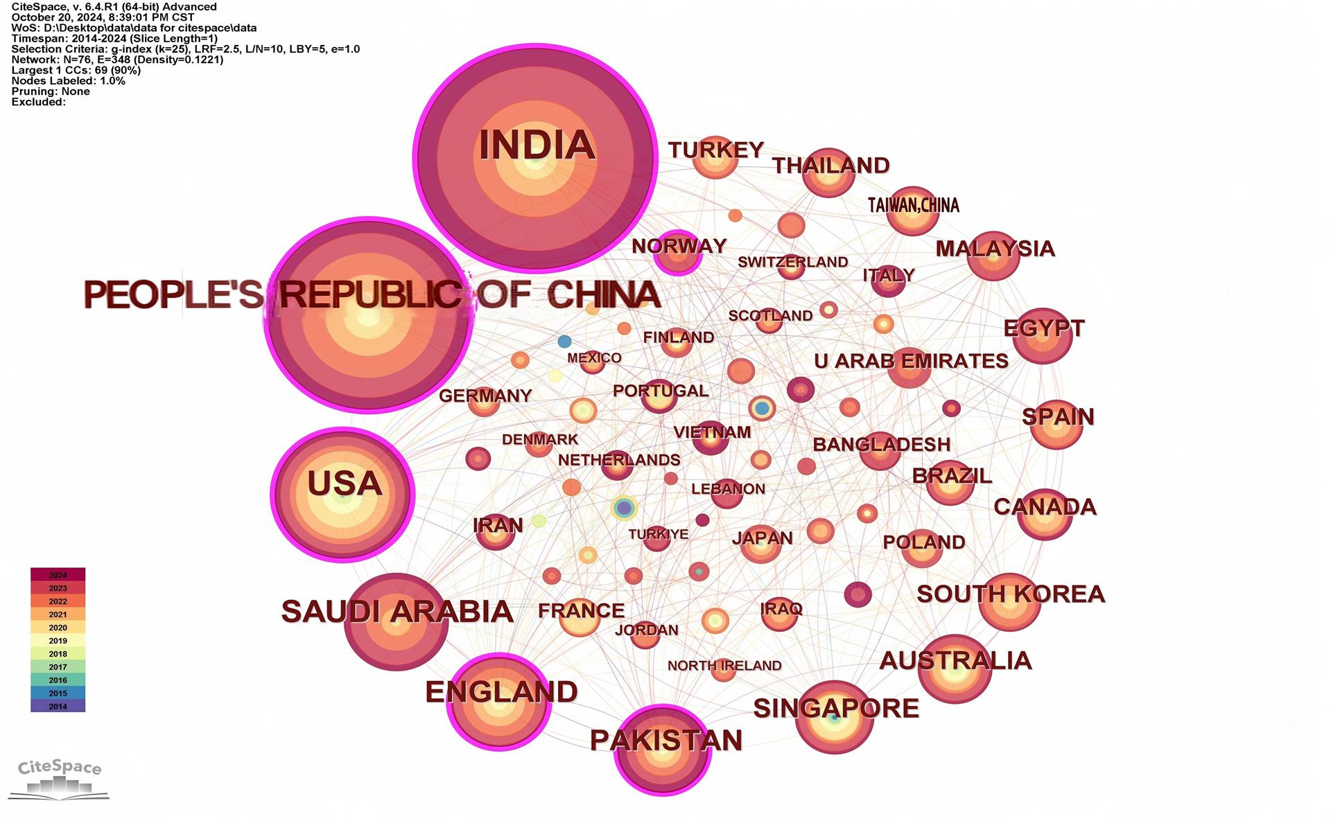
19、‘CiteSpace Home’, CiteSpace. Accessed: Oct. 29, 2024. [Online]. Available: https://citespace.podia.com/‘CiteSpace Home’, CiteSpace. Accessed: Oct. 29, 2024. [Online]. Available: https://citespace.podia.com/
20、%E2%80%98Free%20Online%20Spreadsheet%20Software%3A%20Excel%20%7C%20Microsoft%20365%E2%80%99.%20Accessed%3A%20Oct.%2030%2C%202024.%20%5BOnline%5D.%20Available%3A%20https%3A%2F%2Fwww.microsoft.com%2Fen-us%2Fmicrosoft-365%2Fexcel%3Fmsockid%3D192cdc8e90eb6ff63357ce8f91a86e7a%E2%80%98Free%20Online%20Spreadsheet%20Software%3A%20Excel%20%7C%20Microsoft%20365%E2%80%99.%20Accessed%3A%20Oct.%2030%2C%202024.%20%5BOnline%5D.%20Available%3A%20https%3A%2F%2Fwww.microsoft.com%2Fen-us%2Fmicrosoft-365%2Fexcel%3Fmsockid%3D192cdc8e90eb6ff63357ce8f91a86e7a
21、Gulshan V, Peng L, Coram M, et al. Development and validation of a deep learning algorithm for detection of diabetic retinopathy in retinal fundus photographs. JAMA. 2016, 316(22): 2402-2410. DOI: 10.1001/jama.2016.17216.Gulshan V, Peng L, Coram M, et al. Development and validation of a deep learning algorithm for detection of diabetic retinopathy in retinal fundus photographs. JAMA. 2016, 316(22): 2402-2410. DOI: 10.1001/jama.2016.17216.
22、Vujosevic S, Aldington SJ, Silva P, et al. Screening for diabetic retinopathy: new perspectives and challenges. Lancet Diabetes Endocrinol. 2020, 8(4): 337-347. DOI: 10.1016/S2213-8587(19)30411-5. Vujosevic S, Aldington SJ, Silva P, et al. Screening for diabetic retinopathy: new perspectives and challenges. Lancet Diabetes Endocrinol. 2020, 8(4): 337-347. DOI: 10.1016/S2213-8587(19)30411-5.
23、Li J, Guan Z, Wang J, et al. Integrated image-based deep learning and language models for primary diabetes care. Nat Med. 2024, 30(10): 2886-2896. DOI: 10.1038/s41591-024-03139-8.Li J, Guan Z, Wang J, et al. Integrated image-based deep learning and language models for primary diabetes care. Nat Med. 2024, 30(10): 2886-2896. DOI: 10.1038/s41591-024-03139-8.
24、D. C. Division (DCD), ‘HIPAA & Your Health Rights’. Accessed: Mar. 23, 2025. [Online]. Available: https://www.hhs.gov/programs/hipaa/index.htmlD. C. Division (DCD), ‘HIPAA & Your Health Rights’. Accessed: Mar. 23, 2025. [Online]. Available: https://www.hhs.gov/programs/hipaa/index.html
25、‘General Data Protection Regulation (GDPR) – Legal Text’, General Data Protection Regulation (GDPR). Accessed: Mar. 23, 2025. [Online]. Available: https://gdpr-info.eu/‘General Data Protection Regulation (GDPR) – Legal Text’, General Data Protection Regulation (GDPR). Accessed: Mar. 23, 2025. [Online]. Available: https://gdpr-info.eu/
1、This work was supported by the National Natural Science Foundation of China 62402009), and the Science and Technology Development Fund of Macao under Grant 0013-2024-ITP1.This work was supported by the National Natural Science Foundation of China 62402009), and the Science and Technology Development Fund of Macao under Grant 0013-2024-ITP1. ( )
上一篇
下一篇
目录

点击右上角菜单,浏览器打开下载

我知道了